Review
. 2015:2015:293271.
doi: 10.1155/2015/293271. Epub 2015 Nov 29.

Oxidative Stress in Placenta: Health and Diseases

Affiliations
Review

Oxidative Stress in Placenta: Health and Diseases

Fan Wu et al. Biomed Res Int. 2015.

Abstract

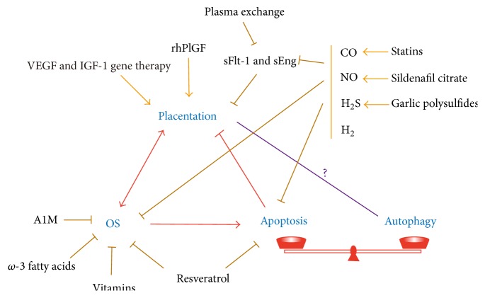
During pregnancy, development of the placenta is interrelated with the oxygen concentration. Embryo development takes place in a low oxygen environment until the beginning of the second trimester when large amounts of oxygen are conveyed to meet the growth requirements. High metabolism and oxidative stress are common in the placenta. Reactive oxidative species sometimes harm placental development, but they are also reported to regulate gene transcription and downstream activities such as trophoblast proliferation, invasion, and angiogenesis. Autophagy and apoptosis are two crucial, interconnected processes in the placenta that are often influenced by oxidative stress. The proper interactions between them play an important role in placental homeostasis. However, an imbalance between the protective and destructive mechanisms of autophagy and apoptosis seems to be linked with pregnancy-related disorders such as miscarriage, preeclampsia, and intrauterine growth restriction. Thus, potential therapies to hold oxidative stress in leash, promote placentation, and avoid unwanted apoptosis are discussed.


Figures

Figure 1
Figure 1
Mechanisms of autophagy. In macroautophagy, the phagophore is induced by the ULK1/2-Atg13-FIP200 (focal adhesion kinase family-interacting protein of 200 kDa) complex and nucleated by class III phosphatidylinositol 3-kinase (PtdIns3K) complex, which is composed of Vps34 (vacuolar protein sorting 34), Beclin1, p150, and Atg14. Thereafter, Atg4 interacts with LC3 (the microtubule-associated protein light chain 3) to form LC3-I (LC3-Gly) [42, 46, 47]. The Atg12-Atg5-Atg16 complex together with Atg3 and Atg7 stimulates LC3-I to bind with phosphatidylethanolamine (PEA) to produce the LC3-PEA complex (LC3-II), during which the autophagosomal membrane begins to extend and enclose to form an integrated autophagosome [42, 46, 47]. Atg9, like a transport cart, circulates to carry membrane materials for the elongation and expansion of the autophagosomal vesicle [46, 47]. In the end, the mature autophagosome docks and fuses with the lysosome where all of its contents are degraded by acid hydrolases [42, 46, 47]. Macroautophagy can sometimes selectively clear ubiquitinated proteins linked with p62, since p62 works with LC3-II to entrap these long-lived proteins into autophagosomes [48]. In other types of autophagy, such as chaperone-mediated autophagy (CMA), lysosomes selectively degrade cytoplasmic proteins with the KFERQ-related motif, which can be recognized by chaperone HSC70 (heat shock cognate protein 70) [42]. LAMP-2A (lysosome-associated membrane protein 2A) then mediates their entry into lysosomes [42].
Figure 2
Figure 2
Autophagic signaling pathways associated with oxidative stress. External OS and other stimuli can activate the PI3K-Akt-mTORC1, Ras-MEK-ERK1/2, and ASK- JUK axis [42, 49, 50]. Activated PI3K recruits PDK1 (phosphoinositide-dependent kinase 1) and phosphorylates Akt, inactivating the TSC1/2 (tuberous sclerosis complex 1/2) and leading to mTORC1 activation [42]. mTORC1 is an energy/redox sensor, the function of which can be blocked by rapamycin [42]. It represses autophagy by phosphorylating Atg13 and separating it from the ULK kinase complex [42]. Elevated cytoplasmic ROS/RNS can be sensed by AMPK, ATM kinase, and PARP-1 [, –53]. AMPK activates autophagy by inhibiting mammalian target of rapamycin complex 1 (mTORC1) and directly activating Unc-51-like kinase 1/2 (ULK1/2) [42, 51]. ATM or PARP-1 are able to activate the LKB1 (liver kinase B1)-AMPK-TSC1/2 metabolic pathway to repress mTORC1 or directly activate ULK1/2 through phosphorylation of its serine groups, upregulating autophagy [52, 53]. As well as OS levels, AMPK also responds to cellular AMP/ATP levels, endoplasmic reticulum (ER) stress, and CaMKKβ (calmodulin-dependent protein kinase kinase-β)-related signaling [42, 46, 51]. p53 exhibits paradoxical autophagic regulation in OS, as nuclear p53 positively enhances autophagy through TSC1/2-dependent pathways, whereas cytoplasmic p53 seems to do the opposite [98]. DAPK, BNIP, Bax, and Puma all function to break down the interaction between Bcl-2/Bcl-xL and Beclin-1, normalizing the essential formation of the class III PtdIns3K complex [64, 101, 102]. UVRAG (ultraviolet irradiation resistance-associated gene) also positively regulates this PtdIns3K complex, while Rubicon counteracts its effect [42].
Figure 3
Figure 3
Crosstalk between autophagy and apoptosis. Many lines of evidence have proven the antiautophagic role of apoptosis. Cytoplasmic p53 inhibits the translation and activation of Atgs, and apoptotic caspases cleave Atgs to render them nonfunctional [97, 98]. However, proapoptotic BH3-only proteins like Bax disrupt the antiautophagic role of Bcl-2/Bcl-xL, resulting in induction of autophagy [64, 101]. Similarly, proapoptotic Mtd/Bok is a powerful autophagy inducer by countering the antiautophagic role of Mcl-1 [103]. Autophagy is thought to be a prosurvival function, but overactivated autophagy accelerates apoptosis by excessively degrading cellular substances [66]. Moreover, Atgs are also said to provide the platform for activation of caspases [100]. In a state of OS, apoptosis and autophagy may occur simultaneously. They may be stimulated separately by OS and at the same time be induced by each other. Thus, their relationship may influence cell fate and lead to different pathophysiological outcomes.
Figure 4
Figure 4
Therapies for OS-related disorders. Placentation and OS are closely interrelated. OS induced apoptosis may destroy normal placentation. At the same time, apoptosis and autophagy are maintained in a delicate balance, although the exact relationship between autophagy and placentation needs further verification. Chemicals or drugs with the potential to rescue pregnancy-related pathologies are listed. Some of them may have overlapping effects.

Similar articles

Cited by

References

    1. Ďuračková Z. Some current insights into oxidative stress. Physiological Research. 2010;59(4):459–469. - PubMed
    1. Burton G. J., Jauniaux E. Oxidative stress. Best Practice and Research: Clinical Obstetrics and Gynaecology. 2011;25(3):287–299. doi: 10.1016/j.bpobgyn.2010.10.016. - DOI - PMC - PubMed
    1. Dröse S., Brandt U. Mitochondrial Oxidative Phosphorylation. Vol. 748. New York, NY, USA: Springer; 2012. Molecular mechanisms of superoxide production by the mitochondrial respiratory chain; pp. 145–169. (Advances in Experimental Medicine and Biology). - DOI - PubMed
    1. Sánchez-Aranguren L. C., Prada C. E., Riaño-Medina C. E., Lopez M. Endothelial dysfunction and preeclampsia: role of oxidative stress. Frontiers in Physiology. 2014;5, article 372 doi: 10.3389/fphys.2014.00372. - DOI - PMC - PubMed
    1. Jiang F., Zhang Y., Dusting G. J. NADPH oxidase-mediated redox signaling: roles in cellular stress response, stress tolerance, and tissue repair. Pharmacological Reviews. 2011;63(1):218–242. doi: 10.1124/pr.110.002980. - DOI - PubMed

Substances

LinkOut - more resources