. 2016 Jul;9(4):1051-64.
doi: 10.1038/mi.2015.122. Epub 2015 Dec 23.

The reproductive cycle is a pathogenic determinant during gonococcal pelvic inflammatory disease in mice

Affiliations

The reproductive cycle is a pathogenic determinant during gonococcal pelvic inflammatory disease in mice

E A Islam et al. Mucosal Immunol. 2016 Jul.

Abstract

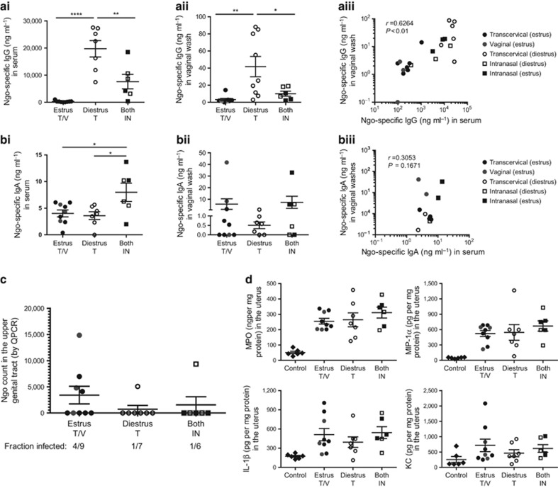
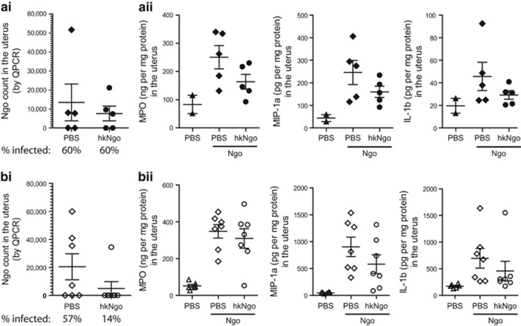
Women with asymptomatic Neisseria gonorrhoeae infection are at risk of developing pelvic inflammatory disease (PID) if the bacteria ascend from the endocervix into the uterus and oviducts. Factors that affect disease severity, ranging from mild discomfort to severe inflammation, pain, and infertility, remain elusive. Herein we perform direct transcervical inoculation of N. gonorrhoeae into the uterus of mice to establish an infection that leads to PID. Profoundly different disease outcomes were apparent at different stages of the reproductive cycle. Mice that were infected during the diestrus stage of the reproductive cycle displayed extensive gonococcal penetration into the submucosa, severe inflammation, and clinical signs reflecting discomfort. Meanwhile, infection during the intervening estrus stage showed only modest effects. Furthermore, a gonococcal-specific humoral response was only elicited following the penetrative upper genital tract (UGT) infection during diestrus but not estrus. Strikingly, the potential for antibodies to contribute to protection during re-infection also depends upon the reproductive stage, as antigonococcal antibodies within the genital tract were markedly higher when mice were in diestrus. Combined, this work establishes a robust new model reflecting gonococcal PID in humans and reveals how the reproductive cycle determines the pathogenic outcome of gonococcal infections of the UGT.


Figures

Figure 1
Figure 1
Differences in gonococcal localization upon transcervical instillation of Neisseria gonorrhoeae (Ngo) into the uterus of female mice. (a) Visualization of inoculums delivered vaginally vs. transcervically using India Ink immediately after delivery. (b) Recovery of Ngo from the upper and lower genital tracts of mice infected either vaginally or transcervically with 108 mix of clinical gonococcal isolates. Mice were either naturally cycling at estrus and diestrus stages for the duration of the infection, or estrusH and diestrusH were induced by administration of β-estradiol and DepoProvera, respectively. Bacterial loads quantified by quantitative PCR (QPCR) amplification of gonococcal DNA 24 h after infection. (c) Comparison of gross anatomy of the genital tract 24 h after transcervically administering phosphate-buffered saline (PBS) and 108 clinical Ngo. Region of interest (dashed line) in the middle panel is magnified in the bottom panel. Black arrows indicate nodes of inflammation. (d) Bacterial localization in infected estrusH and diestrusH tissues at 4 and 24 h after infection. Ngo is stained with an antigonococcal antibody (red) while nuclei are counterstained with DAPI (4,6-diamidino-2-phenylindole; blue). Region of interest (white box) in the top panel is magnified in the middle panel. White dotted line traces the outline of the epithelia, and asterisks denote the lumen. White arrows point at Ngo. Images were representative of at least three animals per time point. (e) Groups of ⩾5 mice were transcervically infected with 107 clinical Ngo isolates during diestrusH, and viable bacterial count was obtained from homogenized upper genital tract and lower genital tract tissues for 3 days. Bars represent mean±s.e.m. CFU, colony-forming unit.
Figure 2
Figure 2
Local and systemic cytokine induction during gonococcal infections. Naturally cycling mice at estrus or diestrus were infected transcervically with 107 MS11 Neisseria gonorrhoeae (Ngo) for 6 and 18 h. Levels of cytokines in the upper genital tract (UGT) and lower genital tract (LGT) homogenates and sera samples were detected by LUMINEX muliplex assays. (a) Heat map showing fold change normalized to uninfected mice (UN) phosphate-buffered saline (PBS) control. (b) Bar graphs showing the levels of select cytokines in the uterus illustrating differences between estrus and diestrus levels at 6 h and 18 h. Mean±s.e.m. plotted on graph, n=3. *P<0.05, ***P<0.001, or ****P<0.0001 as determined by two-way analysis of variance using the GraphPad PRISM 5.0 software. GM-CSF, granulocyte-macrophages colony-stimulating factor; IL, interleukin; IFN, interferon; IP-10, interferon gamma inducible protein 10; KC, keratinocyte chemoattractant; MCP, monocyte chemotactic protein; MIG, monokine induced by gamma-interferon; MIP, macrophage-inflammatory protein; TNF, tumor necrosis factor; VEGF, vascular endothelial growth factor.
Figure 3
Figure 3
Recruitment of neutrophils into the infected tissues and their role in gonococcal clearance. (ai) Myeloperoxidase (MPO) enzyme-linked immunosorbent assays (ELISAs) performed on uterine homogenates as a read out for neutrophil infiltration 24 h after infection with clinical isolates. EstrusH or diestrusH mice received phosphate-buffered saline (PBS) or the indicated dose of Neisseria gonorrhoeae (Ngo) either vaginally or transcervically. Cohort sizes are indicated on graphs. Mean±s.e.m. is plotted for each group. *P<0.05, **P<0.01 in one-way analysis of variance comparing each column to PBS control using Bonferroni's multiple comparison test. (aii) Comparison of MPO levels in the uteri of transcervically infected mice at estrusH or diestrusH using the same data sets as in ai. (b) Mice, synchronized at estrusH or diestrusH by hormone administration, were inoculated transcervically with either PBS or increasing doses of clinical Ngo isolates. Immunohistological staining was performed on uterine and vaginal sections 24 h after infection using anti-Gr-1 antibody to visualize neutrophils (brown). Nuclei are counterstained purple. Images were taken under × 20 magnification. Effect of neutrophil depletion on mice synchronized at (c) estrusH and (d) diestrusH. Mice were either injected with PBS (+PMN) or PMN depleted (−PMN) using anti-Gr-1 antibody clone RB6-8C5. Neutrophil depletion was confirmed by MPO ELISA on homogenized uterine tissue on the left panel of c and d. Representative data from one of two independent experiments has been graphed. Mean±s.e.m. is plotted. **P<0.01 or ***P<0.001, respectively, as determined by Mann–Whitney test on infected samples (uninfected groups were excluded from statistical analysis owing to group sizes). Effect of neutrophil depletion of gonococcal load is plotted on the right panel of c and d. Mice were infected transcervically with 107 clinical Ngo and bacterial load 24 h after infection was determined by quantitative PCR (QPCR). Horizontal bars represent mean±s.e.m. *P<0.1 by Mann–Whitney test. All statistical analyses were performed using the GraphPad PRISM 5.0 software. NS, not significant. PMN, polymorphonuclear neutrophils.
Figure 4
Figure 4
Schematic showing the inflammatory response in the upper and lower genital tracts during different stages of the estrous cycle upon infection with Neisseria gonorrhoeae (Ngo). Early pathological outcome upon gonococcal infection is summarized diagrammatically. The color of the stroma indicates the level of tissue inflammation, where beige is not inflamed and red denotes the presence of inflammatory cytokines. During upper genital tract infections occurring at estrus, Ngo localizes within the uterine lumen with little tissue penetration and mild levels of inflammation. During diestrus, Ngo penetrate into the stroma in large numbers, eliciting a potent inflammatory response that is characterized by neutrophil recruitment. In the lower genital tract, Ngo is found mainly within the vaginal lumen at both estrus and diestrus but is only able to persist during estrus, presumably owing to the lack of inflammation at this stage.
Figure 5
Figure 5
Immunoglobulin G (IgG) response during upper genital tract gonococcal infections. (a) Schematic representation of the experimental setup. The estrous cycle was monitored for 5 days. Mice that entered the first day of estrus or second day of diestrus received 107 clinical Neisseria gonorrhoeae (Ngo) vaginally, transcervically, or intranasally. Serum and vaginal washes were sampled for 21 days, after which all the groups received DepoProvera and were subsequently infected with 107 bacteria. (b) Induction of Ngo-specific IgG in sera (top panel) and vaginal washes (bottom panel) as determined by whole-bacterial enzyme-linked immunosorbent assays for 21days after 1o infection. Mean±s.e.m. plotted on graph. ****P<0.0001 as determined by two-way analysis of variance using Bonferroni post-hoc method comparing means of each treatment to the Vaginal (estrus) group for that particular time point. Group sizes are indicated in the legend. All statistical analyses were performed using the GraphPad PRISM 5.0 software.
Figure 6
Figure 6
Effect of previous gonococcal exposure during recurrent upper genital tract infection. Detection of Neisseria gonorrhoeae (Ngo)-specific (a) immunoglobulin G (IgG) and (b) IgA from previously infected mice prior to secondary challenge (collected on day 33, according to Figure 5a). All mice received DepoProvera treatment to induce diestrusH and were grouped according to stage and method of primary challenge. Estrus T/V, transcervically or vaginally infected during estrus; Diestrus T, transcervically infected during diestrus; Both IN, intranasally infected during estrus or diestrus. Antibody levels in (i) sera and (ii) vaginal washes are shown. Mean±s.e.m. *P<0.05, **P<0.01, and ***P<0.0001, respectively, using one-way analysis of variance and Bonferroni's post-hoc test comparing each group to all other groups. (iii) Scatter plots showing correlation of Ngo-specific Ig in sera to levels found in vaginal washes. Pearson's r correlation values are shown on the graphs. (c) Quantitative PCR (QPCR) quantification of Ngo load in the uterus 24 h after 2o transcervical challenge with 107 of the same strains of Ngo that was used for 1o infection. Mean±s.e.m. is plotted, and the percentage of infected for each group is shown at the bottom. (d) Cytokine levels in homogenized uteri 24 h after 2o infection. Control group contains a mix of previously infected mice that received phosphate-buffered saline (PBS) during 2o challenge. Mean±s.e.m. is plotted. All statistical analyses were performed using the GraphPad PRISM 5.0 software. IL, interleukin; KC, keratinocyte chemoattractant; MCP, monocyte chemotactic protein; MIP, macrophage-inflammatory protein.
Figure 7
Figure 7
Local and systemic immunoglobulin (Ig) responses during parenteral immunization with heat-inactivated gonococci. (a) Schematic representation of the experimental setup. Hormone treatment for inducing estrusH or diestrusH was initiated 2 or 5 days prior to infection, respectively. (b) Neisseria gonorrhoeae (Ngo)-specific IgG, detected by whole-bacterial enzyme-linked immunosorbent assays, following parenteral immunization with 107 heat-inactivated clinical Ngo strains (hk Ngo) in sera (top panel) and vaginal washes (bottom panel). Total number of immunized mice, n=20; phosphate-buffered saline (PBS) controls, n=20. For every time point, mice are grouped according to the stage of the estrous cycle on the sampling day. Group sizes ranged from n=4 to n=16. Mean±s.e.m. plotted. *P<0.05, ***P<0.001, respectively, as calculated by two-way analysis of variance (ANOVA) with Bonferroni post-hoc test comparing each group with every other group. (c) Ngo-specific IgG and (d) IgA in sera (top panel) and vaginal washes (bottom panel) sampled on day 33 after administering β-estradiol or DepoProvera to synchronize mice to estrusH or diestrusH, respectively. Bars represent mean±s.e.m., n=7 for each group. ***P<0.001 using one-way ANOVA with Bonferroni's multiple comparison test. Statistical analysis was performed out using the GraphPad PRISM 5.0 software. NS, not significant.
Figure 8
Figure 8
Effect of parenteral immunization during upper genital tract infections. Mice were immunized with 107 heat-inactivated clinical Neisseria gonorrhoeae (hkNgo) or phosphate-buffered saline (PBS), according to the schematic in Figure 7a. (ai) Bacterial load, (aii) myeloperoxidase (MPO), macrophage-inflammatory protein (MIP)-1α, and interleukin (IL)-β levels 24 h postinfection in the uterus of mice challenged during estrusH. (bi) Bacterial load, (bii) MPO, MIP-1α, and IL-β levels 24 h postinfection in the uterus of mice challenged during diestrusH. Mean±s.e.m. is plotted, n=⩾5 for each group. Bonferroni's multiple comparison test was performed using the GraphPad PRISM 5.0 software. Changes were not statistically significant. QPCR, quantitative PCR.

Similar articles

Cited by

References

    1. Wira, C.R., Fahey, J.V., Sentman, C.L., Pioli, P.A. & Shen, L. Innate and adaptive immunity in female genital tract: cellular responses and interactions. Immunol. Rev. 206, 306–335 (2005). - PubMed
    1. Burgener, A. et al. A systems biology examination of the human female genital tract shows compartmentalization of immune factor expression. J. Virol. 87, 5141–5150 (2013). - PMC - PubMed
    1. Givan, A.L. et al. Flow cytometric analysis of leukocytes in the human female reproductive tract: comparison of fallopian tube, uterus, cervix, and vagina. Am. J. Reprod. Immunol. 38, 350–359 (1997). - PubMed
    1. Pudney, J., Quayle, A.J. & Anderson, D.J. Immunological microenvironments in the human vagina and cervix: mediators of cellular immunity are concentrated in the cervical transformation zone. Biol. Reprod. 73, 1253–1263 (2005). - PubMed
    1. Hook, E.W. 3rd & Holmes, K.K. Gonococcal infections. Ann. Intern. Med. 102, 229–243 (1985). - PubMed

Substances