. 2016 Jun;5(1):111-20.
doi: 10.1007/s40123-015-0043-1. Epub 2015 Dec 22.

Nd:YAG Laser Hyaloidotomy for the Treatment of Acute Subhyaloid Hemorrhage: A Comparison of Two Cases

Affiliations

Nd:YAG Laser Hyaloidotomy for the Treatment of Acute Subhyaloid Hemorrhage: A Comparison of Two Cases

Jens Heichel et al. Ophthalmol Ther. 2016 Jun.

Abstract

Introduction: Subhyaloid hemorrhage, whether spontaneous or in the context of a Valsalva maneuver, can lead to an acute decrease in vision when located in the premacular region. Nd:YAG laser hyaloidotomy (NYLH) is a minimally invasive treatment option.

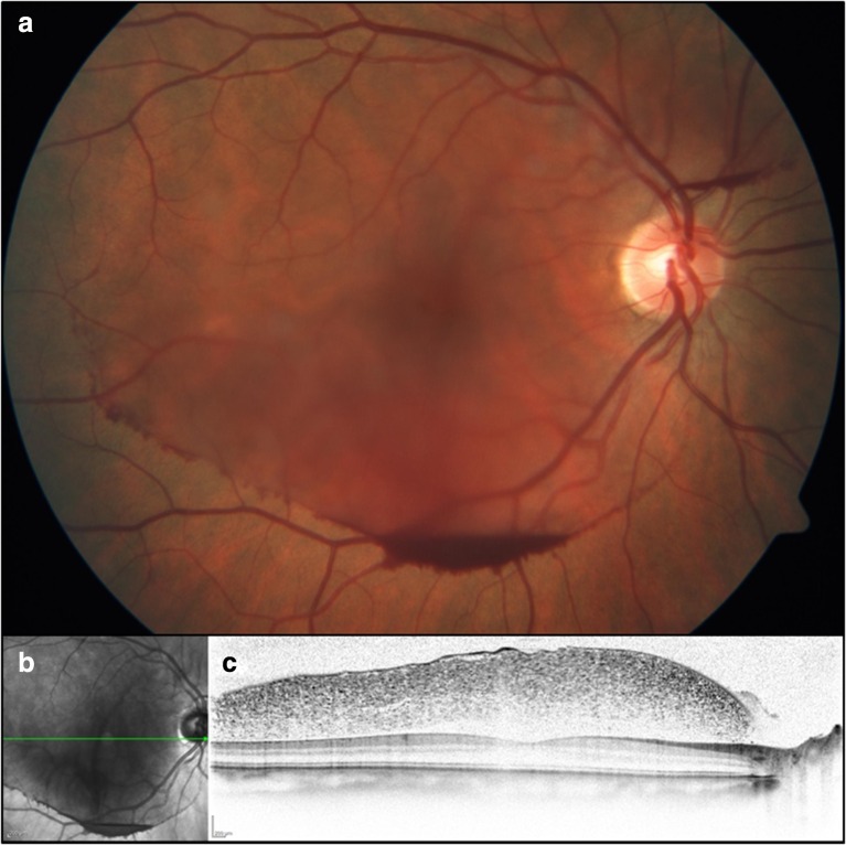
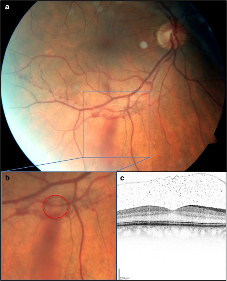
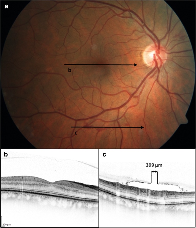
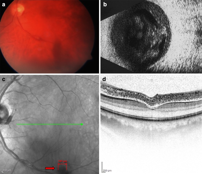
Methods: We examined two different clinical courses based on two case reports of NYLH. One case report described a 52-year-old female patient who presented with a painless decrease of vision to 20/200. The fundoscopy verified a subhyaloid premacular hemorrhage. The precipitating event for the hemorrhage could not be determined, and a NYLH was performed 5 days after the event. The other patient was a 48-year-old man who suffered an acute visual decrease (hand motion) after developing a migraine with vomiting. Fundoscopy showed a dense subhyaloid premacular hemorrhage. NYLH was performed 1 day after the hemorrhage. These clinical courses were documented based on fundus photographs, ultrasounds, and spectral-domain optical coherence tomography (SD-OCT).

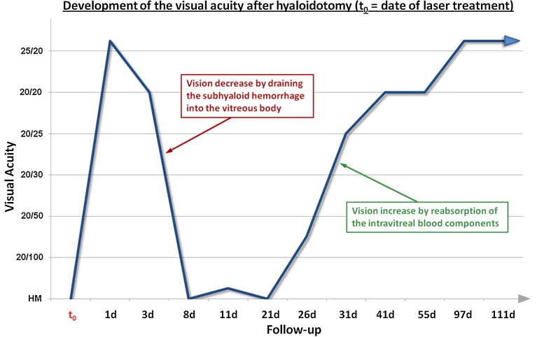
Results: In both cases, there was an effect with approximately 2.2 mJ of laser energy. In the female patient, we observed a gradual but constant increase in vision. After 4 weeks, her vision improved to 20/20. In the male patient, the vision increased to 25/20 1 day after treatment. However, his vision returned to hand motion as he developed a diffuse vitreous opacification. Because of delayed reabsorption, vitrectomy was considered. Since the optical axis was clear with good vision, we decided against this surgery. Complete reabsorption took more than 3 months.

Conclusion: After NYLH for subhyaloid hemorrhage, pronounced vitreous body opacification could develop despite a rapid increase in vision, and requires close monitoring by the surgeon. Fundus photography and SD-OCT are suitable means for clinical course evaluations.

Keywords: Nd:YAG laser; Nd:YAG laser hyaloidotomy; Premacular subhyaloidal bleeding; Valsalva maneuver; Vitreous hemorrhage.


Figures

Fig. 1
Fig. 1
Central fundus at first visit. a Fundus photography: typical “blood level” in the area of the inferior temporal vascular arch. b Infrared photograph (green line marks the respective localization of the SD-OCT image of the c). c Spectral-domain optical coherence tomography shows a subhyaloid accumulation of blood. The macula shows a normal configuration
Fig. 2
Fig. 2
Inferior temporal vascular arch 3 days after laser treatment. a Fundus photography shows a reduction in the premacular hemorrhage as well as remaining streaks of blood at the inferior temporal vessel arch. b Magnified fundus photograph with representation of the hyaloidotomy (red circle). c Spectral-domain optical coherence tomography of the macula: clear reduction of the subhyaloid opacification
Fig. 3
Fig. 3
Central fundus 4 weeks after laser treatment. a Fundus photography: discrete bleeding remained within the area of the inferior temporal vascular arch (the lines mark the respective localizations of the SD-OCT images of the b and c. b SD-OCT of the macula: no subhyaloid opacifications were detectable. c SD-OCT of the hyaloidotomy: a 399-µm rupture of the posterior hyaloid membrane is recognizable. SD-OCT spectral-domain optical coherence tomography
Fig. 4
Fig. 4
Image documentations 1 week after surgery. a Fundus photography: streaky intravitreal blood can be detected that originated from the inferior temporal vascular arch. b Ultrasound B image: hemorrhage with subhyaloidal and intravitreal portions. c Infrared image: in the area of the laser application, a rupture of approximately 747 µm in size is seen; green line shows the localization of the SD-OCT image of Fig. 5d. d SD-OCT of the foveal region: a dense subhyaloidal hemorrhage is recognizable, and the retina appears to be normal. SD-OCT spectral-domain optical coherence tomography
Fig. 5
Fig. 5
Development of visual acuity over the course of time after hyaloidotomy. Operation at time t 0; afterwards, visual acuity increased rapidly. After approximately 1 week, the visual acuity dropped again to hand motions (HM), since the blood had drained into the vitreous body space. In the further 4 weeks, constant reabsorption of the blood with a corresponding increase in vision was seen. The presumed initial vision of 25/20 was only reached after approximately 3 months

Similar articles

Cited by

References

    1. Cleary PE, Kohner EM, Hamilton AM, Bird AC. Retinal macroaneurysms. Br J Ophthalmol. 1975;59:355–361. doi: 10.1136/bjo.59.7.355. - DOI - PMC - PubMed
    1. O’Hanley GP, Canny CL. Diabetic dense premacular hemorrhage. A possible indication for prompt vitrectomy. Ophthalmology. 1985;92:507–511. doi: 10.1016/S0161-6420(85)34014-9. - DOI - PubMed
    1. Iijima H, Satoh S, Tsukahara S. Nd:YAG laser photodisruption for preretinal hemorrhage due to retinal macroaneurysm. Retina. 1998;18:430–434. doi: 10.1097/00006982-199805000-00008. - DOI - PubMed
    1. Rennie CA, Newman DK, Snead MP, Flanagan DW. Nd:YAG laser treatment for premacular subhyaloid haemorrhage. Eye (Lond) 2001;15:519–524. doi: 10.1038/eye.2001.166. - DOI - PubMed
    1. Duane TD. Valsalva hemorrhagic retinopathy. Trans Am Ophthalmol Soc. 1972;70:298–313. - PMC - PubMed

LinkOut - more resources