Review
. 2015 Dec 18;20(12):22718-56.
doi: 10.3390/molecules201219865.

Unfolded Protein Response and Macroautophagy in Alzheimer's, Parkinson's and Prion Diseases

Affiliations
Review

Unfolded Protein Response and Macroautophagy in Alzheimer's, Parkinson's and Prion Diseases

Irina Milisav et al. Molecules. .

Abstract

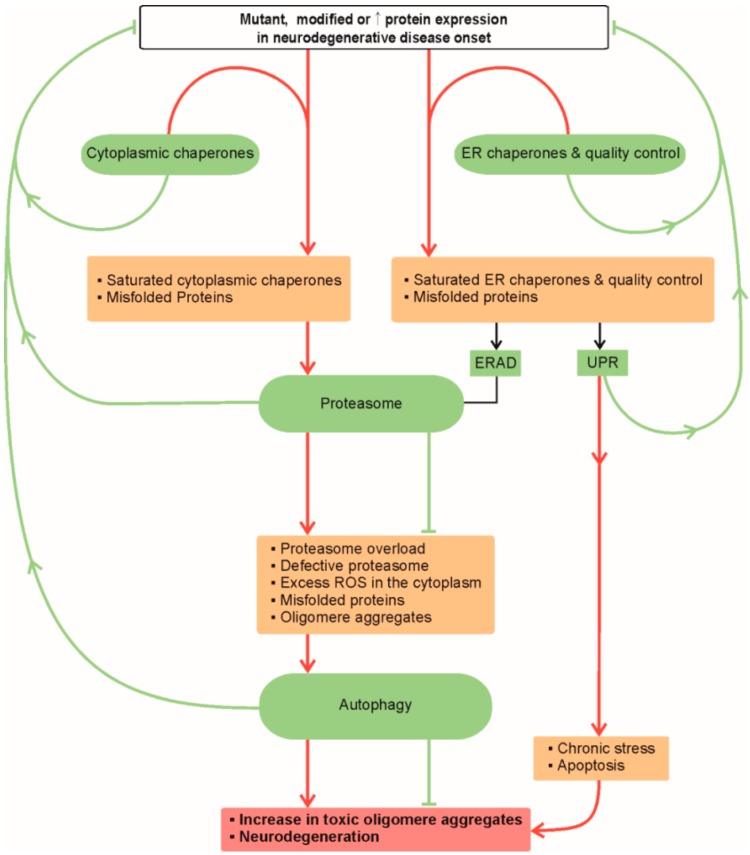
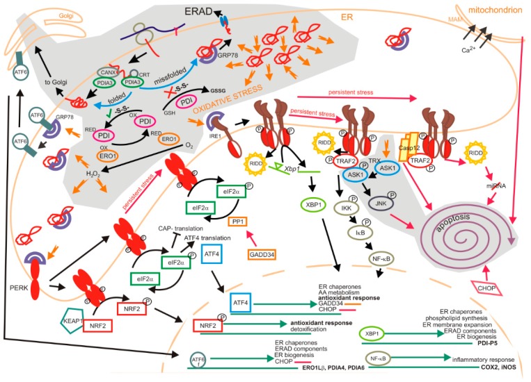
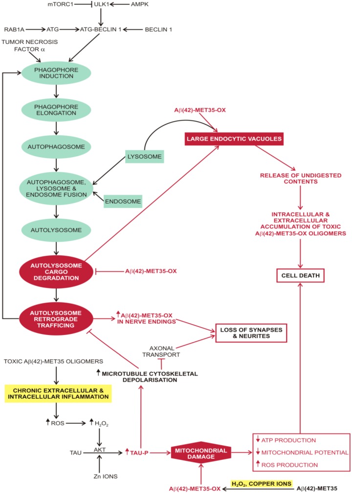
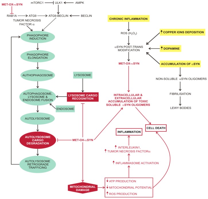
Proteostasis are integrated biological pathways within cells that control synthesis, folding, trafficking and degradation of proteins. The absence of cell division makes brain proteostasis susceptible to age-related changes and neurodegeneration. Two key processes involved in sustaining normal brain proteostasis are the unfolded protein response and autophagy. Alzheimer's disease (AD), Parkinson's disease (PD) and prion diseases (PrDs) have different clinical manifestations of neurodegeneration, however, all share an accumulation of misfolded pathological proteins associated with perturbations in unfolded protein response and macroautophagy. While both the unfolded protein response and macroautophagy play an important role in the prevention and attenuation of AD and PD progression, only macroautophagy seems to play an important role in the development of PrDs. Macroautophagy and unfolded protein response can be modulated by pharmacological interventions. However, further research is necessary to better understand the regulatory pathways of both processes in health and neurodegeneration to be able to develop new therapeutic interventions.

Keywords: Alzheimer’s disease; Parkinson’s disease; autophagy; neurodegeneration; prion diseases; proteostasis; unfolded protein response.


Conflict of interest statement

The authors declare no conflict of interest.

Figures

Figure 1
Figure 1
Proteostasis in neurodegeneration. Red lines and arrows indicate progressive failure of proteostasis ultimately leading to neurodegeneration. Green arrows and lines indicate appropriate response of proteostasis to altered proteins that prevents or slow-downs the progress of neurodegeneration. Abbreviations: ER (endoplasmic reticulum); ERAD (endoplasmic reticulum associated protein degradation); ROS (reactive oxygen species); UPR (unfolded protein response).
Figure 2
Figure 2
Basic mechanisms of unfolded protein response. There are three phases of UPR. The adaptive, transition (black arrows) and the late phase (red arrows); the last two phases occur if the stress is not resolved and there is prolonged stress. Protein load in the ER is decreased during the early adaptive phase. ATF6, PERK and IRE1 are the three classes of main ER stress sensors that respond to the levels of unfolded/misfolded proteins in the ER. They are activated by the dissociation of GRP78/BiP upon increased amounts of misfolded proteins or direct binding of unfolded proteins in the case of yeast Ire1p and possibly IRE1β. Then the cell survival or cell death pathways are conveyed through complex parallel and convergent signal transduction pathways. Impact of oxidative stress on protein folding and unfolded protein response is depicted on the grey background. Protein disulphate isomerases (PDIs) assist in folding of nascent chains by catalization of disulphide bond formation (top left). For example, PDIA3 or Erp57 is expressed in many tissues, including liver, pancreas, kidney, placenta and lungs and to lower extent in heart, skeletal muscle and brain.. Its activity is enhanced when it is in complex with calnexin (CANX) and calreticulin (CRT). The proper formation of disulphide bonds between two cysteine residues in nascent proteins is necessary for correct protein folding. Two cysteines in the active site of PDI accept two electrons from the cysteines of the folding polypeptide. PDI is then oxidized by oxidoreduction 1 (ERO1) proteins that subsequently transfer the electrons to oxygen to produce H2O2. Improperly paired disulphide bonds are reduced by PDI, while producing the oxidized glutathione. Misfolded proteins are exported to the cytosol to be degraded by the proteasome in the process ER-associated degradation (ERAD). ROS produced during protein folding and the decrease of GSH upon the reduction of improperly paired disulphide bonds of misfolded proteins may shift the redox balance towards the oxidative stress. Likewise, oxidative stress is the consequence of excess of misfolded proteins. Upon oxidative stress, all three ER stress sensors, ATF6, PERK and IRE1α are activated. Apoptosis signal-regulating kinase (ASK1) is activated by oxidative stress, ER stress and inflammation (e.g., TNFα). It dissociates from (oxidized) thioredoxin (Trx) and binds TRAF2 to convey the apoptosis signal through JNK activation.
Figure 3
Figure 3
Macroautophagy and oxidated Aβ. Oxidised Aβ peptides attenuate macroautophagy and mitochondrial function. This toxic effect is further exacerbated by the formation of soluble Aβ oligomers that stimulate chronic inflammation with increased production of ROS. A positive feedback loop develops between chronic inflammation and the production of oxidised Aβ peptides, leading to loss of synapses and neurites and ultimately cell death. Abbreviations: ⊥ (attenuation); ↓ (decreased); ↑ (increased); Aβ42-MET35 (soluble amyloid β-peptide with a single methionine residue at position 35); Aβ42-MET35-OX (soluble amyloid β-peptide with a single oxidised methionine residue at position 35); AKT (protein kinase B); AMPK (5’ AMP-activated protein kinase); ATG (autophagy-related protein); ATP (adenosine triphosphate); Copper ions (copper ions in different oxidative states from +1 to +4); mTORC1 (mammalian target of rapamycin complex 1); RAB1A (Ras-related protein Rab-1A); ROS (reactive oxygen species); TAU (tau protein); TAU-P (phosphorylated tau protein); TNFα (tumour necrosis factor alfa/cachexin); ULK1 (serine/threonine-protein kinase).
Figure 4
Figure 4
Macroautophagy and oxidised αSYN. Several processes, e.g., chronic inflammation, copper ion deposition, accumulation of dopamine or even just intracellular accumulation of αSYN promote oxidation of αSYN. Oxidised αSYN attenuates macroautophagy and mitochondrial function. This toxic effect is further exacerbated by the formation of soluble and autophagy resistant αSYN oligomers that stimulate chronic inflammation with increased production of ROS. A positive feedback loop develops between chronic inflammation and the production of soluble αSYN oligomers, leading to cell death. Abbreviations: ⊥ (attenuation); ↓ (decreased); ↑ (increased); αSYN (alpha-synuclein); ATG (autophagy-related protein); ATP (adenosine triphosphate production); Copper ions (copper ions in different oxidative states); MET-OX-αSYN (alpha-synuclein oxidised on methionine residues); mTORC1 (mammalian target of rapamycin complex 1); POST-TRANS (post-translational) RAB1A (Ras-related protein Rab-1A); ROS (reactive oxygen species); TNFα (tumour necrosis factor alfa/cachexin); ULK1 (serine/threonine-protein kinase).
Figure 5
Figure 5
Macroautophagy of oxidated PrPSc. The predisposing factors for the transformation of the native prion protein (PrPc) into the infectious, self-propagating prion protein form (PrPSc) are chronic brain inflammation and copper ion deposition. They promote many post translational modifications of PrPc, including oxidation of its numerous methionine residues, to the prion protein in molten globule form (met-ox-PrPc). Further post translational modifications of met-ox-PrPc can also include a transformation into an oxidised and self-propagating infectious isoform of prion protein (met-ox-PrPSc). The met-ox-PrPSc is resistant to autophagy; attenuates autolysosome cargo degradation and also promotes the formation of large endocytic vacuoles that tend to release their undigested contents, including enzymes, into the cytosol. An intracellular increase in met-ox-PrPSc, due to its resistance to removal by autophagy and propensity for self-propagation, leads to an intracellular reduction of PrPc. The intracellular reduction of PrPc changes the cell’s global oxidative-redox balance which is reflected in mitochondrial damage, a contributing factor to the met-ox-PrPSc induced apoptosis. Although chronic brain inflammation seems to be important for initiating the process of PrPSc production, it is not necessary to sustain it, since the PrPSc only needs the PrPc molecules for its propagation. Abbreviations: ⊥ (attenuation); ↓ (decreased); ↑ (increased); AMPK (5′ AMP-activated protein kinase); ATG (autophagy-related protein); ATP (adenosine triphosphate); Copper ions (copper ions in different oxidative states from +1 to +4); MET-OX-PRPc (oxidised prion protein in molten globule form); MET-OX-PRPSc (oxidised and self-propagating prion protein); mTORC1 (mammalian target of rapamycin complex 1); POST-TRANS-M (post-translational modification); POST-TRANS-MM (post-translational modifications); PrPc (normal form of prion protein); PrPSc (infectious isoform of prion protein); ROS (reactive oxygen species); SIRT1 (NAD-dependent deacetylase sirtuin-1); ULK1 (serine/threonine-protein kinase).

References

    1. Hetz C., Mollereau B. Disturbance of endoplasmic reticulum proteostasis in neurodegenerative diseases. Nat. Rev. Neurosci. 2014;15:233–249. doi: 10.1038/nrn3689. - DOI - PubMed
    1. Brown M.K., Naidoo N. The endoplasmic reticulum stress response in aging and age-related diseases. Front. Physiol. 2012;3:263. doi: 10.3389/fphys.2012.00263. - DOI - PMC - PubMed
    1. Lipinski M.M., Zheng B., Lu T., Yan Z., Py B.F., Ng A., Xavier R.J., Li C., Yankner B.A., Scherzer C.R., et al. Genome-wide analysis reveals mechanisms modulating autophagy in normal brain aging and in Alzheimer’s disease. Proc. Natl. Acad. Sci. USA. 2010;107:14164–14169. doi: 10.1073/pnas.1009485107. - DOI - PMC - PubMed
    1. Roussel B.D., Kruppa A.J., Miranda E., Crowther D.C., Lomas D.A., Marciniak S.J. Endoplasmic reticulum dysfunction in neurological disease. Lancet Neurol. 2013;12:105–118. doi: 10.1016/S1474-4422(12)70238-7. - DOI - PubMed
    1. Heiseke A., Aguib Y., Schatzl H.M. Autophagy, prion infection and their mutual interactions. Curr. Issues Mol. Biol. 2010;12:87–97. - PubMed

Publication types