. 2016 Apr;37(4):371-84.
doi: 10.1002/humu.22948. Epub 2016 Jan 14.

Sporadic and Familial Congenital Cataracts: Mutational Spectrum and New Diagnoses Using Next-Generation Sequencing

Affiliations

Sporadic and Familial Congenital Cataracts: Mutational Spectrum and New Diagnoses Using Next-Generation Sequencing

Alan S Ma et al. Hum Mutat. 2016 Apr.

Abstract

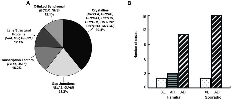
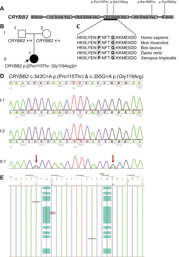
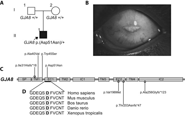
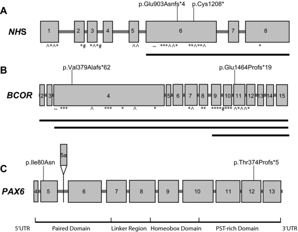
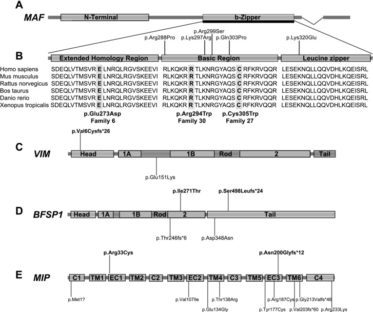
Congenital cataracts are a significant cause of lifelong visual loss. They may be isolated or associated with microcornea, microphthalmia, anterior segment dysgenesis (ASD) and glaucoma, and there can be syndromic associations. Genetic diagnosis is challenging due to marked genetic heterogeneity. In this study, next-generation sequencing (NGS) of 32 cataract-associated genes was undertaken in 46 apparently nonsyndromic congenital cataract probands, around half sporadic and half familial cases. We identified pathogenic variants in 70% of cases, and over 68% of these were novel. In almost two-thirds (20/33) of these cases, this resulted in new information about the diagnosis and/or inheritance pattern. This included identification of: new syndromic diagnoses due to NHS or BCOR mutations; complex ocular phenotypes due to PAX6 mutations; de novo autosomal-dominant or X-linked mutations in sporadic cases; and mutations in two separate cataract genes in one family. Variants were found in the crystallin and gap junction genes, including the first report of severe microphthalmia and sclerocornea associated with a novel GJA8 mutation. Mutations were also found in rarely reported genes including MAF, VIM, MIP, and BFSP1. Targeted NGS in presumed nonsyndromic congenital cataract patients provided significant diagnostic information in both familial and sporadic cases.

Keywords: congenital cataract; eye; microcornea; microphthalmia; next-generation sequencing.


Figures

Figure 1
Figure 1
Genes and inheritance patterns identified with NGS in apparently nonsyndromic sporadic and familial congenital cataract cases. A: Mutations were found in 17 different genes in apparently nonsyndromic sporadic and familial congenital cataract cases. These were in genes that encoded crystallins, gap junction, transcription factor, other structural proteins, and X‐linked syndromal proteins. The relative proportions are illustrated in this diagram. B: Mutation‐positive cases in familial and sporadic cases and the inheritance patterns. The 16 mutation‐positive familial cases comprised two X‐linked (XL, dotted), three autosomal‐recessive (AR, horizontal stripes), and 11 autosomal‐dominant (AD, diagonal stripes) diagnoses. In the sporadic cases with mutations, the inheritance pattern was revised to two cases of de novo X‐linked mutations in BCOR, and 15 de novo AD cases, including the father from Family 6 who had a separate genetic answer (mutation in MAF) to his two affected sons with NHS.
Figure 2
Figure 2
Two novel missense CRYBB2 mutations on the one allele in proband in Family 35, and other CRYBB2 mutations found in this study. A: This schematic shows the encoded domain structure of CRYBB2 (NM_000496.2). This protein contains four “Greek Key” domains between the N and C terminals. Mutations found in this study are illustrated above the schematic, with the two novel missense p.(Pro115Thr) and p.(Gly119Arg) found in Family 35, p.(Ser186Pro) found in Family 13, and p.(Trp195Gly) in Family 37. B: Pedigree from Family 35, with proband highlighted in II:1. C: Protein sequence alignments show that both affected residues, highlighted in gray, are in a highly conserved part of the third Greek Key domain. D: The proband had two de novo missense CRYBB2 mutations, confirmed on Sanger sequencing (red arrows). E: Next‐generation reads reveal that the variants (highlighted in blue) were both consistently in the same reads, showing that they are in cis.
Figure 3
Figure 3
GJA8 mutation in Family 45 caused a severe cataract, sclerocornea, microphthalmia phenotype, and other GJA8 mutations found in this study. A: Pedigree of family 45, with the proband (II:1) highlighted. He was found to have a heterozygous missense mutation in the first extracellular domain (EC1) of GJA8, p.(Asp51Asn), and has the most severe GJA8 ocular phenotype reported to date. B: Left eye image of the proband in Family 45, showing his severe microphthalmia and sclerocornea. C: The schematic shows the encoded domain structure of GJA8 (NM_005267.4). This protein spans the cellular membrane and has a signal peptide (SP), four transmembrane (TM), two extracellular (EC), and two intracellular (IC) domains. Mutations found in GJA8 in this study are shown above the schematic, including the p.(Asp51Asn) mutation in Family 45 in the EC1 domain. The p.(Ile31Hisfs*18) recessive mutation found in Family 14 is the earliest frameshift mutation yet reported in this gene. Other recessive mutations reported in the literature are shown below the gene, and the star highlights recessive mutations. The p.(Ala40Val) and p.(Trp45Ser) heterozygous mutations were found in Families 2 and 20, respectively. D: Protein sequence alignments show that the affected residue in the proband of Family 45, highlighted in gray, lies in a highly conserved region of the EC1 domain.
Figure 4
Figure 4
Phenotypic variability of the cataracts and associated ocular phenotypes in this study. This figure shows images of congenital cataracts and other ocular features found in this study and highlights the diverse heterogeneity of congenital cataracts, as well as the importance of accurate phenotyping, fundoscopy, and electrophysiology to clarify the diagnosis. Images are from families diagnosed with mutations in NHS, Families 6(A) and 40(B‐E); PAX6, Families 10(F) and 18(G‐R); MAF, father from Family 6(S&T); MIP, Family 32(U); and VIM, family 25(V). Image A displays the right sided cortical cataract from the proband (II:1) from Family 6, diagnosed with NHS. The other family with NHS (Family 40) is represented in images BE. B: Right eye from proband (III:3, Family 40), with postsurgical changes following cataract extraction. The variable features demonstrated in obligate carrier females from this family are demonstrated in C, with subcapsular cataract from the mother (II:3) and D (right) and E (left) coralliform cataracts in the sister (III:1). Image F: Cortical congenital cataracts of proband (II:1) from Family 10, diagnosed with a missense PAX6 mutation in the paired domain. Images from Family 18, also diagnosed with a PAX6 mutation, are represented in images GR, revealing the panocular features of PAX6‐related disease. These are from the proband (III:3) showing: G: anterior polar cataract; J: mild limbal stem cell failure with peripheral corneal pannus; M: altered macular reflex (*); P: OCT of foveal hypoplasia (red bracket) with loss of the foveal pit. The mother in Family 18 (II:2) also demonstrates multiple PAX6‐associated abnormalities with: H: limbal stem cell failure with increased corneal pannus; K: mild iris hypoplasia and mild ectropion uvea (arrow); N: reduced macular reflex (*); Q: OCT showing foveal hypoplasia with loss of the foveal pit (red bracket). The sister in Family 18 (III:2) also has: I: anterior polar cataract; L: ectropion uvea (arrow); O: reduced macular reflex (*); and R: OCT showing loss of foveal pit (red bracket). S (right) and T (left): Cerulean blue dot cataracts from the father (I:1) from Family 6, found to have a novel missense MAF mutation. U: Lamellar cataract of proband (II:1) in Family 32, found to have a novel frameshift mutation in MIP. V: The image demonstrates bilateral total cataracts as they appeared on presentation at 11 months of age in the proband of Family 25 (II:1), found to have a novel frameshift mutation in VIM.
Figure 5
Figure 5
Mutations in NHS and BCOR revealed syndromic conditions, and PAX6 mutations indicated more complex ocular phenotypes. A: Exonic structure of NHS (NM_198270.3) with previously reported mutations marked by symbols below the gene. KEY: ^, nonsense; *, frameshift; #, splice site; ∼, missense mutations. The solid black lines delineate reported exonic deletions. Mutations found in this study are demonstrated above the gene, from Families 6 and 40. B: Exonic structure of BCOR (NM_001123385.1) with previously reported mutations marked by symbols below the gene. KEY: ^, nonsense; *, frameshift; #, splice site; ∼. missense mutations. The solid black lines delineate reported exonic deletions. Mutations found in this study are demonstrated above the gene, from Families 44 and 7. C: Exonic and protein domain structure of PAX6 (NM_0001604.4). The exons are numbered, with the alternatively spliced exon 5a highlighted above the gene. Below the gene, the protein domains are highlighted (UTR, untranslated region; PST, Proline, Serine, Threonine rich domain). The two mutations found in this study, from Families 18 and 10, are marked above the gene. Due to the large number of mutations found in this gene, they are not illustrated in the figure. A curated database of PAX6 mutations can be found at LOVD (http://www.lovd.nl/PAX6).
Figure 6
Figure 6
Novel mutations identified in MAF, VIM, BFSP1, and MIP. A: Schematic showing the encoded structure of MAF (NM_001031804.2). MAF has an N‐terminal and b‐zipper domain, and the detailed section of b‐ZIP reveals three evolutionarily conserved subdomains with the EHR, basic and leucine zipper regions. B: The b‐zipper domain is expanded to reveal these three subregions. Previously reported mutations in the basic and leucine zipper regions are demonstrated above the figure. The extensive amino acid conservation of the b‐zipper region is revealed, and the highly conserved three new mutations found in this study in MAF, highlighted in gray, from Families 6, 30, and 27. C: Schematic of encoded structure of VIM (NM_003380.3), which comprises a head, rod, and tail domain. The rod domain has three coil subdomains; 1A, 1B, and 2. A novel frameshift mutation was found in the proband of Family 25 (II:1), in the head domain (labeled above head domain). The only other mutation, a heterozygous missense mutation in the rod domain, is also marked, below the rod domain. D: Schematic of encoded structure of BFSP1 (NM_001195.4), which comprises a head, rod, and tail domain. Similar to VIM, the rod domain of BFSP1 also has coil subdomains 1A, 1B, and 2. Compound heterozygous mutations were found in the proband from Family 46, highlighted above the second coil and tail domains. The other mutations reported in the literature are labeled below the gene; a recessive frameshift mutation in the rod domain, and a heterozygous missense mutation in the tail domain in an autosomal‐dominant family. E: Schematic of encoded structure of MIP (NM_012064.3), which is a protein with four cytoplasmic (C) domains, six transmembrane (TM) domains, and three extracellular (EC) domains. The two mutations found in this study from Families 28 and 32 are illustrated above the gene, along with other mutations reported in the literature to date below the gene.

Similar articles

Cited by

References

    1. Abecasis GR, Auton A, Brooks LD, DePristo MA, Durbin RM, Handsaker RE, Kang HM, Marth GT, McVean GA. 2012. An integrated map of genetic variation from 1,092 human genomes. Nature 491:56–65. - PMC - PubMed
    1. Adzhubei IA, Schmidt S, Peshkin L, Ramensky VE, Gerasimova A, Bork P, Kondrashov AS, Sunyaev SR. 2010. A method and server for predicting damaging missense mutations. Nat Methods 7:248–249. - PMC - PubMed
    1. Bateman JB, von‐Bischhoffshaunsen FR, Richter L, Flodman P, Burch D, Spence MA. 2007. Gene conversion mutation in crystallin, beta‐B2 (CRYBB2) in a Chilean family with autosomal dominant cataract. Ophthalmology 114:425–432. - PubMed
    1. Bennett TM, Mackay DS, Knopf HL, Shiels A. 2004. A novel missense mutation in the gene for gap‐junction protein alpha3 (GJA3) associated with autosomal dominant "nuclear punctate" cataracts linked to chromosome 13q. Mol Vis 10:376–382. - PubMed
    1. Beyer EC, Ebihara L, Berthoud VM. 2013. Connexin mutants and cataracts. Front Pharmacol 4:43. - PMC - PubMed

Publication types

MeSH terms