. 2016 Feb 19;291(8):3821-36.
doi: 10.1074/jbc.M115.680983. Epub 2015 Dec 22.

Cyclic AMP-elevating Agents Promote Cumulus Cell Survival and Hyaluronan Matrix Stability, Thereby Prolonging the Time of Mouse Oocyte Fertilizability

Affiliations

Cyclic AMP-elevating Agents Promote Cumulus Cell Survival and Hyaluronan Matrix Stability, Thereby Prolonging the Time of Mouse Oocyte Fertilizability

Monica Di Giacomo et al. J Biol Chem. .

Abstract

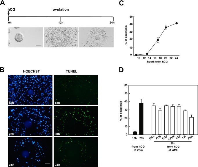
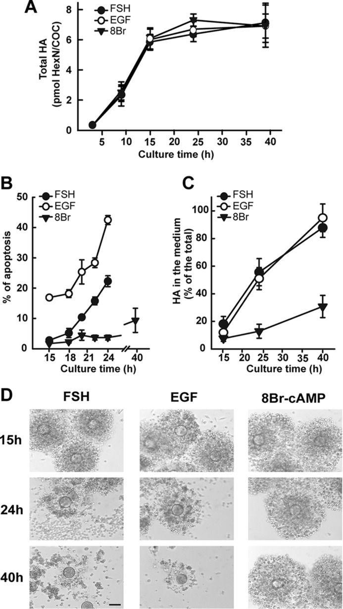
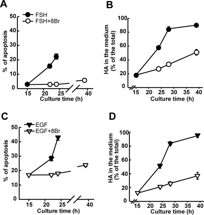
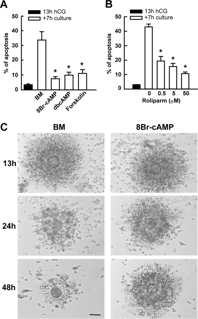
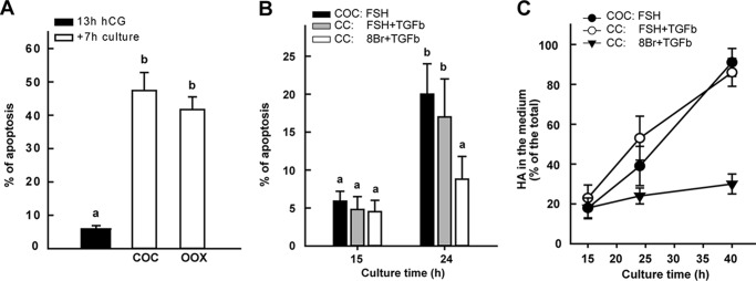
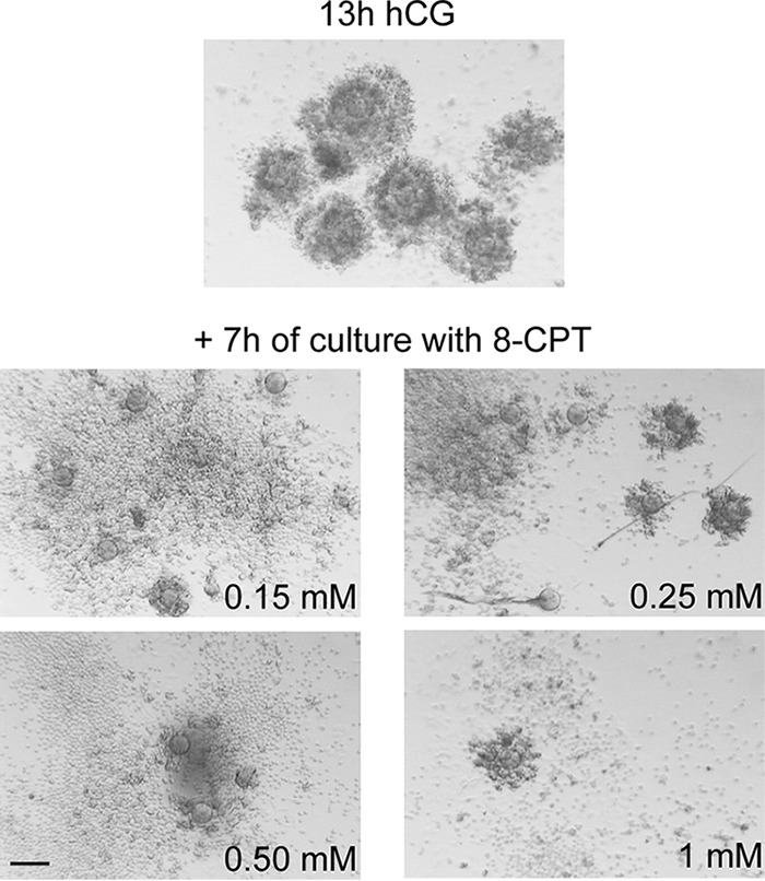
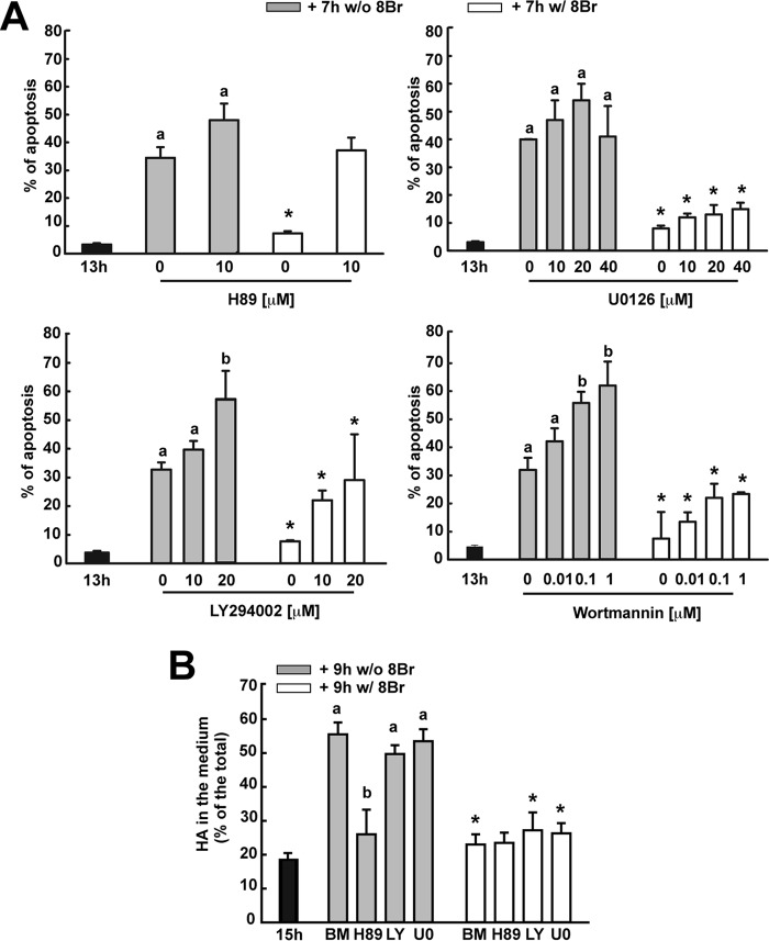
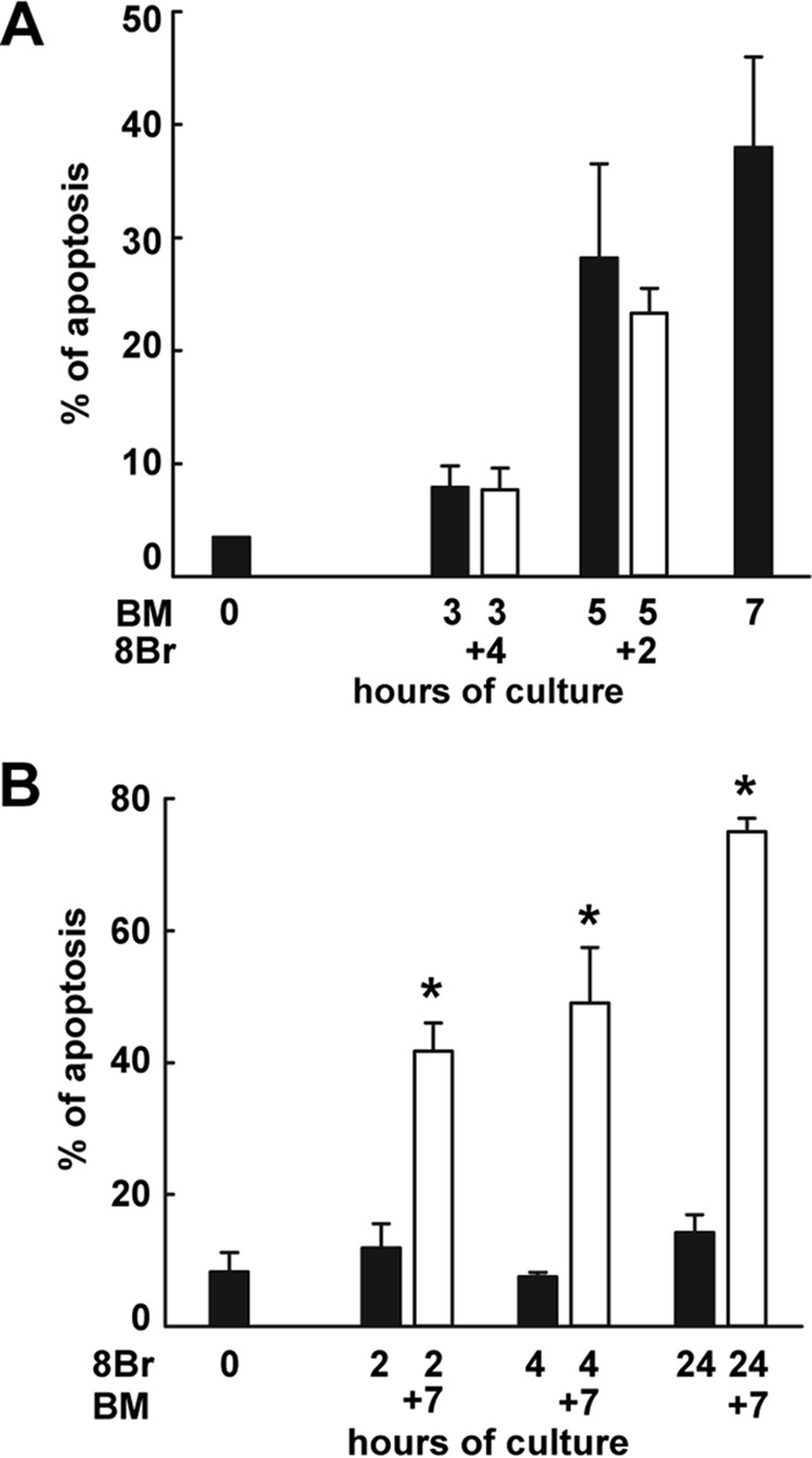
Cumulus cells sustain the development and fertilization of the mammalian oocyte. These cells are retained around the oocyte by a hyaluronan-rich extracellular matrix synthesized before ovulation, a process called cumulus cell-oocyte complex (COC) expansion. Hyaluronan release and dispersion of the cumulus cells progressively occur after ovulation, paralleling the decline of oocyte fertilization. We show here that, in mice, postovulatory changes of matrix are temporally correlated to cumulus cell death. Cumulus cell apoptosis and matrix disassembly also occurred in ovulated COCs cultured in vitro. COCs expanded in vitro with FSH or EGF underwent the same changes, whereas those expanded with 8-bromo-adenosine-3',5'-cyclic monophosphate (8-Br-cAMP) maintained integrity for a longer time. It is noteworthy that 8-Br-cAMP treatment was also effective on ovulated COCs cultured in vitro, prolonging the vitality of the cumulus cells and the stability of the matrix from a few hours to >2 days. Stimulation of endogenous adenylate cyclase with forskolin or inhibition of phosphodiesterase with rolipram produced similar effects. The treatment with selective cAMP analogues suggests that the effects of cAMP elevation are exerted through an EPAC-independent, PKA type II-dependent signaling pathway, probably acting at the post-transcriptional level. Finally, overnight culture of ovulated COCs with 8-Br-cAMP significantly counteracted the decrease of fertilization rate, doubling the number of fertilized oocytes compared with control conditions. In conclusion, these studies suggest that cAMP-elevating agents prevent cumulus cell senescence and allow them to continue to exert beneficial effects on oocyte and sperm, thereby extending in vitro the time frame of oocyte fertilizability.

Keywords: apoptosis; cumulus cells; cyclic AMP (cAMP); extracellular matrix; fertilization; hyaluronan.


Figures

FIGURE 1.
FIGURE 1.
Cumulus cells undergo apoptosis after ovulation. Mice were injected with PMSG to promote the formation of antral follicles and, after 48 h, were injected with hCG to induce ovulation. COCs were isolated at different times after the administration of hCG. A, morphological changes of COCs before and after ovulation. Light micrographs of compact (0 h), expanded (12 h), and dissociated (24 h) COCs isolated from the ovary and from the oviducts at the indicated hours after hCG injection. Bar, 100 μm. B, representative images of apoptotic staining at fluorescent microscopy. Fixed COCs were individually transferred on a slide and gently pressed under a coverslip until cumulus cells, scattered in the muco-elastic matrix, lay on the same focal plane. Nuclear morphology was examined in ovulated COCs by Hoechst 33258 staining (blue), and DNA fragmentation was assessed by TUNEL (green). Bar, 100 μm. C, time course and quantitative analysis of apoptosis. Apoptosis was determined by assessment of nuclear morphology using Hoechst 33258, as reported under “Experimental Procedures.” Total and apoptotic cells were counted in six random fields of each COC through a ×400 magnification in fluorescent mode, and the percentage of apoptotic cells was calculated for each COC. 7–10 COCs for each experimental sample were analyzed. The data represent the mean ± S.E. (error bars) of at least three independent experiments. D, influence of serum, growth factors, and gonadotropins on apoptosis of in vitro cultured ovulated COCs. Ovulated COCs were collected from the oviducts at 13 and 20 h after the administration of hCG (solid bars) or collected at 13 h and cultured for 7 h (total of 20 h after hormone stimulation) (empty bars) with 1 mg/ml BSA alone or with 5% FCS, 10 ng/ml EGF, 30 ng/ml basic FGF (bFGF), 100 ng/ml insulin-like growth factor (IGF), 500 ng/ml LH, or 100 ng/ml FSH. The values are expressed as a percentage of apoptotic cells and represent the mean ± S.E. of at least three independent experiments. None of the treatments produced a rate of apoptosis significantly different from the control (BSA) (p > 0.05).
FIGURE 2.
FIGURE 2.
Temporal pattern of apoptosis and matrix disassembly in COC matured in vitro. Compact COCs were collected from PMSG-primed mice and induced to expand in vitro with 20 ng/ml FSH, 1 ng/ml EGF, or 1 mm 8-Br-cAMP in the presence of 5% FCS. A, time course of HA synthesis. The amount of HA (in hexosamine equivalents) was determined, as described under “Experimental Procedures,” at different culture times from 3 to 40 h. The values represent the mean ± S.E. (error bars) of three independent experiments. B, COCs were fixed at the time of cumulus expansion completion (15 h) and at different times following, and the apoptotic rate was evaluated as described in the legend to Fig. 1. The values are expressed as a percentage of apoptotic cells and represent the mean ± S.E. of at least three independent experiments. C and D, disassembly of the HA-rich matrix synthesized during expansion was assessed at 24 and 40 h of culture by morphological analysis at the inverted microscope (D) (bar, 100 μm) and quantified by calculating the proportion of the total HA that was released from the matrix in the medium, following metabolic labeling (C), as described under “Experimental Procedures.” The values represent the mean ± S.E. of five independent experiments.
FIGURE 3.
FIGURE 3.
Onset of spontaneous apoptosis and matrix disassembly in FSH- and EGF-stimulated COCs are counteracted by 8-Br-cAMP at an early pre-apoptotic stage. Compact COCs were cultured with 20 ng/ml FSH (A and B) or 1 ng/ml EGF (C and D) in the presence of 5% FCS. In some cultures, 8-Br-cAMP was added at 15 h at the final concentration of 1 mm. The apoptotic rate (A and C) and the HA release (B and D) were analyzed in all conditions at 15 h and at different times following. For HA metabolic labeling, [3H]glucosamine was added at the beginning of culture. The values are mean ± S.E. (error bars) of at least three independent experiments.
FIGURE 4.
FIGURE 4.
Elevation of cAMP level prolongs in vitro cell viability and integrity of ovulated COCs. Ovulated COCs were collected at 13 h after hCG, and the apoptotic rate was evaluated at 7 h of culture (total 20 h after hCG) in the absence (basal medium (BM)) or in the presence of 1 mm 8-Br-cAMP, 1 mm dbcAMP, or 10 μm forskolin (A) or with different concentrations of rolipram (B). The values are the mean ± S.E. (error bars) of at least three independent experiments. *, statistically significant difference from control (BM or 0). C, representative micrographs of ovulated COCs collected at 13 h after hCG and cultured in the presence of 8-Br-cAMP for 11 and 35 h (i.e. for a total of 24 and 48 h after hCG). Micrographs were taken by an inverted microscope. Bar, 100 μm.
FIGURE 5.
FIGURE 5.
The oocyte does not influence the pattern of apoptosis and matrix disassembly in COCs after ovulation. A, ovulated COCs were collected at 13 h after hCG, and the enclosed oocyte was punctured with a needle under the microscope. The resulting oocytectomized complexes (OOX) consisted of a zona pellucida surrounded by the expanded cumulus. Intact COCs and oocytectomized complexes were cultured for 7 h (total 20 h after hCG) with 1 mg/ml BSA. The values are mean ± S.E. (error bars) of four experiments. B and C, compact COCs were collected 48 h after PMSG injection, and some of them were mechanically dissected, and the oocyte was removed. To induce the synthesis of HA-rich expanded matrix, COCs were cultured with 5% FCS, 20 ng/ml FSH, or 1 mm 8-Br-cAMP, and cumulus cells (CCs) were cultured in the same conditions plus 5 ng/ml TGFβ, which mimics the oocyte action on cumulus expansion (33). Apoptotic rate (B) and HA release (C) were analyzed at the indicated culture times. The values are the mean ± S.E. of three independent experiments. In A and B, multiple comparison analysis (one-way analysis of variance) was performed, and significant differences are indicated by different superscript letters (p < 0.05).
FIGURE 6.
FIGURE 6.
Effects of a selective EPAC activator cAMP analogue on dissociation of ovulated COCs cultured in vitro. Shown are representative micrographs of COCs collected at 13 h after hCG and cultured for 7 h in the presence of different concentrations of 8-pCPT-2′-O-Me-cAMP, a selective EPAC activator. The 8-pCPT-2′-O-Me-cAMP was not able to preserve the matrix. Note that cumulus cells were shed in the medium during the culture at all concentrations tested. Bar, 200 μm.
FIGURE 7.
FIGURE 7.
Signaling pathways involved in the protective action of cAMP on apoptosis and matrix disassembly. A, effect of PKA inhibitor H89, PI3K inhibitors LY294002 and wortmannin, and MAPKK inhibitor UO126 on apoptosis. Ovulated COCs were preincubated for 1 h with the aforementioned inhibitors and then cultured for 7 h in the presence of inhibitors with or without 1 mm 8-Br-cAMP. The data represent the mean ± S.E. (error bars) percentage of apoptotic cumulus cells/COC of at least four independent experiments. B, effect of H89, LY294002, and UO126 on disassembly of the HA-rich matrix synthesized during in vitro expansion. Compact COCs were expanded in vitro with 20 ng/ml FSH plus 5% FCS. At 15 h of culture, the inhibitors were added to the culture medium, and after 1 h, vehicle or 8-Br-cAMP was added, and the cells cultured for an additional 9 h. The proportion of the total HA that was released from the matrix in the medium was assessed as described under “Experimental Procedures.” The values represent the mean ± S.E. of five independent experiments. Multiple-comparison analysis (one-way analysis of variance) was performed among samples untreated and treated with different concentrations of inhibitors alone, and significant differences are indicated by different superscript letters (p < 0.05). *, significance of treatment with inhibitors and 8-Br-cAMP as compared with inhibitors alone at the same concentration.
FIGURE 8.
FIGURE 8.
Prevention of spontaneous apoptosis by 8-Br-cAMP is protein synthesis-independent. Ovulated COCs were preincubated for 1 h with different concentrations of cycloheximide, an inhibitor of protein synthesis, and cultured for 7 h in the presence of the inhibitor without (solid bars) or with (empty bars) 1 mm 8-Br-cAMP. The data represent the mean ± S.E. (error bars) percentage of apoptotic cumulus cells/COC of at least four independent experiments. Multiple comparison analysis (one-way analysis of variance) was performed among samples untreated and treated with different concentrations of cycloheximide alone, and significant differences are indicated by different superscript letters (p < 0.05). *, significance of treatment with the cycloheximide and 8-Br-cAMP as compared with cycloheximide alone at the same concentration. The dissociation index of the COC, reported at the top, was based on the morphological evaluation of cumulus cell shedding and expressed in arbitrary units (AU) from 0 (intact COC) to 4 (denuded oocyte).
FIGURE 9.
FIGURE 9.
The cAMP agonist 8-Br-cAMP exerts a rapid and reversible action on cumulus cell apoptosis. A, ovulated COCs were cultured in vitro with 1 mg/ml BSA (basal medium (BM)), and apoptosis was evaluated at 0, 3, 5, and 7 h of culture (solid bars). In another series of cultures, 8-Br-cAMP was added to the medium at 3 or 5 h, and COCs were cultured for an additional 4 and 2 h, respectively (i.e. for a total of 7 h of culture), before apoptosis evaluation (empty bars). Note that the apoptosis rate at total 7-h culture (3 + 4 or 5 +2) was not significantly different from that at the time of the 8-Br-cAMP addition (3 or 5 h) (p = 0.09). B, ovulated COCs were cultured in the presence of 1 mm 8-Br-cAMP, and apoptosis was evaluated at 2, 4, and 24 h of culture (solid bars). In another series of cultures, 8-Br-cAMP was washed out at 2, 4, and 24 h, and COCs were cultured for an additional 7 h before apoptosis evaluation (empty bars). Apoptosis was significantly increased 7 h after 8-Br-cAMP removal at each time culture (*, p < 0.01). Error bars, S.E.

Similar articles

Cited by

References

    1. Conti M., Hsieh M., Zamah A. M., and Oh J. S. (2012) Novel signaling mechanisms in the ovary during oocyte maturation and ovulation. Mol. Cell Endocrinol. 356, 65–73 - PMC - PubMed
    1. Chian R. C., Lim J. H., and Tan S. L. (2004) State of the art in in-vitro oocyte maturation. Curr. Opin. Obstet. Gynecol. 16, 211–219 - PubMed
    1. Chang H. C., Liu H., Zhang J., Grifo J., and Krey L. C. (2005) Developmental incompetency of denuded mouse oocytes undergoing maturation in vitro is ooplasmic in nature and is associated with aberrant Oct-4 expression. Hum. Reprod. 20, 1958–1968 - PubMed
    1. Luciano A. M., Lodde V., Beretta M. S., Colleoni S., Lauria A., and Modina S. (2005) Developmental capability of denuded bovine oocyte in a co-culture system with intact cumulus-oocyte complexes: role of cumulus cells, cyclic adenosine 3′,5′-monophosphate, and glutathione. Mol. Reprod. Dev. 71, 389–397 - PubMed
    1. Chen J., Torcia S., Xie F., Lin C. J., Cakmak H., Franciosi F., Horner K., Onodera C., Song J. S., Cedars M. I., Ramalho-Santos M., and Conti M. (2013) Somatic cells regulate maternal mRNA translation and developmental competence of mouse oocytes. Nat. Cell Biol. 15, 1415–1423 - PMC - PubMed

LinkOut - more resources