. 2015 Dec 21;211(6):1121-30.
doi: 10.1083/jcb.201503135.

In vivo single-particle imaging of nuclear mRNA export in budding yeast demonstrates an essential role for Mex67p

Affiliations

In vivo single-particle imaging of nuclear mRNA export in budding yeast demonstrates an essential role for Mex67p

Carlas Smith et al. J Cell Biol. .

Abstract

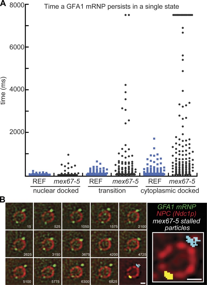
Many messenger RNA export proteins have been identified; yet the spatial and temporal activities of these proteins and how they determine directionality of messenger ribonucleoprotein (mRNP) complex export from the nucleus remain largely undefined. Here, the bacteriophage PP7 RNA-labeling system was used in Saccharomyces cerevisiae to follow single-particle mRNP export events with high spatial precision and temporal resolution. These data reveal that mRNP export, consisting of nuclear docking, transport, and cytoplasmic release from a nuclear pore complex (NPC), is fast (∼ 200 ms) and that upon arrival in the cytoplasm, mRNPs are frequently confined near the nuclear envelope. Mex67p functions as the principal mRNP export receptor in budding yeast. In a mex67-5 mutant, delayed cytoplasmic release from NPCs and retrograde transport of mRNPs was observed. This proves an essential role for Mex67p in cytoplasmic mRNP release and directionality of transport.


Figures

Figure 1.
Figure 1.
REF strain design and characterization. (A) Schematic displaying features of the yeast strains used to monitor mRNP export. Upon transcription, the GFA1 mRNA that carries 24xPP7 loops in the 3′ UTR is bound by the PP7-CP-3xYFP appearing as particles that can be tracked in relation to NPCs that are marked by Ndc1-tdTomato. (B) Fluorescent images of the Ndc1-tdTomato signal in REF cells using identical image acquisition settings showing the improvement in image quality after removal of the yeast cell wall. Examples of both raw and Laplacian filtered images are shown. Bar, 1 µm. (C) Dot plot displaying the localization precision (pixel = 96 nm) obtained when tracking mRNP particles in cells with (−sphero; n = 86) and without a cell wall (+sphero; n = 156), with the mean denoted by a black line for each.
Figure 2.
Figure 2.
GFA1 mRNP export and NE scanning in REF strain. (A) Merged and registered images show consecutive frames of a successful export event based upon tracking of the tagged GFA1 mRNP across the NE in the REF strain (see Video 2). (B and C) Selected nonconsecutive frames show local (B) and distributive (C) NE scanning interactions between an mRNP and the NE (see Videos 5 and 6, respectively). For all panels, cells were imaged at 26°C and 67 Hz with the time from the start of the event given in the bottom right of each image in milliseconds. The last image and inset show an overlay of the mRNP path that is color coded based on position (white, nuclear docked; yellow, transition; blue, cytoplasmic docked/cytoplasm). Each green arrowhead and star denotes mRNP positions at the beginning and end of the track, respectively. Bars, 1 µm.
Figure 3.
Figure 3.
Prolonged GFA1 mRNP interactions with the NE in mex67-5. (A) Dot plot displaying the length of time a GFA1 mRNP persists in a single state (nuclear docked, transition, or cytoplasmic docked) in REF (n = 49, 82, and 108) and mex67-5 (n = 34, 94, and 151) strains. Note that the data presented here use all trace data where a particle interacted with the NE independent of the trace resulting in mRNP export. (B) Selected nonconsecutive frames show the persistent interaction of GFA1 mRNPs with the NE in mex67-5 cells (see Video 7). Cells were imaged at 26°C and 67 Hz with the time from the start of the event given in the bottom right of each image in milliseconds. The last image and inset show an overlay of the mRNP path that is color coded based on position (gray, nuclear docked; yellow, transition; blue, cytoplasmic docked/cytoplasm). Bars, 1 µm.
Figure 4.
Figure 4.
mRNP export kinetics and retrograde transport in mex67-5. (A) Merged and registered images show select frames of a successful export event in a mex67-5 cell (see Video 8). (B) Dot plot showing the distribution of GFA1 mRNP export times in REF (n = 43) and mex67-5 (n = 9) cells, with the mean denoted by a black line. (C) Selected nonconsecutive frames show a retrograde transport event in which an mRNP on the cytoplasmic side of the NE returns to the nucleus (see Video 10). For A and C, cells were imaged at 26°C and 67 Hz with the time from the start of the event given in the bottom right of each image in milliseconds. The last image and inset show an overlay of the mRNP path that is color coded based on position (white, nuclear docked; yellow, transition; blue, cytoplasmic docked/cytoplasm). Each green arrowhead and star denotes mRNP positions at the beginning and end of the track, respectively. Bars, 1 µm.

Similar articles

Cited by

References

    1. Alber F., Dokudovskaya S., Veenhoff L.M., Zhang W., Kipper J., Devos D., Suprapto A., Karni-Schmidt O., Williams R., Chait B.T., et al. . 2007. The molecular architecture of the nuclear pore complex. Nature. 450:695–701. 10.1038/nature06405 - DOI - PubMed
    1. Alcázar-Román A.R., Bolger T.A., and Wente S.R.. 2010. Control of mRNA export and translation termination by inositol hexakisphosphate requires specific interaction with Gle1. J. Biol. Chem. 285:16683–16692. 10.1074/jbc.M109.082370 - DOI - PMC - PubMed
    1. Azimi M., Bulat E., Weis K., and Mofrad M.R.K.. 2014. An agent-based model for mRNA export through the nuclear pore complex. Mol. Biol. Cell. 25:3643–3653. 10.1091/mbc.E14-06-1065 - DOI - PMC - PubMed
    1. Bachi A., Braun I.C., Rodrigues J.P., Panté N., Ribbeck K., von Kobbe C., Kutay U., Wilm M., Görlich D., Carmo-Fonseca M., and Izaurralde E.. 2000. The C-terminal domain of TAP interacts with the nuclear pore complex and promotes export of specific CTE-bearing RNA substrates. RNA. 6:136–158. 10.1017/S1355838200991994 - DOI - PMC - PubMed
    1. Baejen C., Torkler P., Gressel S., Essig K., Söding J., and Cramer P.. 2014. Transcriptome maps of mRNP biogenesis factors define pre-mRNA recognition. Mol. Cell. 55:745–757. 10.1016/j.molcel.2014.08.005 - DOI - PubMed

Publication types

MeSH terms