. 2015 Dec 21;211(6):1157-76.
doi: 10.1083/jcb.201502001.

Physical and functional interaction between the α- and γ-secretases: A new model of regulated intramembrane proteolysis

Affiliations

Physical and functional interaction between the α- and γ-secretases: A new model of regulated intramembrane proteolysis

Allen C Chen et al. J Cell Biol. .

Abstract

Many single-transmembrane proteins are sequentially cleaved by ectodomain-shedding α-secretases and the γ-secretase complex, a process called regulated intramembrane proteolysis (RIP). These cleavages are thought to be spatially and temporally separate. In contrast, we provide evidence for a hitherto unrecognized multiprotease complex containing both α- and γ-secretase. ADAM10 (A10), the principal neuronal α-secretase, interacted and cofractionated with γ-secretase endogenously in cells and mouse brain. A10 immunoprecipitation yielded γ-secretase proteolytic activity and vice versa. In agreement, superresolution microscopy showed that portions of A10 and γ-secretase colocalize. Moreover, multiple γ-secretase inhibitors significantly increased α-secretase processing (r = -0.86) and decreased β-secretase processing of β-amyloid precursor protein. Select members of the tetraspanin web were important both in the association between A10 and γ-secretase and the γ → α feedback mechanism. Portions of endogenous BACE1 coimmunoprecipitated with γ-secretase but not A10, suggesting that β- and α-secretases can form distinct complexes with γ-secretase. Thus, cells possess large multiprotease complexes capable of sequentially and efficiently processing transmembrane substrates through a spatially coordinated RIP mechanism.


Figures

Figure 1.
Figure 1.
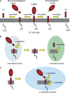
Models of APP processing by the various secretases. (A) Processing of APP by α-, β-, and γ-secretases. (B) Current model of γ-secretase substrate processing in which the ectodomain shedding and the intramembrane cleavages are assumed to be separated spatially and temporally. (C) Proposed new model of γ-secretase processing based on all data herein in which the principal sheddase (α-/β-secretase) exists in an HMW complex with γ-secretase that accepts full-length substrates for rapid sequential processing.
Figure 2.
Figure 2.
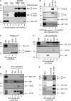
The α-secretases A10 and TACE interact with γ-secretase at overexpressed levels and at the plasma membrane. (A) CHAPSO-solubilized lysates of γ-30 CHO cells were immunoprecipitated for Aph-1 or PAA as a control in the absence or presence of the metalloprotease inhibitor 1,10-phenanthroline. Immunoprecipitates were blotted to probe for coIP of A10 or TACE, APP, and for the γ components NCT and Pen-2. (B) γ-30 lysates were immunoprecipitated for either A10 or NPR A as a control. Resulting immunoprecipitates were probed for the coIP of PS1 CTF. (C) Lysates from cells that express A10 only, human γ-secretase only, neither, or both (lanes 1, 2, 3, and 4, respectively) were specifically pooled to form mix 1 (A10 only + γ-secretase only) or mix 2 (neither + both) and then immunoprecipitated for γ-secretase with an M2 resin targeting the Flag tag on Pen-2. Immunoprecipitates were probed for the coIP of A10 and the γ components PS1 NTF and Pen-2. (D) γ-30 cells were treated with a nonpermeable biotinylation reagent at 4°C to label cell surface proteins. Lysates were immunoprecipitated for Aph-1, and the resultant immunoprecipitates were eluted and pulled down with streptavidin to enrich for cell surface interactors of γ-secretase, followed by blotting for α- and γ-secretases as in A. im, immature; m, mature.
Figure 3.
Figure 3.
The association between A10 and γ-secretase is observed at endogenous expression levels in cells and WT mouse brain. (A) 7W cells were lysed in 1% CHAPSO in the absence or presence of 1,10-phenanthroline, immunoprecipitated for TACE or just with PAA, and then probed for coIP of PS1 CTF. (B) M17D cells were lysed in CHAPSO with 1,10-phenanthroline. Lysates were immunoprecipitated for TACE or A10 and probed for coIP of PS1 CTF. (C) Microsomes prepared from WT mouse brains were immunoprecipitated for A10 with two different antibodies (19026 and 422751) and blotted for the coIP of PS1 CTF. (D and E) Mouse brain microsomes were immunoprecipitated for TACE, A10, or ADAM9 (A9) and controls of PAA only or TFR and probed for coIP of PS1 CTF, TFR, TACE, and A10. The asterisks indicate nonspecific bands. (F) Mouse brain microsomes were immunoprecipitated for PS1 CTF and blotted for coIP of A10, PS1, and APH-1. im, immature; m, mature.
Figure 4.
Figure 4.
A10 exists with γ-secretase in an HMW complex isolated from normal brain by SEC. (A) WT mouse brain microsomes were fractionated on a Superose 6 column and blotted with antibodies to the indicated proteins. (B) Mouse brain microsome fractions from a Superose 6 column were pooled into HMW or LMW fractions and immunoprecipitated for A10 and then probed for A10 and coIP of NCT and PS1 CTF. The asterisks indicate nonspecific bands. (C) S20 cells were lysed in 0.25% DDM, loaded onto a BN-PAGE, and probed for A10 and γ-secretase. (D) 7W cells transfected with control or A10 siRNA were analyzed as in C. im, immature; m, mature.
Figure 5.
Figure 5.
The coimmunoprecipitated α–γ-complex contains both α- and γ-secretase proteolytic activities. (A) S20 lysates were immunoprecipitated for A10, PS1, or just PAA. The immunoprecipitates were subjected to a γ-secretase activity assay (see Activity assays in Materials and methods) using a C100-Flag APP substrate. γ-Secretase activity was documented by generation of both AICD and Aβ via WB (left). The AICD generation was quantified (right). (B) S20 lysates were immunoprecipitated for γ-secretase using M2 (to Flag-Pen2) or 3F10 (to Aph-1-HA) and as controls TFR or M2 preabsorbed with Flag peptide. α-Secretase activity assays were performed on the resin using a fluorogenic peptide substrate (left) and quantified (right). A one-way ANOVA with Tukey’s posttest for multiple comparisons was used. *, P < 0.05. n = 6. After the activity assays, the immunoprecipitates were eluted and blotted for coIP of A10, NCT, and PS1 NTF. Error bars indicate the SD. im, immature; m, mature.
Figure 6.
Figure 6.
Confocal microscopy and SIM of A10 and γ-secretase in 7W cells. (A) A 1-μm optical section of 7W cells stained for A10 (green), PS1 (red), and Hoechst by confocal microscopy (left). Individual channel image for A10 (green, top right) and PS1 (red, bottom right). (B) Orthogonal view of SIM image showing colocalization (yellow) of some A10 (green) and PS1 (red) immunoreactive puncta in 7W cells. (C) Superresolution SIM images of A10 (green) and PS1 (red) immunoreactive puncta in 7W cells. (D) Magnified image of inset in C. (E) 3D-rendered image of D by Imaris processing. Bars: (A) 20 µm; (B) 2 µm; (C) 4 µm; (D and E) 3 µm.
Figure 7.
Figure 7.
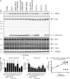
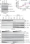
Multiple structurally diverse γ-secretase inhibitors regulate processing by α- and β-secretase. (A) γ-30 cells were treated with various γ inhibitors for APP, and the CM and lysates were analyzed for the indicated proteins. (B) Western blot quantification of APPs-α was performed on CM samples from A, normalized to total APP levels in the lysate, and then normalized to DMSO-treated control samples. A one-way ANOVA with Dunnett’s posttest using DMSO as the control was performed. n = 6. (C) APPs-β ELISA was performed and quantified. All samples were equalized for APP levels, normalized to the DMSO-alone sample, and then analyzed by the same statistical test as in B. n = 4. (D) Levels of APP CTFs in lysates were plotted against APPs-α levels in CM (from Fig. 8 A) and fitted with a linear regression line. n = 36 from two independent experiments. *, P < 0.05; **, P < 0.01; ***, P < 0.001; ****, P < 0.0001.
Figure 8.
Figure 8.
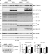
Inhibition of γ-secretase activity increases the cell surface presentation of APP and BACE1. (A) Top: 7W cells were treated with DAPT, and cell surface proteins were labeled with a non–cell permeable biotinylation reagent. Lysates from the biotinylated cells were pulled down with streptavidin. Whole lysates (left) and cell surface proteins (right) were probed for the indicated proteins. (B) CM from A were analyzed for APPs-α and total APPs by WB. (C) Surface levels of APP and BACE were quantitated from A and normalized to total APP or BACE levels in the lysate, respectively. A two-way ANOVA with a Sidak’s posttest was performed. ****, P < 0.0001. n = 8. (D) Total lysate levels of APP and BACE were quantitated from A and normalized to total protein concentration. A two-way ANOVA with a Sidak’s posttest was performed. ***, P < 0.001. n = 8. im, immature; m, mature.
Figure 9.
Figure 9.
TSPAN12 and 17 are required for A10 and γ-secretase association and contribute to the α–β-secretase activity regulation by γ inhibition. (A) 7W cells were transfected with siRNA targeting TSPAN5 + 14, TSPAN12 + 17, or the control. CM were analyzed for APPs-α and lysates for A10 and PS1. (B) S20 cells were transfected with siRNA as in A, and the resultant lysates were immunoprecipitated for γ-secretase by pull-down of Flag–Pen-2 (via an M2 resin). The resulting immunoprecipitates were probed for the coIP of A10 and other γ components. (C) 7W cells were transfected with control siRNA or siRNA targeting both TSPAN12 and 17 and treated in duplicate with increasing amounts of DAPT (3 nM–1 µM). The resulting CM was probed for APPs-α and lysates for A10, PS1, and APP CTFs. (D) CM from C was analyzed by an MSD ELISA for APPs-α and APPs-β. Data are represented as a ratio of APPs-α/APPs-β. A two-way ANOVA with a Sidak’s posttest was performed. *, P < 0.05; ***, P < 0.001. n = 6. CON, control; im, immature; m, mature.
Figure 10.
Figure 10.
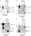
BACE1 also interacts with γ-secretase but not with A10. (A) CHAPSO-solubilized mouse brain microsomes were immunoprecipitated for PS1 CTF or using control resin. Immunoprecipitates were probed for PS1 NTF and for the coIP of BACE1. (B) Mouse brain microsomes solubilized in 1% CHAPSO were immunoprecipitated for BACE1 or TFR and probed for the coIP of PS1 CTF. (C and D) Mouse brain microsomes were immunoprecipitated for A10 (C) or BACE1 (D) and probed for the coIP of BACE1 or A10, respectively. Con, control; im, immature; m, mature.

References

    1. Arduise C., Abache T., Li L., Billard M., Chabanon A., Ludwig A., Mauduit P., Boucheix C., Rubinstein E., and Le Naour F.. 2008. Tetraspanins regulate ADAM10-mediated cleavage of TNF-α and epidermal growth factor. J. Immunol. 181:7002–7013. 10.4049/jimmunol.181.10.7002 - DOI - PubMed
    1. Arimon M., Takeda S., Post K.L., Svirsky S., Hyman B.T., and Berezovska O.. 2015. Oxidative stress and lipid peroxidation are upstream of amyloid pathology. Neurobiol. Dis. 84:109–119. 10.1016/j.nbd.2015.06.013 - DOI - PMC - PubMed
    1. Brown M.S., Ye J., Rawson R.B., and Goldstein J.L.. 2000. Regulated intramembrane proteolysis: a control mechanism conserved from bacteria to humans. Cell. 100:391–398. 10.1016/S0092-8674(00)80675-3 - DOI - PubMed
    1. Buxbaum J.D., Liu K.N., Luo Y., Slack J.L., Stocking K.L., Peschon J.J., Johnson R.S., Castner B.J., Cerretti D.P., and Black R.A.. 1998. Evidence that tumor necrosis factor α converting enzyme is involved in regulated α-secretase cleavage of the Alzheimer amyloid protein precursor. J. Biol. Chem. 273:27765–27767. 10.1074/jbc.273.43.27765 - DOI - PubMed
    1. Cacquevel M., Aeschbach L., Osenkowski P., Li D., Ye W., Wolfe M.S., Li H., Selkoe D.J., and Fraering P.C.. 2008. Rapid purification of active γ-secretase, an intramembrane protease implicated in Alzheimer’s disease. J. Neurochem. 104:210–220. - PubMed

Publication types

Substances