

The effects of airway pressure release ventilation on respiratory mechanics in extrapulmonary lung injury
- PMID: 26694915
- PMCID: PMC4688284
- DOI: 10.1186/s40635-015-0071-0
The effects of airway pressure release ventilation on respiratory mechanics in extrapulmonary lung injury
Abstract
Background: Lung injury is often studied without consideration for pathologic changes in the chest wall. In order to reduce the incidence of lung injury using preemptive mechanical ventilation, it is important to recognize the influence of altered chest wall mechanics on disease pathogenesis. In this study, we hypothesize that airway pressure release ventilation (APRV) may be able to reduce the chest wall elastance associated with an extrapulmonary lung injury model as compared with low tidal volume (LVt) ventilation.
Methods: Female Yorkshire pigs were anesthetized and instrumented. Fecal peritonitis was established, and the superior mesenteric artery was clamped for 30 min to induce an ischemia/reperfusion injury. Immediately following injury, pigs were randomized into (1) LVt (n = 3), positive end-expiratory pressure (PEEP) 5 cmH2O, V t 6 cc kg(-1), FiO2 21 %, and guided by the ARDSnet protocol or (2) APRV (n = 3), P High 16-22 cmH2O, P Low 0 cmH2O, T High 4.5 s, T Low set to terminate the peak expiratory flow at 75 %, and FiO2 21 %. Pigs were monitored continuously for 48 h. Lung samples and bronchoalveolar lavage fluid were collected at necropsy.
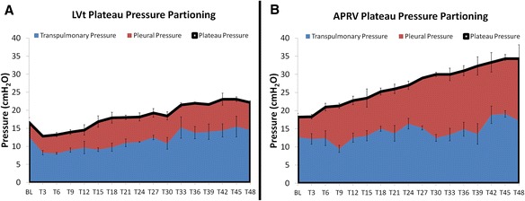
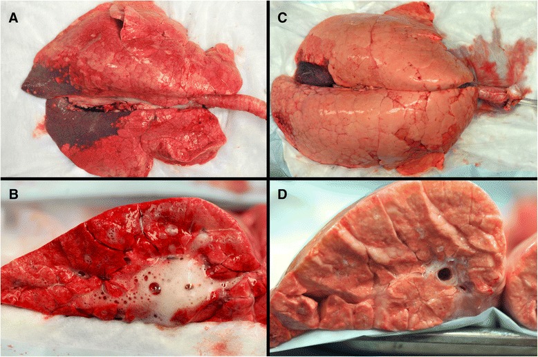
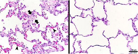
Results: LVt resulted in mild acute respiratory distress syndrome (ARDS) (PaO2/FiO2 = 226.2 ± 17.1 mmHg) whereas APRV prevented ARDS (PaO2/FiO2 = 465.7 ± 66.5 mmHg; p < 0.05). LVt had a reduced surfactant protein A concentration and increased histologic injury as compared with APRV. The plateau pressure in APRV (34.3 ± 0.9 cmH2O) was significantly greater than LVt (22.2 ± 2.0 cmH2O; p < 0.05) yet transpulmonary pressure between groups was similar (p > 0.05). This was because the pleural pressure was significantly lower in LVt (7.6 ± 0.5 cmH2O) as compared with APRV (17.4 ± 3.5 cmH2O; p < 0.05). Finally, the elastance of the lung, chest wall, and respiratory system were all significantly greater in LVt as compared with APRV (all p < 0.05).
Conclusions: APRV preserved surfactant and lung architecture and maintenance of oxygenation. Despite the greater plateau pressure and tidal volumes in the APRV group, the transpulmonary pressure was similar to that of LVt. Thus, the majority of the plateau pressure in the APRV group was distributed as pleural pressure in this extrapulmonary lung injury model. APRV maintained a normal lung elastance and an open, homogeneously ventilated lung without increasing lung stress.
Keywords: Airway pressure release ventilation (APRV); Chest wall elastance; Low tidal volume ventilation; Lung injury; Transpulmonary pressure.
Figures
References
LinkOut - more resources
Full Text Sources
Other Literature Sources
