

Seamless Combination of Fluorescence-Activated Cell Sorting and Hanging-Drop Networks for Individual Handling and Culturing of Stem Cells and Microtissue Spheroids
- PMID: 26694967
- PMCID: PMC7610554
- DOI: 10.1021/acs.analchem.5b03513
Seamless Combination of Fluorescence-Activated Cell Sorting and Hanging-Drop Networks for Individual Handling and Culturing of Stem Cells and Microtissue Spheroids
Abstract
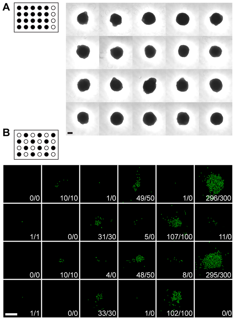
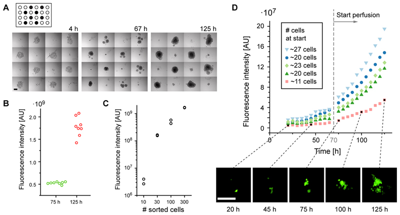
Open microfluidic cell culturing devices offer new possibilities to simplify loading, culturing, and harvesting of individual cells or microtissues due to the fact that liquids and cells/microtissues are directly accessible. We present a complete workflow for microfluidic handling and culturing of individual cells and microtissue spheroids, which is based on the hanging-drop network concept: The open microfluidic devices are seamlessly combined with fluorescence-activated cell sorting (FACS), so that individual cells, including stem cells, can be directly sorted into specified culturing compartments in a fully automated way and at high accuracy. Moreover, already assembled microtissue spheroids can be loaded into the microfluidic structures by using a conventional pipet. Cell and microtissue culturing is then performed in hanging drops under controlled perfusion. On-chip drop size control measures were applied to stabilize the system. Cells and microtissue spheroids can be retrieved from the chip by using a parallelized transfer method. The presented methodology holds great promise for combinatorial screening of stem-cell and multicellular-spheroid cultures.
Conflict of interest statement
The authors declare no competing financial interest.
Figures


Similar articles
-
Fabrication and Operation of Microfluidic Hanging-Drop Networks.Methods Mol Biol. 2018;1771:183-202. doi: 10.1007/978-1-4939-7792-5_15. Methods Mol Biol. 2018. PMID: 29633214 Free PMC article.
-
96-well format-based microfluidic platform for parallel interconnection of multiple multicellular spheroids.J Lab Autom. 2015 Jun;20(3):274-82. doi: 10.1177/2211068214564056. Epub 2014 Dec 18. J Lab Autom. 2015. PMID: 25524491
-
Reconfigurable microfluidic hanging drop network for multi-tissue interaction and analysis.Nat Commun. 2014 Jun 30;5:4250. doi: 10.1038/ncomms5250. Nat Commun. 2014. PMID: 24977495
-
Spheroids as a Type of Three-Dimensional Cell Cultures-Examples of Methods of Preparation and the Most Important Application.Int J Mol Sci. 2020 Aug 28;21(17):6225. doi: 10.3390/ijms21176225. Int J Mol Sci. 2020. PMID: 32872135 Free PMC article. Review.
-
Application of Single Cell Type-Derived Spheroids Generated by Using a Hanging Drop Culture Technique in Various In Vitro Disease Models: A Narrow Review.Cells. 2024 Sep 14;13(18):1549. doi: 10.3390/cells13181549. Cells. 2024. PMID: 39329734 Free PMC article. Review.
Cited by
-
Microfluidic tools for lipid production and modification: a review.Environ Sci Pollut Res Int. 2019 Dec;26(35):35482-35496. doi: 10.1007/s11356-019-05833-4. Epub 2019 Jul 20. Environ Sci Pollut Res Int. 2019. PMID: 31327140 Review.
-
Fabrication and Operation of Microfluidic Hanging-Drop Networks.Methods Mol Biol. 2018;1771:183-202. doi: 10.1007/978-1-4939-7792-5_15. Methods Mol Biol. 2018. PMID: 29633214 Free PMC article.
-
Selective control of the contact and transport between droplet pairs by electrowetting-on-dielectric for droplet-array sandwiching technology.Sci Rep. 2021 Jun 11;11(1):12355. doi: 10.1038/s41598-021-91219-x. Sci Rep. 2021. PMID: 34117288 Free PMC article.
-
Reconfigurable microfluidic device with discretized sidewall.Biomicrofluidics. 2017 May 3;11(3):034103. doi: 10.1063/1.4983148. eCollection 2017 May. Biomicrofluidics. 2017. PMID: 28503247 Free PMC article.
-
Recent Advances in the Analysis of Single Cells.Anal Chem. 2017 Jan 3;89(1):2-21. doi: 10.1021/acs.analchem.6b04255. Epub 2016 Dec 7. Anal Chem. 2017. PMID: 28105840 Free PMC article. Review. No abstract available.
References
Publication types
MeSH terms
Grants and funding
LinkOut - more resources
Full Text Sources
Other Literature Sources
Medical
