. 2016 May 15;193(10):1111-22.
doi: 10.1164/rccm.201510-2058OC.

Haptoglobin Preserves Vascular Nitric Oxide Signaling during Hemolysis

Affiliations

Haptoglobin Preserves Vascular Nitric Oxide Signaling during Hemolysis

Christian A Schaer et al. Am J Respir Crit Care Med. .

Abstract

Rationale: Hemolysis occurs not only in conditions such as sickle cell disease and malaria but also during transfusion of stored blood, extracorporeal circulation, and sepsis. Cell-free Hb depletes nitric oxide (NO) in the vasculature, causing vasoconstriction and eventually cardiovascular complications. We hypothesize that Hb-binding proteins may preserve vascular NO signaling during hemolysis.

Objectives: Characterization of an archetypical function by which Hb scavenger proteins could preserve NO signaling during hemolysis.

Methods: We investigated NO reaction kinetics, effects on arterial NO signaling, and tissue distribution of cell-free Hb and its scavenger protein complexes.

Measurements and main results: Extravascular translocation of cell-free Hb into interstitial spaces, including the vascular smooth muscle cell layer of rat and pig coronary arteries, promotes vascular NO resistance. This critical disease process is blocked by haptoglobin. Haptoglobin does not change NO dioxygenation rates of Hb; rather, the large size of the Hb:haptoglobin complex prevents Hb extravasation, which uncouples NO/Hb interaction and vasoconstriction. Size-selective compartmentalization of Hb functions as a substitute for red blood cells after hemolysis and preserves NO signaling in the vasculature. We found that evolutionarily and structurally unrelated Hb-binding proteins, such as PIT54 found in avian species, functionally converged with haptoglobin to protect NO signaling by sequestering cell-free Hb in large protein complexes.

Conclusions: Sequential compartmentalization of Hb by erythrocytes and scavenger protein complexes is an archetypical mechanism, which may have supported coevolution of hemolysis and normal vascular function. Therapeutic supplementation of Hb scavengers may restore vascular NO signaling and attenuate disease complications in patients with hemolysis.

Keywords: PIT54; extravasation; haptoglobin; hemoglobin; hemolysis.


Figures
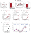
Figure 1.
Figure 1.
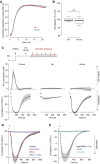
Haptoglobin (Hp) enhances nitric oxide (NO)-mediated arterial dilation during Hb exposure. (A, left panel) Change in mean arterial blood pressure (MAP) of rats after injection of 35 mg cell-free Hb with or without Hp. Data show the mean ± SEM, n = 4. (A, right panel) Comparison of maximum MAP changes (*P < 0.01). (B, left panel) Ten minutes after the initial Hb ± Hp injection, the animals were treated with l-NG-nitroarginine methyl ester (l-NAME), and MAP was monitored for a further 20 minutes to estimate the “NO reserve.” (B, right panel) Comparison of maximum l-NAME–induced changes in MAP in Hb ± Hp-pretreated rats (*P < 0.01). (C) Relative contractile response of porcine coronary arteries pretreated with prostaglandin F2α (PGF2α) (±N5-​[1-​iminoethyl]-​l-​ornithine [l-NIO]) in response to a range of concentrations of Hb or Hb:Hp complexes (mean ± SEM of n = 11–15 vascular rings) (*P < 0.001). (D, E) PGF2α precontracted porcine coronary arteries were dilated with either the intracellular NO donor nitroglycerin (NTG) (D) or the extracellular NO donor DETA-NONOate (DETA) (E). Each NO donor was applied at two different concentrations, and the contractile response to various concentrations of Hb and Hb:Hp was measured (mean ± SEM of n = 15 vascular rings) (*P < 0.001). (F) Relative contraction of PGF2α precontracted and nitroglycerin (10 μM)-dilated porcine coronary arteries in response to a range of concentrations of ferrous HbO2 (Fe2+) or ferric Hb (Fe3+) (mean ± SEM of n = 24 and 9 vascular rings for ferrous and ferric Hb, respectively) (*P < 0.001). (G) Original tension traces of three arterial rings (prepared from different porcine hearts) from an experiment as shown in DF. At the end of the Hb dose response, albumin or Hp were added at a concentration of 32 μM. Black arrows mark the serial addition of the indicated compounds in half-log10 steps. For all arterial ring tension experiments, the baseline tension before PGF2α treatment is considered 0%, and the tension at maximum PGF2α response is considered 100%. Responses are therefore indicated as % PGF2α. n.s. = not significant.
Figure 2.
Figure 2.
Haptoglobin (Hp) uncouples nitric oxide (NO) depletion in solution and NO–cyclic guanosine 3′,5′-monophosphate (cGMP) signaling function in the vascular wall during Hb exposure. (A) An example of a stopped-flow experiment of the reaction of HbO2 ± Hp with NO. The traces show the normalized absorption changes at 406 nm (red: free Hb; blue: Hb:Hp complex), with HbO2 in excess at pH 7.4. (B) NO depletion after injection with either Hb or Hb:Hp into an air-tight, oxygen-free reaction chamber in which NO was continuously produced by DETA-NONOate decay in phosphate-buffered saline (pH 7.4). NO was measured online in the carrier gas phase with an ANTEK chemiluminescence NO detector. Depletion by Hb was set at 100% and was not statistically different from depletion by Hb:Hp. (C) The NO-mediated dilatory response of prostaglandin F2α (PGF2α) precontracted porcine coronary artery segments was measured after injection of MAHMA-NONOate boluses (30 nM) into the immersion buffer. Experiments were performed in buffer only, with Hb (32 μM) or with Hb:Hp (32 μM). The traces show mean ± SEM of at least 15 averaged responses recorded in three independent experiments. In parallel to the vascular response, the NO concentration was measured in vessel immersion buffer using an NO-sensitive microelectrode. The red arrows indicate addition of NO donor. (D) MAHMA-NONOate induced vascular dilation in the presence of intact red blood cells (RBCs) (32 μM Hb). No dilation was observed after lysis of RBCs (32 μM Hb) or with intact RBCs in the presence of the guanylate-cyclase inhibitor 1H-[1,2,4]oxadiazolo[4,3-a]quinoxalin-1-one (ODQ) (10 μM). The experiment was identical to that in C. Red arrow indicates the time point of MAHMA-NONOate injection. (E) MAHMA-NONOate induced vascular dilation in the presence of lysed RBCs (32 μM Hb) + Hp. The cGMP inhibitors ODQ (10 μM) and NS2028 (10 μM) blocked the NO dilatory response. The experiment was identical to that in C. The red arrow indicates the time point of MAHMA-NONOate injection. Traces in D and E represent mean ± SEM of at least 15 averaged responses recorded in three independent experiments. n.s. = not significant.
Figure 3.
Figure 3.
Large molecular-sized protein complex formation restores dilatory nitric oxide (NO) signaling during Hb exposure. (A) Dilatory response after in situ polymerization of Hb. The plot shows the dilatory response of porcine coronary arteries that were sequentially pretreated with prostaglandin F2α (PGF2α), nitroglycerin (NTG), and Hb, resulting in an Hb-mediated constricted state. At this point (time = 0 s) the albumin component was added. Hb polymerization and dilation were only observed when both protein components were click-functionalized to participate in the tetrazine-trans-cyclooctene (TCO) ligation reaction. The plot represents averaged tension traces of six experiments ± SEM (gray lines). (B) Identical experiment as in A, with the exception that haptoglobin (Hp) was added instead of (functionalized) albumin at time 0 s, resulting in an identical dilatory response. (C) Dose-dependent contractile response of PGF2α- and NTG-pretreated porcine coronary artery segments during exposure to increasing concentrations of unmodified and glutaraldehyde-polymerized bovine Hbs. The number indicates the degree of polymerization (glutaraldehyde to protein ratio), whereas T/R indicates the Hb conformational state. Data represent mean ± SEM of at least 13 independent dose–response experiments per compound (*P < 0.001). (D) Correlation plot of the molecular size of glutaraldehyde-polymerized Hb versus [1/(maximum contraction at Hb 10−5 M) × 100]. An analysis of variance for this data set can be found in the online supplement. n.s. = not significant.
Figure 4.
Figure 4.
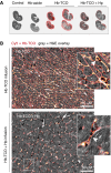
Extravascular cell-free Hb translocation in rats. Rats were infused with Hb-trans-cyclooctene (TCO), and Hb was visualized on tissue slides by the Cy5-tetrazine-TCO ligation reaction. (A) Renal sections. Red indicates the intensity of Cy5 fluorescence in Hb-TCO or Hb-TCO-haptoglobin (Hp)–exposed kidneys. Gray shows a superimposed bright-field image. (B) Myocardial sections from Hb-TCO ± Hp–infused animals. Red-yellow illustrates the intensity of the Cy5 channel (Hb); the gray image shows a superimposed bright-field image of the hematoxylin and eosin (H&E)-stained tissue. The white arrowhead in the zoom images indicate a capillary with Hb-TCO in plasma and nonfluorescent red blood cells (original magnification, ×250).
Figure 5.
Figure 5.
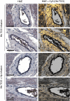
Cell-free Hb translocation in the smooth muscle layer of rat coronary arteries. Coronary artery sections from Hb-trans-cyclooctene (TCO) ± haptoglobin (Hp)-treated animals. The different images pertain to two different animals per treatment. Images on the left show hematoxylin and eosin (H&E) staining of tissues, whereas those on the right show the superimposed Cy5 channel intensity (Hb-TCO) acquired on the identical sections (original magnification, ×100).
Figure 6.
Figure 6.
Extravascular cell-free Hb translocation in porcine coronary arteries. (A) Porcine hearts (n = 4) were perfused with buffer with or without Hb and haptoglobin (Hp). Coronary artery segments were placed inside-out into glass tubes. Images show a view of the endothelial surface with slight red discoloration of Hb-perfused, but not of control or Hb:Hp–perfused, arteries. The lower panel shows a color-deconvoluted image, whereby the most discriminating color is extracted and quantified. The right panel shows quantitative data from the color-deconvoluted image. (B) Detection of fluorescent Hb-trans-cyclooctene (TCO) in lysates from coronary artery segments after reaction with Cy5-tetrazine and polyacrylamide gel electrophoresis separation. Blue fluorescence channel indicates total protein; yellow-orange indicates the Cy5-tetrazine (Hb) signal. Right: Quantitative analysis of Hb-TCO detection in lysates from coronary arteries. The box plots represent data from 12 perfused porcine hearts per condition. Cy5 channel intensities were corrected for total protein intensities. (C) Visualization of Hb-TCO in porcine coronary arteries after conjugation with Cy5-tetrazine (yellow-orange). The white color indicates the orcein positive staining of the internal (*) and external (**) elastic membranes. The scale bar is 200 μm; original magnification ×100. (D) Quantitative image analysis of the intensity of Cy5 fluorescence. Data represent four different coronary arteries with 10 slides per artery. (E) Exemplary electron paramagnetic resonance traces of coronary arteries from control (ctrl) and Hb ± Hp–perfused porcine hearts showing a typical ferric Hb(Fe3+) signal. AU = arbitrary units.
Figure 7.
Figure 7.
PIT54 sequesters chicken Hb in a large protein complex and restores the nitric oxide (NO) dilatory vascular response during Hb exposure. (A) Gene organization of PIT54 on chicken chromosome 4. The soluble protein consists of four scavenger receptor cysteine-rich domains. (B) Binding of human Hb (hHb) and chicken Hb (cHb) to PIT54 or human Hp immobilized on a Proteon SPR chip. (C) Size exclusion high-performance liquid chromatography shows that cHb and PIT54 form a large protein complex eluting at 17.8 minutes when mixed under physiologic conditions. (D) An example of a stopped-flow experiment of the reaction of chicken HbO2 ± PIT54 with NO. The curves show the normalized absorption changes at 406 nm (red: free cHb; blue: cHb:PIT54 complex). (E) The NO-mediated dilatory response of prostaglandin F2α (PGF2α) precontracted porcine coronary artery segments was measured after injection of MAHMA-NONOate boluses into the immersion buffer. Experiments were performed in buffer only, or with 10 μM cHb, with or without 15 μM PIT54. The PIT54-dependent NO dilatory response was blocked by the cGMP inhibitor 1H-[1,2,4]oxadiazolo[4,3-a]quinoxalin-1-one (ODQ). The traces show mean ± SEM of averaged responses obtained with nine arterial rings from three porcine hearts. The red arrows indicate addition of NO donor. (F) Scatterdot plot of dilatory responses (maximum dilation per response) across all experimental conditions. DMBT1 = deleted in malignant brain tumors 1; Hp = haptoglobin.

Comment in

Similar articles

Cited by

References

    1. Schaer DJ, Buehler PW, Alayash AI, Belcher JD, Vercellotti GM. Hemolysis and free hemoglobin revisited: exploring hemoglobin and hemin scavengers as a novel class of therapeutic proteins. Blood. 2013;121:1276–1284. - PMC - PubMed
    1. Rother RP, Bell L, Hillmen P, Gladwin MT. The clinical sequelae of intravascular hemolysis and extracellular plasma hemoglobin: a novel mechanism of human disease. JAMA. 2005;293:1653–1662. - PubMed
    1. Schaer DJ, Buehler PW. Cell-free hemoglobin and its scavenger proteins: new disease models leading the way to targeted therapies. Cold Spring Harb Perspect Med. 2013;3:3. - PMC - PubMed
    1. Vermeulen Windsant IC, de Wit NC, Sertorio JT, van Bijnen AA, Ganushchak YM, Heijmans JH, Tanus-Santos JE, Jacobs MJ, Maessen JG, Buurman WA. Hemolysis during cardiac surgery is associated with increased intravascular nitric oxide consumption and perioperative kidney and intestinal tissue damage. Front Physiol. 2014;5:340. - PMC - PubMed
    1. Donadee C, Raat NJ, Kanias T, Tejero J, Lee JS, Kelley EE, Zhao X, Liu C, Reynolds H, Azarov I, et al. Nitric oxide scavenging by red blood cell microparticles and cell-free hemoglobin as a mechanism for the red cell storage lesion. Circulation. 2011;124:465–476. - PMC - PubMed

Publication types