Review
. 2015;37(2):237-50.

The Role of Innate Immunity in Alcoholic Liver Disease

Affiliations
Review

The Role of Innate Immunity in Alcoholic Liver Disease

Laura E Nagy. Alcohol Res. 2015.

Abstract

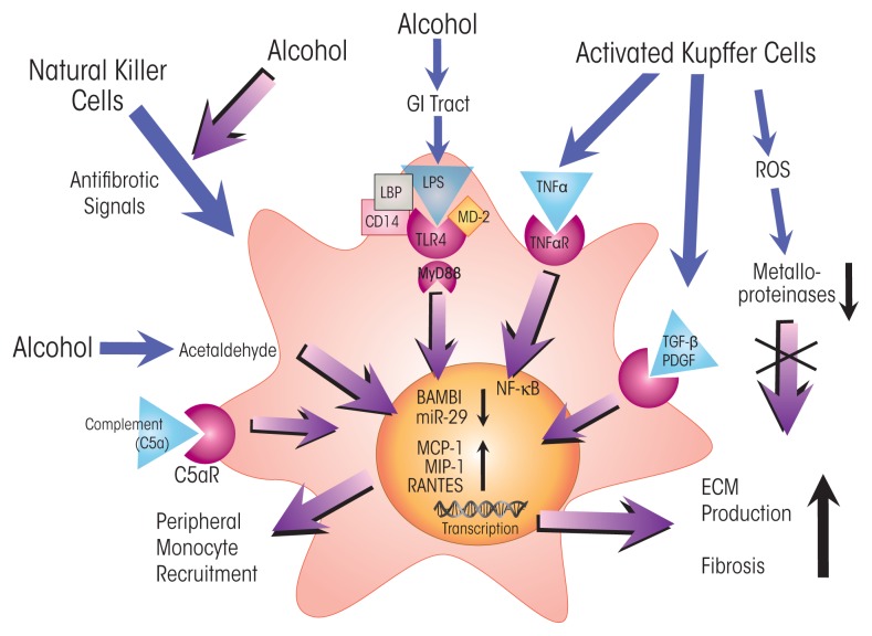
The innate immune system represents the first-line response to invading microbes, tissue damage, or aberrant cell growth. Many of the proteins and cells involved in innate immunity are produced by, and reside in, the liver. This abundance in immune cells and proteins reflects the liver's adaptation to various immune challenges but also makes the organ particularly vulnerable to alcohol's effects. Heavy alcohol consumption may produce leakage of microbes and microbial products from the gastrointestinal tract, which quickly reach the liver via the portal vein. Exposure to these immune challenges and to alcohol and its breakdown products dysregulates the liver's normally fine-tuned immune signaling pathways, leading to activation of various cellular sensors of pathogen- or damage-associated molecular patterns. The ensuing expression of pro-inflammatory cytokines (e.g., tumor necrosis factor a [TNFα], interleukin [IL]-8, and IL-1b) results in cellular dysfunction that contributes to alcoholic liver disease (ALD). Investigations into the roles of the various components of liver innate immunity in ALD have begun to uncover the molecular basis of this disease. Further progress in this area may help inform the development of interventions targeting the innate system to augment current treatments of ALD. These treatments could include antibodies against pro-inflammatory cytokines, use of anti-inflammatory cytokines, or suppression of alcohol-induced epigenetic regulators of innate immunity.


Figures

Figure 1
Figure 1
The role of innate immunity in the natural history of alcoholic liver disease (ALD). Heavy alcohol consumption causes release of bacterial products (i.e., lipopolysaccharides [LPSs]) from the gut into the bloodstream. These LPSs lead to activation of liver innate immunity by stimulating Toll-like receptor 4 (TLR 4) signaling on Kupffer cells and hepatocytes. The damaging effects of alcohol and its metabolism on cells trigger additional immune responses. Steatosis and inflammation in hepatocytes represent the early stages of ALD; continued alcohol-induced inflammation leads to apoptosis/necroptosis in hepatocytes. Downregulation of BMP and activin membrane–bound inhibitor (BAMBI) and increased transforming growth factor β (TGF-β) signaling activate hepatic stellate cells, which differentiate into myofibroblasts causing fibrosis. About 10 to 20 percent of patients with ALD (about 70 percent of patients with alcoholic hepatitis) progress to cirrhosis. Differentiation and proliferation of precancerous liver cells present in cirrhosis lead to cancer in about 10 percent of cirrhosis patients. Acute alcohol-induced inflammation (i.e., alcoholic hepatitis), characterized by high levels of pro-inflammatory cytokines (e.g., interleukin [IL]-17 and IL-8), may occur at any stage of ALD and, in severe cases, may cause death in about 50 percent of patients.
Figure 2
Figure 2
Alcohol’s effects on pro-inflammatory pathways in liver macrophages (i.e., Kupffer cells). Excessive alcohol consumption increases the permeability of the gastrointestinal (GI) tract, exposing Kupffer cells to bacterial endotoxin (i.e., lipopolysaccharide [LPS]). LPS is bound by LPS-binding protein (LBP), enabling engagement with Toll-like receptor 4 (TLR 4) and activating the myeloid differentiation primary response (MyD) 88–independent signaling pathway involving interferon regulatory factor 3 (IRF3) and TIR domain–containing adapter-inducing interferon-β (TRIF). IRF3–TRIF signaling induces production of reactive oxygen species (ROS) by nicotinamide adenine dinucleotide phosphate (NADPH) oxidases and activates nuclear factor κB (NF-κB) and histone acetylation, which trigger transcription of genes for several pro-inflammatory cytokines (i.e., tumor necrosis factor α [TNFα] and interleukin [IL]-17). Alcohol’s breakdown to acetaldehyde and acetate also stimulates ROS signaling and cytokine production. In addition, IRF3–TRIF signaling and detection of damage-associated molecular patterns (DAMPs or alarmins) released from hepatocytes after alcohol exposure stimulate the inflammasome, a multiprotein complex containing caspase 1, which cleaves and thus activates another pro-inflammatory cytokine, IL-1β. Alcohol activates complement, generating anaphylatoxins C3a and C5a, which dock with their cognate receptor on Kupffer cells, further stimulating cytokine production. NOTES: ADH = alcohol dehydrogenase; ALDH = acetaldehyde dehydrogenase; C3a/C5a R = C3a/C5a receptor; CD14 = cluster of differentiation 14 protein; MD-2 = myeloid differentiation 2 protein; NOD-like R = nucleotide-binding oligomerization domain–like receptor.
Figure 3
Figure 3
Alcohol’s direct effects on activity and viability of parenchymal liver cells (i.e., hepatocytes) and on immune-cell signaling to hepatocytes. Excessive alcohol consumption increases the permeability of the gastrointestinal (GI) tract, exposing hepatocytes to bacterial endotoxin (i.e., lipopolysaccharide [LPS]). LPS is bound by LPS-binding protein (LBP), enabling engagement with Toll-like receptor 4 (TLR4) and activation of pro-inflammatory signaling pathways. TLR4 signaling activates expression of nuclear factor κB (NF-κB), which, along with reactive oxygen species (ROS) generated in mitochondria (as a result of exposure to the toxic alcohol-breakdown product acetaldehyde) and Kupffer cells, activates transcription of pro-inflammatory cytokines (i.e., IL-8). Tumor necrosis factor α (TNFα) produced by activated Kupffer cells stimulates sterol regulatory element–binding protein 1c (SREBP-1c), which triggers expression of genes in lipid synthesis, in turn initiating the development of abnormal fat deposition (i.e., steatosis). The combined action of lipid synthesis and upregulated expression of pro-inflammatory cytokines may spur programmed cell death (i.e., apoptosis) and necrosis, resulting in alcohol-induced loss of hepatocytes from tissues NOTES: ADH = alcohol dehydrogenase; CD14 = cluster of differentiation 14 protein; MD-2 = myeloid differentiation 2 protein; TNFαR = TNFα receptor.
Figure 4
Figure 4
Alcohol’s effects on fibrogenic pathways in hepatic stellate cells (HSCs). HSCs are quiescent liver cells that, on stimulation by pro-inflammatory proteins and other agents, differentiate into myofibroblasts to repair damaged tissues. Excessive alcohol consumption increases the permeability of the gastrointestinal (GI) tract, exposing HSCs to endotoxin (i.e., lipopolysaccharide [LPS]). LPS is bound by LPS-binding protein (LBP), enabling engagement with Toll-like receptor 4 (TLR 4) and activating the myeloid differentiation primary response (MyD88)-dependent pathway. MyD88 signaling decreases expression of BMP and activin membrane–bound inhibitor (BAMBI), a pseudoreceptor that suppresses responses to transforming growth factor β (TGF-β; secreted by activated Kupffer cells). Thus, alcohol-induced TLR4–MyD88 signaling increases the HSCs’ responsiveness to TGF-β. microRNA 29 (miR-29) inhibits the production of extracellular matrix (ECM), and its downregulation by MyD88 signaling therefore increases ECM deposition. TLR4–MyD88 signaling in HSCs—along with complement 5a and exposure to the alcohol-breakdown product acetaldehyde and platelet-derived growth factor (PDGF) and tumor necrosis factor α (TNFα) secreted from activated Kupffer cells—upregulates the expression of various chemokines (i.e., monocyte chemotactic protein [MCP-1], macrophage inflammatory protein 1 [MIP-1], and regulated on activation, normal T cell expressed and secreted [RANTES]). These chemokines recruit macrophages (i.e., Kupffer cells and scar-associated macrophages) and other immune cells to the site where HSCs reside (i.e., the liver perisinusoidal space or space of Disse). These signals spur the differentiation of HSCs into myelofibroblasts that produce and secrete ECM, leading to liver fibrosis. In addition, Kupffer cell–produced ROS inhibit activities of metalloproteinases, which normally degrade ECM and thus inhibit fibrosis. NOTES: C5aR = C5a receptor; CD14 = cluster of differentiation 14 protein; MD-2 = myeloid differentiation 2 protein; TNFαR = TNFα receptor.

Similar articles

Cited by

References

    1. An L, Wang X, Cederbaum AI. Cytokines in alcoholic liver disease. Archives of Toxicology. 2012;86(9):1337–1348. - PubMed
    1. Barnes MA, McMullen MR, Roychowdhury S, et al. Macrophage migration inhibitory factor contributes to ethanol-induced liver injury by mediating cell injury, steatohepatitis, and steatosis. Hepatology. 2013;57(5):1980–1991. - PMC - PubMed
    1. Bode C, Kugler V, Bode JC. Endotoxemia in patients with alcoholic and non-alcoholic cirrhosis and in subjects with no evidence of chronic liver disease following acute alcohol excess. Journal of Hepatology. 1987;4(1):8–14. - PubMed
    1. Cohen JI, Chen X, Nagy LE. Redox signaling and the innate immune system in alcoholic liver disease. Antioxidants & Redox Signaling. 2011;15(2):523–534. - PMC - PubMed
    1. Cohen JI, Nagy LE. Pathogenesis of alcoholic liver disease: Interactions between parenchymal and non-parenchymal cells. Journal of Digestive Diseases. 2011;12(1):3–9. - PMC - PubMed

MeSH terms