Review
. 2015;37(2):331-41, 344-51.

Neuroimmune Function and the Consequences of Alcohol Exposure

Affiliations
Review

Neuroimmune Function and the Consequences of Alcohol Exposure

Fulton T Crews et al. Alcohol Res. 2015.

Abstract

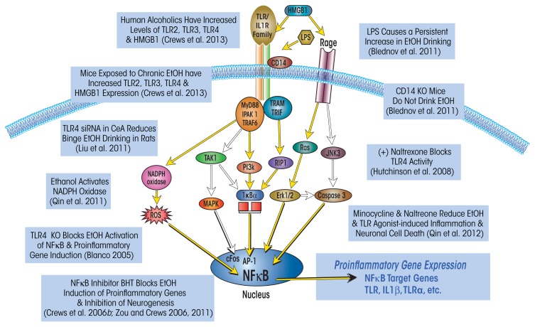
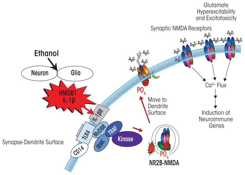
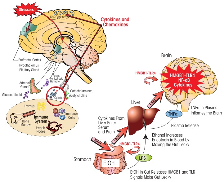
Induction of neuroimmune genes by binge drinking increases neuronal excitability and oxidative stress, contributing to the neurobiology of alcohol dependence and causing neurodegeneration. Ethanol exposure activates signaling pathways featuring high-mobility group box 1 and Toll-like receptor 4 (TLR4), resulting in induction of the transcription factor nuclear factor kappa-light-chain-enhancer of activated B cells, which regulates expression of several cytokine genes involved in innate immunity, and its target genes. This leads to persistent neuroimmune responses to ethanol that stimulate TLRs and/or certain glutamate receptors (i.e., N-methyl-d-aspartate receptors). Alcohol also alters stress responses, causing elevation of peripheral cytokines, which further sensitize neuroimmune responses to ethanol. Neuroimmune signaling and glutamate excitotoxicity are linked to alcoholic neurodegeneration. Models of alcohol abuse have identified significant frontal cortical degeneration and loss of hippocampal neurogenesis, consistent with neuroimmune activation pathology contributing to these alcohol-induced, long-lasting changes in the brain. These alcohol-induced long-lasting increases in brain neuroimmune-gene expression also may contribute to the neurobiology of alcohol use disorder.


Figures

Figure 1
Figure 1
Simplified schematic of the Toll-like receptor (TLR) and the receptor for advanced glycation end products (RAGE) signaling cascades. Stimulation of TLRs with high-mobility group box 1 protein (HMGB1) and other inflammation-inducing agents leads to the generation of reactive oxygen species (ROS) and downstream activation of the transcription factor nuclear factor kappa-light-chain-enhancer of activated B cells (NF)-κB. Similarly, HMGB1 activation of the RAGE receptor results in downstream activation of NF-κB and induction of ROS. Transfer of NF-κB to the nucleus induces proinflammatory gene expression, neuroimmune induction, and cell death. Expression of several TLRs (i.e., TLR2, TLR3, and TLR4) and HMGB1 is upregulated in the postmortem human alcoholic brain and mouse brain following exposure to ethanol (Crews et al. 2013); this is accompanied by an upregulation of NADPH oxidase expression (Qin et al. 2011). Interestingly, blockade of neuroimmune signaling, either genetically (Blanco 2005) or pharmacologically (Crews et al. 2006b; Qin et al. 2012; Zou and Crews 2006, 2011), prevents ethanol-induced neuroimmune-gene induction and neurodegeneration. The neuroimmune system also contributes to alcohol-drinking behavior, because activation (Blednov et al. 2001) or blockade of this system (Blednov et al. 2011; Liu et al. 2011) increases and decreases self-administration, respectively. NOTE: AP-1: activator protein-1; CD14: cluster of differentiation 14; ERK: extracellular signal–regulated kinase; IKK: inhibitor of NF-κB; IRAK 1: interleukin-1 receptor–associated kinase 1; JNK: c-jun N-terminal kinases; LPS: lipopolysaccharide; MAPK: mitogen-activated protein kinase; MyD88: myeloid differentiation primary response gene 88; NADPH oxidase: nicotinamide adenine dinucleotide phosphate-oxidase; PI3K: phosphatidylinositol-4,5-bisphosphate 3-kinase; RIP: receptor interacting protein; TAK1: transforming growth factor beta–activated kinase 1; TRAF: tumor necrosis factor receptor–associated factor; TRAM: TRIF-related adaptor molecule; TRIF: TIR-domain-containing adaptor–inducing interferon-beta. SOURCE: Adapted from Crews et al. 2011, .
Figure 2
Figure 2
Microglial activation, as indicated by expression of the microglial marker Iba-1, is increased in postmortem alcoholic brain. The photomicrographs depict microglia from postmortem brain samples of alcoholics and control subjects. The number of Iba-1–positive microglia (dark stains) is higher in the alcoholic than in the control samples. SOURCE: He and Crews 2008.
Figure 3
Figure 3
Alcohol increases high-mobility group box 1 (HMGB1) expression in mouse brain, and human brain and induces HMGB1 release from rat brain slices. (Left) Chronic ethanol treatment of mice for 10 days increases expression of HMGB1 mRNA and protein. (Middle) Postmortem human alcoholic orbitofrontal cortex (OFC) has significantly more HMGB1-immunoreactive cells than seen in age-matched moderately drinking control subjects. (Right) Ethanol causes the release of HMGB1 into the media from hippocampal-entorhinal cortex (HEC) slice culture. NOTE: ** P < 0.01, relative to the corresponding control group. SOURCE: Adapted from Crews et al. 2013.
Figure 4
Figure 4
Cycles of chronic alcohol consumption lead to persistently increased neuroimmune-gene expression. (Top) Repeated ethanol (EtOH) binges result in increased brain neuroimmune activation (i.e., microglial and astrocytic activation as well as upregulated neuroimmune-gene expression). (Bottom) In humans, lifetime alcohol consumption is positively correlated with neuroimmune signal immunoreactivity. Symbols indicate levels of Toll-like receptor (TLR) 2, TLR3, TLR4, and high-mobility group box 1 (HMGB1) in individual moderate drinkers and alcoholics. Results for moderate drinkers are clustered along the Y-axis because of their low lifetime alcohol consumption and similar neuroimmune expression. Alcoholic subjects show a more than 10-fold variation in lifetime alcohol consumption as well as considerable variation in expression of all four neuroimmune genes. NOTE: Correlations are as follows: TLR2: r = 0.66 (p < 0.01); TLR3: r = 0.83 (P < 0.001); TLR4: r = 0.62 (P < 0.01); HMGB1: r = 0.83 (P < 0.001). SOURCE: Crews et al. 2013.
Figure 5
Figure 5
Simplified schematic depicting how neuroimmune signaling leads to neuronal hyperexcitability and the neurobiology of addiction. Alcohol and stress activate neurons and glia in the central nervous system, resulting in the release of various neuroimmune signals (e.g., high-mobility group box 1 [HMGB1] and interleukin-1beta [IL-1β]) that activate neuroimmune receptors (e.g., Toll-like receptors [TLRs]). Neuroimmune receptor stimulation leads to phosphorylation, and thus activation, of glutamatergic N-methyl-d-aspartate (NMDA) receptors that are transported to the cell surface (Iori et al. 2013; Maroso et al. 2010). The increased number of NMDA receptors increases Ca2+ flux, triggering further induction of neuroimmune genes, and also promotes glutamate hyperexcitability and excitotoxicity.
Figure 6
Figure 6
Neuroimmune signaling integrates central nervous system (CNS) responses to alcohol and stress. (Left) Stressors activate the body’s stress response system, which is comprised of the hypothalamus, pituitary gland, and adrenal glands (i.e., HPA axis) as well as the stress hormones they produce (e.g., adrenocorticotropic hormone and glucocorticoids). Stress also activates the sympathetic nervous system, which secretes catecholamines. These hormones act on various organs and tissues that are part of the immune system. In response, immune cells secrete cytokines that via the blood are transported to the brain. There, these cytokines lead to brain neuroimmune-gene induction that sensitizes stress-response pathways. At the same time, the immune system communicates with the CNS through sensory (afferent) nerves that activate the brain in response to stressful stimuli. This communication pathway involves particularly the vagus nerve and the nucleus tractus solitarius in the brain stem. (Right) Alcohol influences neuroimmune signaling via its effects on the gastrointestinal tract. Consumed ethanol enters the stomach and gut and makes them “leaky” by inducing the release of high-mobility group box 1 (HMGB1), which in turn activates Toll-like receptor 4 (TLR4) in the gut. As a result, bacterial products such as lipopolysaccharide (LPS) can enter the blood and reach the liver. Both LPS and ethanol (which also reaches the liver via the circulation) contribute to inflammatory reactions in the liver, which lead to release of tumor necrosis factor-alpha (TNF-α) and other proinflammatory cytokines from the liver. These proinflammatory cytokines in the blood enter the brain and increase neuroimmune-gene expression. Chronic ethanol also increases expression of HMGB1–TLR4 signaling in the brain, leading to persistent and progressive increases in neuroimmune-gene expression in the brain.

Similar articles

Cited by

References

    1. Acheson SK, Stein RM, Swartzwelder HS. Impairment of semantic and figural memory by acute ethanol: Age-dependent effects. Alcoholism: Clinical and Experimental Research. 1998;22(7):1437–1442. - PubMed
    1. Anderson RI, Varlinskaya EI, Spear LP. Ethanol-induced conditioned taste aversion in male Sprague-Dawley rats: Impact of age and stress. Alcoholism: Clinical and Experimental Research. 2010;34(12):2106–2115. - PMC - PubMed
    1. Brown SA, McGue M, Maggs J, et al. A developmental perspective on alcohol and youths 16 to 20 years of age. Pediatrics. 2008;121(Suppl 4):S290–S310. - PMC - PubMed
    1. Carr LG, Spence JP, Peter Eriksson CJ, et al. AA and ANA rats exhibit the R100Q mutation in the GABAA receptor alpha 6 subunit. Alcohol. 2003;31(1–2):93–107. - PubMed
    1. Coleman LG, Jr, He J, Lee J, et al. Adolescent binge drinking alters adult brain neurotransmitter gene expression, behavior, brain regional volumes, and neurochemistry in mice. Alcoholism: Clinical and Experimental Research. 2011;35(4):671–688. - PMC - PubMed

MeSH terms