. 2015 Dec 23:12:224.
doi: 10.1186/s12985-015-0452-8.

Analysis of the interaction between host factor Sam68 and viral elements during foot-and-mouth disease virus infections

Affiliations

Analysis of the interaction between host factor Sam68 and viral elements during foot-and-mouth disease virus infections

Devendra K Rai et al. Virol J. .

Abstract

Background: The nuclear protein Src-associated protein of 68 kDa in mitosis (Sam68) is known to bind RNA and be involved in cellular processes triggered in response to environmental stresses, including virus infection. Interestingly, Sam68 is a multi-functional protein implicated in the life cycle of retroviruses and picornaviruses and is also considered a marker of virus-induced stress granules (SGs). Recently, we demonstrated the partial redistribution of Sam68 to the cytoplasm in FMDV infected cells, its interaction with viral protease 3C(pro), and found a significant reduction in viral titers as consequence of Sam68-specific siRNA knockdowns. Despite of that, details of how it benefits FMDV remains to be elucidated.

Methods: Sam68 cytoplasmic localization was examined by immunofluorescent microscopy, counterstaining with antibodies against Sam68, a viral capsid protein and markers of SGs. The relevance of RAAA motifs in the IRES was investigated using electromobility shift assays with Sam68 protein and parental and mutant FMDV RNAs. In addition, full genome WT and mutant or G-luc replicon RNAs were tested following transfection in mammalian cells. The impact of Sam68 depletion to virus protein and RNA synthesis was investigated in a cell-free system. Lastly, through co-immunoprecipitation, structural modeling, and subcellular fractionation, viral protein interactions with Sam68 were explored.

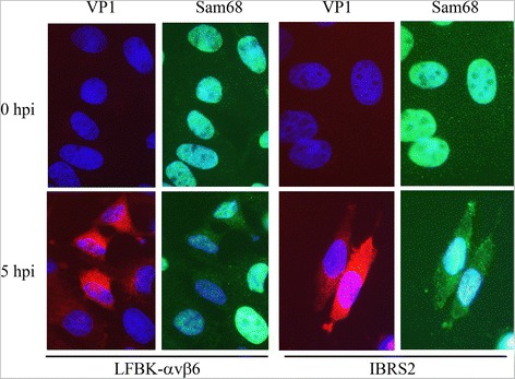
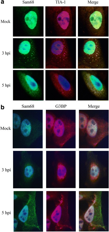
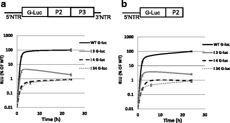
Results: FMDV-induced cytoplasmic redistribution of Sam68 resulted in it temporarily co-localizing with SG marker: TIA-1. Mutations that disrupted FMDV IRES RAAA motifs, with putative affinity to Sam68 in domain 3 and 4 cause a reduction on the formation of ribonucleoprotein complexes with this protein and resulted in non-viable progeny viruses and replication-impaired replicons. Furthermore, depletion of Sam68 in cell-free extracts greatly diminished FMDV RNA replication, which was restored by addition of recombinant Sam68. The results here demonstrated that Sam68 specifically co-precipitates with both FMDV 3D(pol) and 3C(pro) consistent with early observations of FMDV 3C(pro)-induced cleavage of Sam68.

Conclusion: We have found that Sam68 is a specific binding partner for FMDV non-structural proteins 3C(pro) and 3D(pol) and showed that mutations at RAAA motifs in IRES domains 3 and 4 cause a decrease in Sam68 affinity to these RNA elements and rendered the mutant RNA non-viable. Interestingly, in FMDV infected cells re-localized Sam68 was transiently detected along with SG markers in the cytoplasm. These results support the importance of Sam68 as a host factor co-opted by FMDV during infection and demonstrate that Sam68 interact with both, FMDV RNA motifs in the IRES and viral non-structural proteins 3C(pro) and 3D(pol).


Figures

Fig. 1
Fig. 1
Sam68 redistribution from the nucleus to the cytoplasm. Two different FMDV-susceptible cell lines (LFBK-αvβ6, left; and IBRS2, right) were mock-infected or infected with FMDV at a MOI of 10 and fixed at 5 hpi. Cells were examined by IFM probing with rabbit polyclonal anti-Sam68 followed by goat-anti-rabbit-AF488 (green) and mouse monoclonal anti-FMDV VP1 followed by goat-anti-mouse-AF568 (red). Nuclei were stained with DAPI (blue)
Fig. 2
Fig. 2
FMDV-induced cytoplasmic Sam68 co-localizes with TIA-1. LFBK cells were mock-infected or infected with FMDV at a MOI of 10 and were fixed at 3 and 5 hpi. Cells were examined by IFM probing with rabbit polyclonal anti-Sam68 followed by goat-anti-rabbit-AF488 (green) and goat polyclonal anti-TIA-1 (a) or mouse monoclonal anti-G3BP (b) followed by donkey-anti-goat-AF568 (red, a) or goat-anti-mouse-AF568 (red; b). Nuclei were stained with DAPI (blue)
Fig. 3
Fig. 3
Sam68 interacts with FMDV IRES 4. a Cartoon diagram in the upper panel describes the modular structure of FMDV A24 IRES and the location of two unpaired UAAA and a CAAA sequence motifs in domain 4 and 3, respectively. Sam68 potential binding sites are shown as a grey incomplete oval. Lower panel in Fig. 3a shows anti-Sam68 Western blot (rabbit anti-Sam68) of pull-down experiments conducted between Sam68 and selected IRES domains. IRES domains used in the experiment are shown. b Determination Sam68 binding to FMDV IRES RNAs by EMSA. WT probe in the left panel consists of 5′ biotin labeled 65 nt long synthetic RNA representing residues 435–499 of FMDV A24-Cru IRES that spanned at least 20 bases upstream and downstream of the two UAAA sequence motifs present in IRES domain 4. The binding of Sam68 to RNA probe was carried out in the presence of 100-fold excess of tRNA. The concentrations of Sam68 used are indicated in each lane. In the mutant probe in the right panel, the two UAAA motifs aremutated to UACG. c Upper panel depicts cartoon representation of the wild-type and KH-domain deleted Sam68 constructs. Lower panel shows EMSA results with the addition of Sam68-WT (left) and Sam68-delta KH (right). Probe and conditions used were the same as in section (b). d Determination of binding interference by various 5′ NTR RNA segments on the complexes formed between WT probe representing partial FMDV IRES domain 4 and Sam68. The binding of Sam68 to WT probe was performed under similar conditions as mentioned in section (b) but using a 2 μM Sam68 and 30 nM of probe. WT probe-Sam68 binding was competed with 10-fold molar excess of either full-length IRES (lane 3) or miscellaneous RNAs, including the FMDV cre (lane 4), S-fragment (lane 5), and IRES domains 2, 3, 4 (lanes 6, 7, 8, respectively). Lane 1 contains the binding mixture of Sam68 and domain 4 RNA in the absence of competitor RNAs, whereas lane 9 contains a probe alone control. Lane 2 was left blank
Fig. 4
Fig. 4
Effect of Sam68-depletion on FMDV protein and RNA synthesis using cell-free extracts. a Depletion of Sam68 from BHK-21 CFE. BHK-21 CFE was prepared as described in Materials and Methods. The depletion of Sam68 was confirmed by Western blot probing of non-depleted (left lane) and depleted (right lane) extract with anti-Sam68. b Determination of the effect of Sam68-6H addition on the translation of FMDV A24-Cru. FMDV A24-Cru RNA was translated using non-depleted or depleted BHK-21 CFE that were supplemented with 0 or 1 μM Sam68-6H as marked. The reaction was carried out at 32 ° C for 2 h and the products were resolved by SDS-PAGE and Western blot probed for FMDV 3Dpol. c Determination of the effect of Sam68-6H addition on the synthesis of FMDV A24-Cru RNA. FMDV A24-Cru RNA was used for RNA synthesis using non-depleted or depleted BHK-21 CFE that were supplemented with 0–2.5 μM Sam68-6H as marked. The reaction was carried out at 37 ° C for 5 h and the products were SDS-PAGE resolved by dot blotting
Fig. 5
Fig. 5
Replication and translation kinetics of WT G-luc, I 3 G-luc, I 4 G-luc and I 34 G-luc replicon constructs. a Schematic representation of the G-luc replicon construct digested with SwaI restriction enzyme (above). The luciferase activity of transcript RNAs for WT G-luc, I 3 G-luc, I 4 G-luc and I 34 G-luc SwaI digested replicon plasmids. On the Y-axis, G-luc signals are expressed in relative luciferase units (RLU) per nanogram of protein and the error bars represent the standard deviation from two independent experiments. X-axis values indicate the time points of G-luc measurement (hours post-transfection). WT, I 3, I 4 and I 34 G-luc are marked as solid black, solid gray, dotted gray and point gray lines, respectively. b Schematic representation of truncated G-luc replicon produced by digestion with MfeI restriction enzyme (above)
Fig. 6
Fig. 6
Sam68 interacts with FMDV 3Cpro and 3Dpol. a Co-immunoprecipitation of FMDV 3Dpol and Sam68 during FMDV infection. BHK-21 cells either mock-infected or infected with FMDV at a MOI of 10 were lysed and the lysates were immunoprecipitated using either anti-FMDV 3Dpol or anti-Sam68 and the eluates examined by Western blot. One lane in each panel (as indicated below) was not subject to IP as a control. Equal amount of isotype control antibody served as an IP control. The eluates from the anti-3Dpol IP reaction were probed with anti-Sam68 (left panel). Conversely, the Sam68 IP eluates were probed with an anti-3Dpol (right panel). In the left panel, lane 1 corresponds to the isotype IP control, lane 2 is mock-infected cell lysate (1:10 dilution), lane 3 is a FMDV-infected cell lysate (1:10 dilution) that was not IP and lane 4 is the anti-3Dpol IP eluate from FMDV-infected cell lysates. Similarly, in the right panel, lane 1 corresponds to the isotype control, lane 2 is mock-infected cell lysate (1:10 dilution), lane 3 is a FMDV-infected cell lysate (1:10 dilution) that was not IP, and lane 4 is the anti-Sam68 IP eluate from FMDV-infected cell lysates. b The fragments (frag) listed in the table correspond to the amino acid (aa) sequence of FMDV 3Dpol, starting from the N-terminus: frag #1 aa 1–48, frag #2 aa 49–108, frag #3 aa 109–157, frag #4 aa 158–217, frag #5 aa 218–268, frag #6 aa 269–331, frag #7 aa 332–404, and frag #8 aa 405–470. A scrambled peptide was used as a negative control. c Computational prediction of the interaction between FMDV 3Dpol and Sam68. (i) Electrostatic surface representation of FMDV 3Dpol in the docking pose (PDB: 1U09); red color depicts the negatively charged surface, white shows the neutral surface, and blue color shows the positively charged surface. Color intensity is proportional to the surface charge. Areas under dashed lines indicate Sam68 binding interface of FMDV 3Dpol. (ii) Electrostatic surface representation of Sam68 in the docking pose. Surface charge and color annotation are same as section (i). Surface marked with dashed lines indicates FMDV 3Dpol binding interface of Sam68. (iii) Electrostatic representation of Sam68 docked to FMDV 3Dpol. [98] FMDV 3Dpol green docked on Sam68 blue in cartoon representation. The 3Dpol frag-4 residues 193–217 (orange), frag-5 residues 221, 222, 225, 226 (magenta) and frag-8 residues 453–470 (red) form the Sam68 binding interface of 3Dpol (d) LFBK cells were uninfected or infected with FMDV at a MOI of 10, and cells were harvested at 1, 3, and 5 hpi by treatment with versine. Left panel: cell lysates were IP with mouse monoclonal anti-FMDV 3Cpro, and examined by Western blot probing with rabbit polyclonal anti-Sam68 (N-terminus). Right panel: collected cells were lysed and separated into nuclear and cytoplasmic fractions, and the cytoplasmic fractions were examined by Western blot probing with rabbit polyclonal anti-Sam68 (N-terminus). Loading control is indicated confirming equivalent loading per lane

Similar articles

Cited by

References

    1. Rodriguez LL, Gay CG. Development of vaccines toward the global control and eradication of foot-and-mouth disease. Expert Rev Vaccines. 2011;10(3):377–87. doi: 10.1586/erv.11.4. - DOI - PubMed
    1. Grubman MJ, Baxt B. Foot-and-mouth disease. Clin Microbiol Rev. 2004;17(2):465–93. doi: 10.1128/CMR.17.2.465-493.2004. - DOI - PMC - PubMed
    1. Mason PW, Grubman MJ, Baxt B. Molecular basis of pathogenesis of FMDV. Virus Res. 2003;91(1):9–32. doi: 10.1016/S0168-1702(02)00257-5. - DOI - PubMed
    1. Pelletier J, Sonenberg N. Internal initiation of translation of eukaryotic mRNA directed by a sequence derived from poliovirus RNA. Nature. 1988;334(6180):320–5. doi: 10.1038/334320a0. - DOI - PubMed
    1. Jang SK, Krausslich HG, Nicklin MJ, Duke GM, Palmenberg AC, Wimmer E. A segment of the 5′ nontranslated region of encephalomyocarditis virus RNA directs internal entry of ribosomes during in vitro translation. J Virol. 1988;62(8):2636–43. - PMC - PubMed

Publication types

MeSH terms

LinkOut - more resources