Case Reports
. 2015 Dec 22:15:263.
doi: 10.1186/s12883-015-0518-7.

Diagnosing moyamoya syndrome using ultrasound - a case report

Affiliations
Case Reports

Diagnosing moyamoya syndrome using ultrasound - a case report

Tobias Braun et al. BMC Neurol. .

Abstract

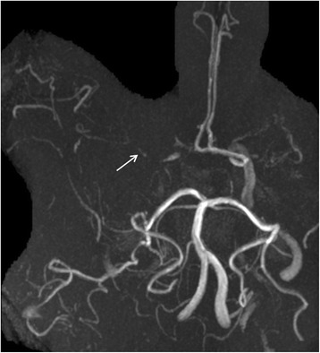
Background: Moyamoya syndrome is a vasculopathy characterised by progressive occlusion of the cerebral arteries resulting in the development of abnormal collateral circulation. To diagnose this syndrome, imaging of the cerebral arteries is required including CT- or MR-angiography and conventional angiography. We present a case of moyamoya disease with typical findings detected in the sonography. The diagnosis was suspected after reviewing the initial ultrasound images of the cerebral arteries with evidence for obliterated intracranial arteries and the detection of an existing collateral circulation network.

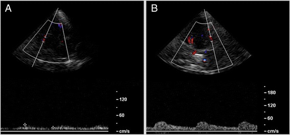
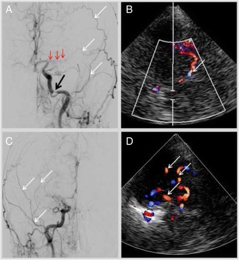
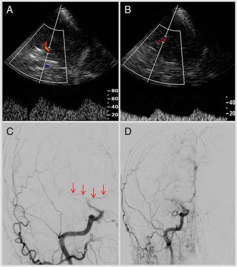
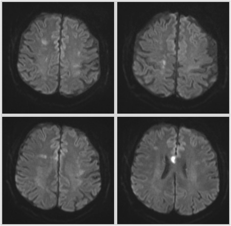
Case presentation: A 62 years old male patient presented in the hospital's emergency department with symptoms indicating a subacute cerebrovascular event. Immediate sonographic studies showed a right-sided pulsatile Doppler-signal in the common and internal carotid arteries, suggestive of distal stenoses. In addition, the transcranial examination indicated obliteration of both middle cerebral arteries. Numerous arterial vessels suggestive of leptomeningeal collateral arteries revealed a strong arterial leptomeningeal flow. At this stage of the diagnostic work-up, the collateral circulation network, characteristic of moyamoya disease, was indicated by sonography. Moyamoya syndrome was verified by conventional angiography. The aetiological work remained empty, so the diagnosis of moyamoya disease was established.

Conclusion: Our case report indicates that sonography can be a useful tool for detecting the vaculopathy in moyamoya syndrome. In case routine procedures, such as the CT- or MR-angiography, with evidence for obliterated intracerebral arteries, ultrasound studies might provide important information regarding an existing collateral network in the scope of a moyamoya syndrome.


Figures

Fig. 1
Fig. 1
Middle cerebral artery left (a); due to obliteration the Doppler-spectrum is low pulsatile, the corresponding detected flow velocities are also low. In contrast flow velocities detected in collaterals are higher (b)
Fig. 2
Fig. 2
Cerebral digital subtraction angiography left (a) in frontal projection. Severe stenosis of the carotid artery (black arrow); the middle cerebral artery is obliterated (red arrows). Branches of the collateral network with leptomeningeal anastomoses are indicated by white arrows. Image b demonstrates a leptomeningeal collateral branch in the duplex-sonography. The digital subtraction angiography right (c) indicates collateral vessels (white arrows) by obliterated middle and anterior cerebral artery; correspondently the transcranial duplex-sonography right (d) demonstrates numerous arterial vessels assembling the collateral network
Fig. 3
Fig. 3
Cerebral MR angiography indicates the obliteration pathology in the anterior circulation; the middle cerebral arteries are bilaterally rudimentary visible (arrows)
Fig. 4
Fig. 4
Transcranial Doppler- and duplex-sonography right, showing deteriorated Doppler-spectrum in the distal portion of the internal carotid artery (a) and proximal portion of the middle cerebral artery (b) indicating the vessel obliteration; in accordance with sonographic findings the conventional angiography (c) confirmed the obliteration of the middle cerebral artery and anterior cerebral artery (red arrows). Image (d) demonstrates the collateral network
Fig. 5
Fig. 5
Brain MRI, diffusion weighted imaging showing multiple bilateral acute ischaemia

References

    1. Burke GM, Burke AM, Sherma AK, Hurley MC, Batjer HH, Bendok BR. Moyamoya disease: a summary. Neurosurg Focus. 2009;26:E11. doi: 10.3171/2009.1.FOCUS08310. - DOI - PubMed
    1. Houkin K, Ito M, Sugiyama T, Shichinohe H, Nakayama N, Kazumata K, et al. Review of past research and current concepts on the etiology of moyamoya disease. Neurol Med Chir (Tokyo) 2012;52:267–77. doi: 10.2176/nmc.52.267. - DOI - PubMed
    1. Kuroda S, Houkin K. Moyamoya disease: current concepts and future perspectives. Lancet Neurol. 2008;7:1056–66. doi: 10.1016/S1474-4422(08)70240-0. - DOI - PubMed
    1. Liu W, Xu G, Liu X. Neuroimaging diagnosis and the collateral circulation in moyamoya disease. Interv Neurol. 2013;1:77–86. doi: 10.1159/000346765. - DOI - PMC - PubMed
    1. Kronenburg A, Braun KP, van der Zwan A, Klijn CJ. Recent advances in moyamoya disease: pathophysiology and treatment. Curr Neurol Neurosci Rep. 2014;14:423–0423. doi: 10.1007/s11910-013-0423-7. - DOI - PubMed

Publication types

LinkOut - more resources