Comparative Study
. 2015 Dec 22:6:257.
doi: 10.1186/s13287-015-0255-2.

Comparison of intraspinal and intrathecal implantation of induced pluripotent stem cell-derived neural precursors for the treatment of spinal cord injury in rats

Affiliations
Comparative Study

Comparison of intraspinal and intrathecal implantation of induced pluripotent stem cell-derived neural precursors for the treatment of spinal cord injury in rats

Takashi Amemori et al. Stem Cell Res Ther. .

Abstract

Background: Stem cell treatment provides a promising therapy for patients with spinal cord injury (SCI). However, the applied stem cells exert their effects in different manners that are dependent on the route used for administration.

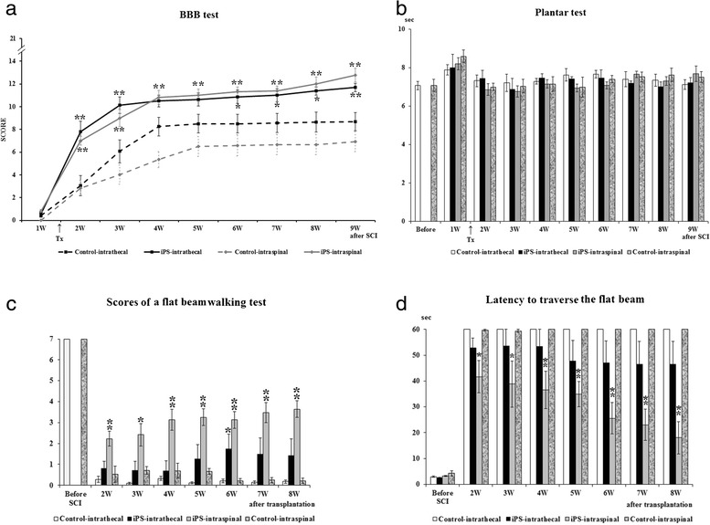
Methods: In the present study, we administered neural precursors derived from induced pluripotent stem cells (iPS-NPs) either intraspinally into the lesion center or intrathecally into the subarachnoid space of rats with a balloon-induced spinal cord compression lesion. Functional locomotor performance, cell survival, astrogliosis, axonal sprouting and the expression of endogenous neurotrophic growth factors were evaluated using behavioral tests (BBB, flat beam test, rotarod, plantar test), morphometric analysis, immunohistochemistry and qPCR.

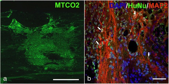
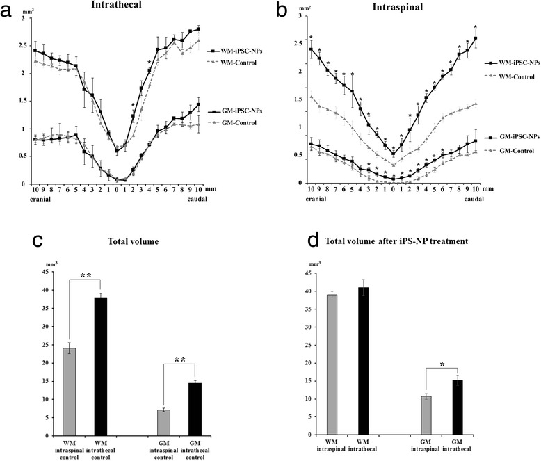
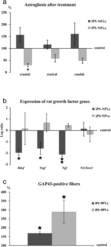
Results: Both treatments facilitated the functional locomotor recovery of rats with SCI. iPS-NPs injected intraspinally survived well for 2 months and were positive for MAP2, while cells grafted intrathecally were undetectable at the site of administration or in the spinal cord tissue. Intraspinal implantation increased gray and white matter sparing and axonal sprouting and reduced astrogliosis, while intrathecal application resulted only in an improvement of white matter sparing and an increase in axonal sprouting, in parallel with no positive effect on the expression of endogenous neurotrophic growth factor genes or glial scar reduction.

Conclusions: Intrathecally grafted iPS-NPs had a moderate therapeutic benefit on SCI through a paracrine mechanism that does not require the cells to be present in the tissue; however, the extended survival of i.s. grafted cells in the spinal cord may promote long-term spinal cord tissue regeneration.


Figures

Fig. 1
Fig. 1
Functional motor recovery evaluated by the BBB test a. Statistical differences between the appropriate control and cell-treated groups are indicated by *p <0.05 and **p <0.01. The sensitivity of the hind paws to thermal stimuli was examined by the Plantar test b. The withdrawal latency before surgery is represented by the two left-most columns in the figure. There were no significant differences between the controls and the iPS-NP-treated groups throughout the experiment. The flat beam scores evaluate the locomotor function of the hind legs c. The maximum score is 7 in a healthy animal. The differences between the i.s. control and cell-treated groups were always significant (**p <0.01 above the gray columns). Locomotor ability to traverse a beam with a flat surface d. The latency decreased week by week in the treated groups when compared with the control groups. Statistical differences between the appropriate control and cell-treated groups are indicated: *p <0.05 and **p <0.01. BBB Basso, Beattie, and Bresnahan, iPS induced pluripotent stem cell, SCI spinal cord injury, W weeks, Tx day of transplantation
Fig. 2
Fig. 2
MTCO2-positive transplanted cells in the host spinal cord 8 weeks after i.s. implantation of iPS-NPs. Scale bar = 500 μm a. HuNu-positive transplanted cells (green) express the neuronal marker MAP2 (red). Nuclei are stained by DAPI (blue). Colocalization is marked by arrows. Scale bar = 50 μm b. DAPI 4',6-diamidino-2-phenylindole, HuNu human nuclei, MAP2 microtubule-associated protein 2
Fig. 3
Fig. 3
Morphometric measurement of the white matter (WM) and gray matter (GM). The cross-sectional area (mm2) is plotted at 1 mm increments in both the cranial and caudal directions from the injury epicenter, which is indicated by the number 0. The WM was significantly spared only 2 and 4 mm caudal to the injury epicenter in the i.t.-treated group a. In contrast, the WM and GM were prominently spared in the i.s. iPS-NP-transplanted group b. The total volume of a 2-mm-long spinal segment, including the injury epicenter, is compared between the i.s. and i.t. control groups c. Statistically significant differences were found in both the volume of the WM and also the volume of the GM (**p <0.01). Total volume of a 2-mm-long spinal segment after iPS-NP treatment d. There is a significant difference in the volume of the GM between i.s. and i.t. administration (*p <0.05). iPSC-NP neural precursors derived from induced pluripotent stem cells
Fig. 4
Fig. 4
GFAP-positive areas are compared in three different parts of the spinal segment: cranial, between 3 and 7 mm cranial; central, between 2 mm cranial and 2 mm caudal; and caudal, between 3 and 7 mm caudal to the injury epicenter a. The average GFAP-positive area around the lesion in the appropriate control group is shown as the baseline of 100 %. Rat gene expression of BDNF, VEGF, nerve growth factor (NGF), and neurotrophin 3 (NT3) analyzed by qPCR b. The level of the control expression is indicated by the number 0. An increase or decrease from this level is shown as positive or negative scale numbers, respectively. The levels of BDNF, VEGF, and NGF in the i.t. iPS-NP-treated group are significantly lower than those in the control animals. There was no significant difference between the i.s. iPS-NP-treated and control groups in terms of gene expression. Axonal sprouting evaluated by GAP43-positive fibers c. The average number of positive fibers in the control group is shown as the baseline of 100 %. Both cell-treated groups show the increased expression of GAP43, which was more pronounced in the i.s. cell-treated animals. iPS-NP neural precursors derived from induced pluripotent stem cells, i.s. intraspinal, i.t. intrathecal

Similar articles

Cited by

References

    1. Angelos MG, Kaufman DS. Pluripotent stem cell applications for regenerative medicine. Curr Opin Organ Transplant. 2015;20:663–70. - PMC - PubMed
    1. Erceg S, Lukovic D, Moreno-Manzano V, Stojkovic M, Bhattacharya SS. Derivation of cerebellar neurons from human pluripotent stem cells. Curr Protoc Stem Cell Biol. 2012;Chapter 1:Unit 1H 5. - PubMed
    1. Hodgetts SI, Edel M, Harvey AR. The state of play with iPSCs and spinal cord injury models. J Clin Med. 2015;4:193–203. doi: 10.3390/jcm4010193. - DOI - PMC - PubMed
    1. Jin X, Lin T, Xu Y. Stem cell therapy and immunological rejection in animal models. Curr Mol Pharmacol. 2015. doi:10.2174/1874467208666150928153511. - PubMed
    1. Lee-Kubli CA, Lu P. Induced pluripotent stem cell-derived neural stem cell therapies for spinal cord injury. Neural Regen Res. 2015;10:10–6. doi: 10.4103/1673-5374.150638. - DOI - PMC - PubMed

Publication types

MeSH terms

Substances