

Age-dependent mesial temporal lobe lateralization in language fMRI
- PMID: 26696589
- PMCID: PMC4749038
- DOI: 10.1111/epi.13258
Age-dependent mesial temporal lobe lateralization in language fMRI
Abstract
Objective: Functional magnetic resonance imaging (fMRI) activation of the mesial temporal lobe (MTL) may be important for epilepsy surgical planning. We examined MTL activation and lateralization during language fMRI in children and adults with focal epilepsy.
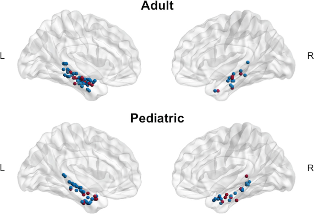
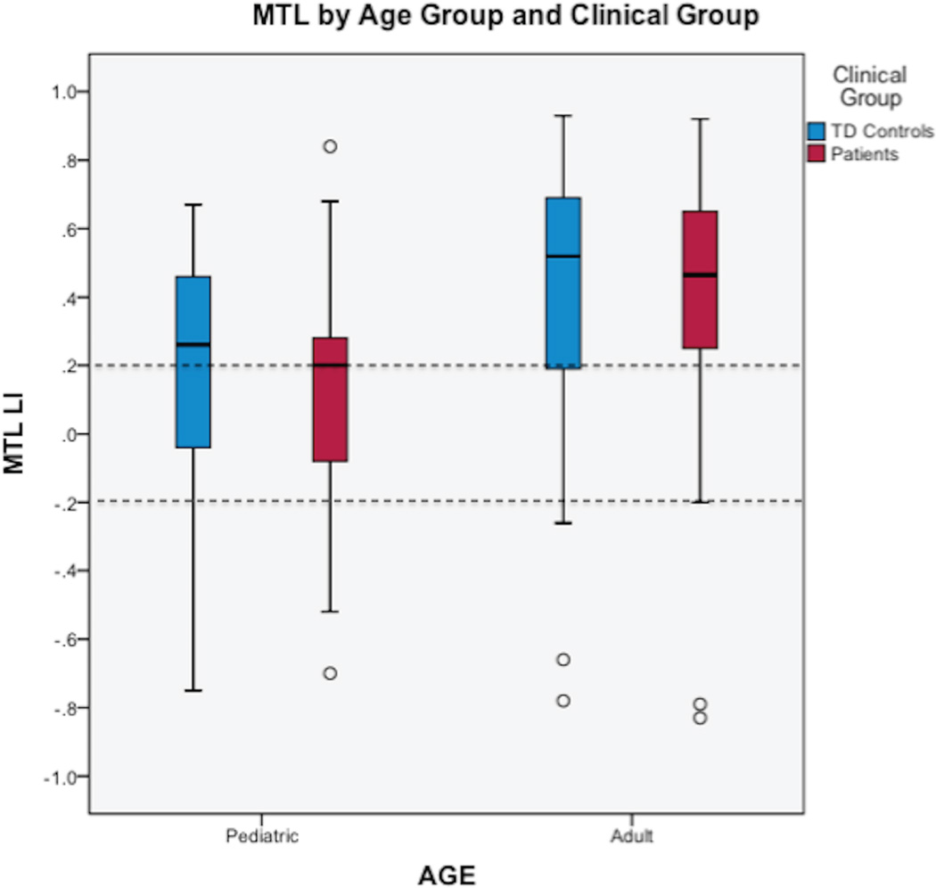
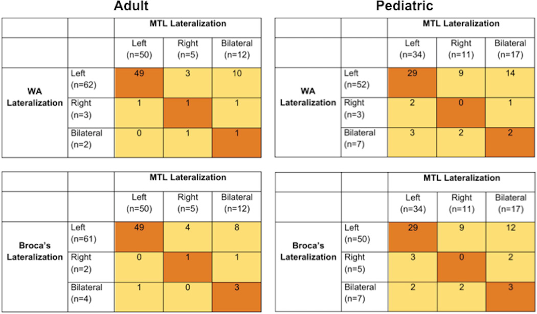
Methods: One hundred forty-two controls and patients with left hemisphere focal epilepsy (pediatric: epilepsy, n = 17, mean age = 9.9 ± 2.0; controls, n = 48; mean age = 9.1 ± 2.6; adult: epilepsy, n = 20, mean age = 26.7 ± 5.8; controls, n = 57, mean age = 26.2 ± 7.5) underwent 3T fMRI using a language task (auditory description decision task). Image processing and analyses were conducted using Statistical Parametric Mapping (SPM8); regions of interest (ROIs) included MTL, Broca's area, and Wernicke's area. We assessed group and individual MTL activation, and examined degree of lateralization.
Results: Patients and controls (pediatric and adult) demonstrated group and individual MTL activation during language fMRI. MTL activation was left lateralized for adults, but less so in children (p's < 0.005). Patients did not differ from controls in either age group. Stronger left-lateralized MTL activation was related to older age (p = 0.02). Language lateralization (Broca's and Wernicke's) predicted 19% of the variance in MTL lateralization for adults (p = 0.001), but for not children.
Significance: Language fMRI may be used to elicit group and individual MTL activation. The developmental difference in MTL lateralization and its association with language lateralization suggests a developmental shift in lateralization of MTL function, with increased left lateralization across the age span. This shift may help explain why children have better memory outcomes following resection compared to adults.
Keywords: Functional neuroimaging; Neuropsychological assessment; Seizures.
Wiley Periodicals, Inc. © 2015 International League Against Epilepsy.
Conflict of interest statement
The remaining authors have no conflicts of interest to report.
Figures
Similar articles
-
Medial temporal fMRI activation reflects memory lateralization and memory performance in patients with epilepsy.Epilepsy Behav. 2008 Apr;12(3):410-8. doi: 10.1016/j.yebeh.2007.11.012. Epub 2007 Dec 26. Epilepsy Behav. 2008. PMID: 18162441
-
Language lateralization correlates with verbal memory performance in children with focal epilepsy.Epilepsia. 2010 Apr;51(4):627-38. doi: 10.1111/j.1528-1167.2009.02406.x. Epub 2009 Dec 2. Epilepsia. 2010. PMID: 19958383
-
FMRI lateralization of expressive language in children with cerebral lesions.Epilepsia. 2006 Jun;47(6):998-1008. doi: 10.1111/j.1528-1167.2006.00572.x. Epilepsia. 2006. PMID: 16822246
-
Beyond Broca's and Wernicke's: Functional Mapping of Ancillary Language Centers Prior to Brain Tumor Surgery.Tomography. 2023 Jun 25;9(4):1254-1275. doi: 10.3390/tomography9040100. Tomography. 2023. PMID: 37489468 Free PMC article. Review.
-
Clinical recommendations for conducting pediatric functional language and memory mapping during the phase I epilepsy presurgical workup.Clin Neuropsychol. 2024 Jul;38(5):1060-1084. doi: 10.1080/13854046.2023.2281708. Epub 2023 Nov 20. Clin Neuropsychol. 2024. PMID: 37985747 Review.
Cited by
-
Imaging episodic memory during development and childhood epilepsy.J Neurodev Disord. 2018 Dec 13;10(1):40. doi: 10.1186/s11689-018-9255-8. J Neurodev Disord. 2018. PMID: 30541437 Free PMC article. Review.
-
Absence of neural speech discrimination in preterm infants at term-equivalent age.Dev Cogn Neurosci. 2019 Oct;39:100679. doi: 10.1016/j.dcn.2019.100679. Epub 2019 Jul 10. Dev Cogn Neurosci. 2019. PMID: 31437736 Free PMC article.
-
Mapping Cognition in Epilepsy: From the Lab to the Clinic.Epilepsy Curr. 2024 Nov 21:15357597241280485. doi: 10.1177/15357597241280485. Online ahead of print. Epilepsy Curr. 2024. PMID: 39582595 Free PMC article. Review.
-
Weaker semantic language lateralization associated with better semantic language performance in healthy right-handed children.Brain Behav. 2018 Nov;8(11):e01072. doi: 10.1002/brb3.1072. Epub 2018 Oct 8. Brain Behav. 2018. PMID: 30298640 Free PMC article.
-
Atypical language representation is unfavorable for language abilities following childhood stroke.Eur J Paediatr Neurol. 2019 Jan;23(1):102-116. doi: 10.1016/j.ejpn.2018.09.007. Epub 2018 Sep 25. Eur J Paediatr Neurol. 2019. PMID: 30314763 Free PMC article.
References
-
- Lah S. Neuropsychological outcome following focal cortical removal for intractable epilepsy in children. Epilepsy Behav. 2004;5:804–817. - PubMed
-
- Golby AJ, Poldrack RA, Illes J, et al. Memory lateralization in medial temporal lobe epilepsy assessed by functional MRI. Epilepsia. 2002;43:855–863. - PubMed
-
- Rabin ML. Functional MRI predicts post-surgical memory following temporal lobectomy. Brain. 2004;127:2286–2298. - PubMed
Publication types
MeSH terms
Substances
Grants and funding
LinkOut - more resources
Full Text Sources
Other Literature Sources
Medical
