

Review
doi: 10.1159/000440656.
Epub 2015 Sep 11.
The Clinical Pictures of Autoimmune Hemolytic Anemia
Affiliations
- PMID: 26696800
- PMCID: PMC4678314
- DOI: 10.1159/000440656
Item in Clipboard
Review
The Clinical Pictures of Autoimmune Hemolytic Anemia
Transfus Med Hemother.
2015 Sep.
Abstract
Autoimmune hemolytic anemia is characterized by shortened red blood cell survival and a positive Coombs test. The responsible autoantibodies may be either warm reactive or cold reactive. The rate of hemolysis and the severity of the anemia may vary from mild to severe and life-threatening. Diagnosis is made in the laboratory by the findings of anemia, reticulocytosis, a positive Coombs test, and specific serologic tests. The prognosis is generally good but renal failure and death sometimes occur, especially in cases mediated by drugs.
Keywords: Agglutinin; Autoimmunity; Direct antiglobulin test; Hemolysin; Hemolysis.
Figures
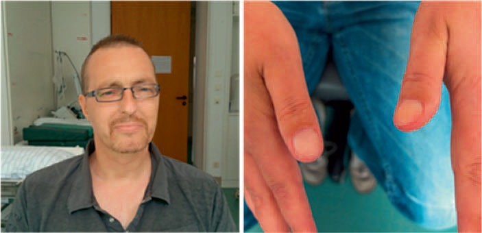
Physical examination in warm antibody AIHA. Skin pallor, icterus and nailbed pallor in a patient with treatment-refractory warm AIHA, hemoglobin 4.8 g/dl (photographs courtesy of Professor A. Salama, with kind permission of the patient).
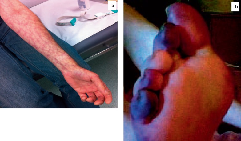
Physical examination in cold antibody AIHA. a Livedo reticularis (photograph courtesy of Professor A. Salama). b Acrocyanosis involving the toes in a patient with a high-titer IgM cold agglutinin (reprinted from Sinha A, Richardson G, Patel RT : Cold agglutinin related acrocyanosis and paroxysmal hemolysis. Eur J Vasc Endovasc Surg, 30:563-565, copyright 2005, with permission from Elsevier).
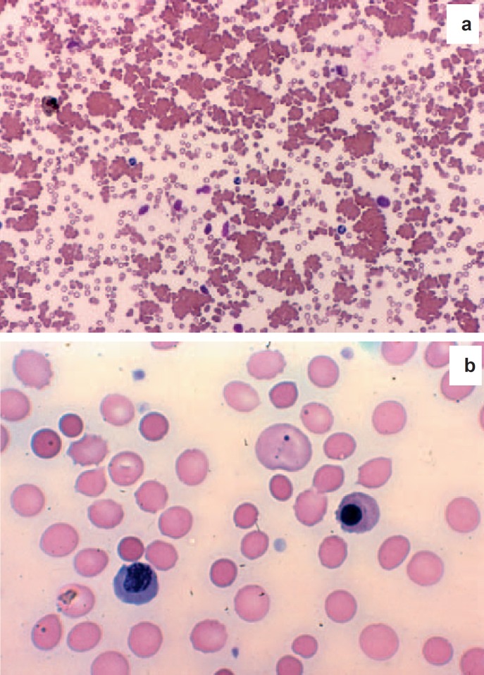
Blood films from patients with AIHA. Used with permission from Lichtman's Atlas of Hematology, www.accessmedicine.com . a Cold agglutinin disease, low power, slide made at room temperature. Note agglutination of RBCs. b Warm antibody AIHA. Note frequent small, round RBCs lacking central pallor, large bluish RBCs (polychromasia), Howell-Jolly body and nucleated RBCs.
References
-
- Packman CH. Hemolytic anemia resulting from immune injury; in Kaushansky K, Lichtman MA, Prchal JT, Levi MM, Press OW, Burns LJ, Caligiuri MA (eds): Williams Hematology, 9th ed. New York, McGraw-Hill, chapter 54, 2016 (in press).
-
- Dacie JV. The Haemolytic Anaemias, vol 3, The Autoimmune Haemolytic Anaemias, 3rd ed. New York, Churchill Livingstone, 1992.
-
- Petz LD, Garratty G. Acquired Immune Hemolytic Anemias. Philadelphia, Churchill Livingstone, 2004.
-
- Mayer B, Yürek S, Kiesewetter H, Salama A. Mixed‐type autoimmune hemolytic anemia: differential diagnosis and a critical review of reported cases. Transfusion. 2008;48:2229–2234. - PubMed
Publication types
LinkOut - more resources
Full Text Sources
Other Literature Sources
Medical
