

Preliminary Evidence of Increased Hippocampal Myelin Content in Veterans with Posttraumatic Stress Disorder
- PMID: 26696852
- PMCID: PMC4667092
- DOI: 10.3389/fnbeh.2015.00333
Preliminary Evidence of Increased Hippocampal Myelin Content in Veterans with Posttraumatic Stress Disorder
Abstract
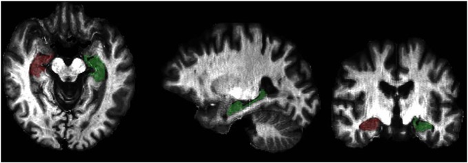
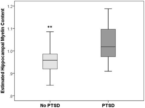
Recent findings suggest the formation of myelin in the central nervous system by oligodendrocytes is a continuous process that can be modified with experience. For example, a recent study showed that immobilization stress increased oligodendrogensis in the dentate gyrus of adult rat hippocampus. Because changes in myelination represents an adaptive form of brain plasticity that has a greater reach in the adult brain than other forms of plasticity (e.g., neurogenesis), the objective of this "proof of concept" study was to examine whether there are differences in myelination in the hippocampi of humans with and without post-traumatic stress disorder (PTSD). We used the ratio of T1-weighted/T2-weighted magnetic resonance image (MRI) intensity to estimate the degree of hippocampal myelination in 19 male veterans with PTSD and 19 matched trauma-exposed male veterans without PTSD (mean age: 43 ± 12 years). We found that veterans with PTSD had significantly more hippocampal myelin than trauma-exposed controls. There was also found a positive correlation between estimates of hippocampal myelination and PTSD and depressive symptom severity. To our knowledge, this is the first study to examine hippocampal myelination in humans with PTSD. These results provide preliminary evidence for stress-induced hippocampal myelin formation as a potential mechanism underlying the brain abnormalities associated with vulnerability to stress.
Keywords: hippocampus; imaging; myelin; plasticity; post-traumatic stress disorder.
Figures
Similar articles
-
Automated measurement of hippocampal subfields in PTSD: Evidence for smaller dentate gyrus volume.J Psychiatr Res. 2017 Dec;95:247-252. doi: 10.1016/j.jpsychires.2017.09.007. Epub 2017 Sep 9. J Psychiatr Res. 2017. PMID: 28923718 Free PMC article.
-
Smaller hippocampal volume as a vulnerability factor for the persistence of post-traumatic stress disorder.Psychol Med. 2015 Oct;45(13):2737-46. doi: 10.1017/S0033291715000707. Epub 2015 May 4. Psychol Med. 2015. PMID: 25936409
-
Regional gray matter oligodendrocyte- and myelin-related measures are associated with differential susceptibility to stress-induced behavior in rats and humans.Transl Psychiatry. 2021 Dec 13;11(1):631. doi: 10.1038/s41398-021-01745-5. Transl Psychiatry. 2021. PMID: 34903726 Free PMC article.
-
Hippocampal volume deficits associated with exposure to psychological trauma and posttraumatic stress disorder in adults: a meta-analysis.Prog Neuropsychopharmacol Biol Psychiatry. 2010 Oct 1;34(7):1181-8. doi: 10.1016/j.pnpbp.2010.06.016. Epub 2010 Jun 21. Prog Neuropsychopharmacol Biol Psychiatry. 2010. PMID: 20600466 Review.
-
[Posttraumatic stress disorder (PTSD) as a consequence of the interaction between an individual genetic susceptibility, a traumatogenic event and a social context].Encephale. 2012 Oct;38(5):373-80. doi: 10.1016/j.encep.2011.12.003. Epub 2012 Jan 24. Encephale. 2012. PMID: 23062450 Review. French.
Cited by
-
Labelfree mapping and profiling of altered lipid homeostasis in the rat hippocampus after traumatic stress: Role of oxidative homeostasis.Neurobiol Stress. 2022 Aug 11;20:100476. doi: 10.1016/j.ynstr.2022.100476. eCollection 2022 Sep. Neurobiol Stress. 2022. PMID: 36032405 Free PMC article.
-
Neuroplasticity in Post-Traumatic Stress Disorder.Rev Neurol. 2025 Jul 23;80(6):33478. doi: 10.31083/RN33478. Rev Neurol. 2025. PMID: 40767105 Free PMC article. Review. English.
-
Preservation of a remote fear memory requires new myelin formation.Nat Neurosci. 2020 Apr;23(4):487-499. doi: 10.1038/s41593-019-0582-1. Epub 2020 Feb 10. Nat Neurosci. 2020. PMID: 32042175 Free PMC article.
-
Regulation of Central Nervous System Myelination in Higher Brain Functions.Neural Plast. 2018 Mar 5;2018:6436453. doi: 10.1155/2018/6436453. eCollection 2018. Neural Plast. 2018. PMID: 29692804 Free PMC article. Review.
-
Myelin plasticity: sculpting circuits in learning and memory.Nat Rev Neurosci. 2020 Dec;21(12):682-694. doi: 10.1038/s41583-020-00379-8. Epub 2020 Oct 12. Nat Rev Neurosci. 2020. PMID: 33046886 Free PMC article. Review.
References
-
- American Psychology Association (2013). Diagnostic and Statistical Manual of Mental Disorders. Washington, D.C.
LinkOut - more resources
Full Text Sources
Other Literature Sources