

Dynamic Functional Brain Connectivity for Face Perception
- PMID: 26696870
- PMCID: PMC4672064
- DOI: 10.3389/fnhum.2015.00662
Dynamic Functional Brain Connectivity for Face Perception
Abstract
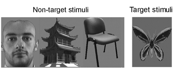
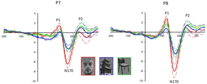
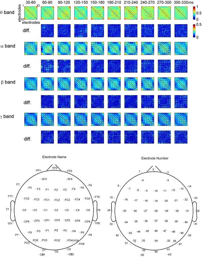
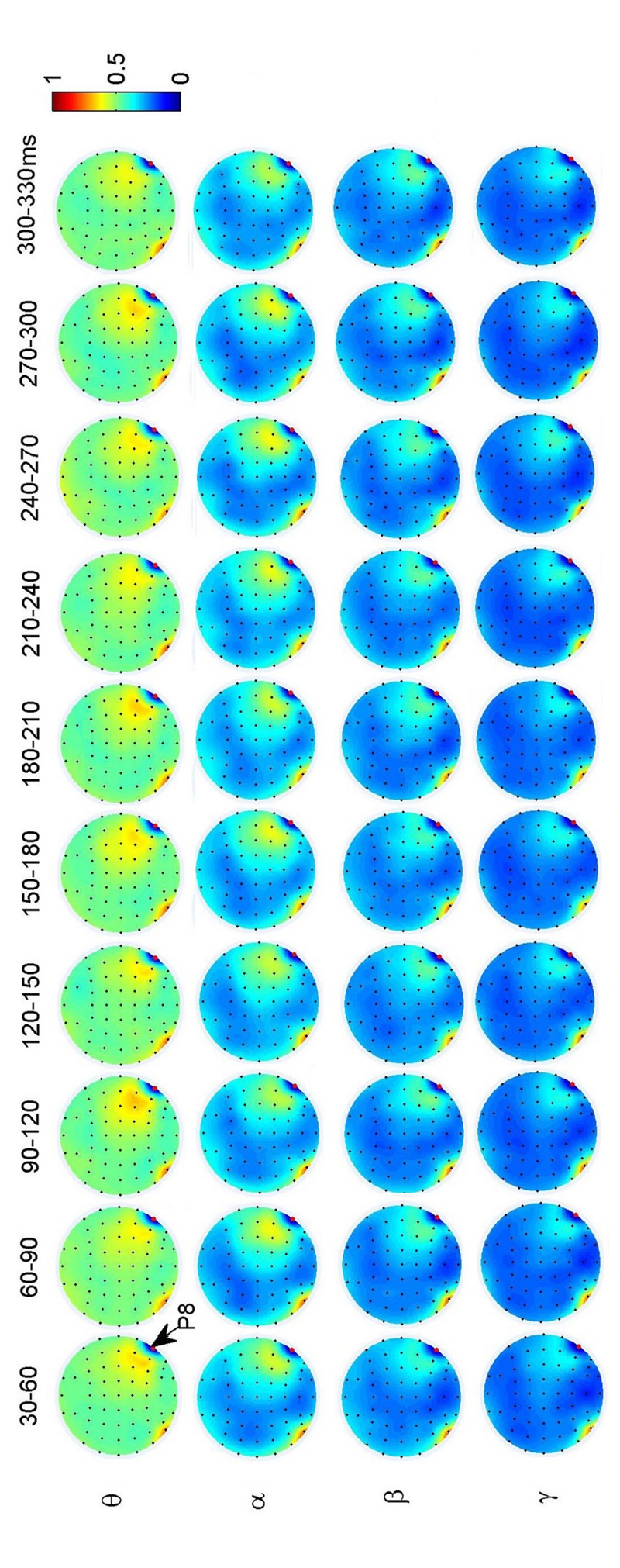
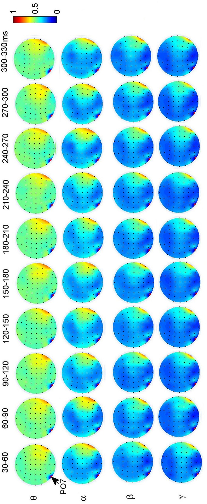
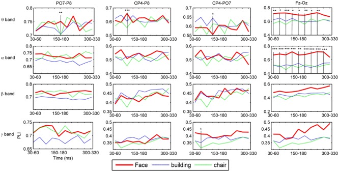
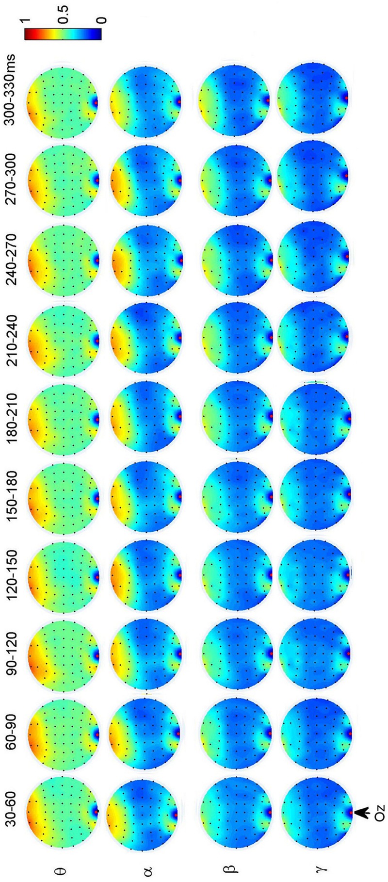
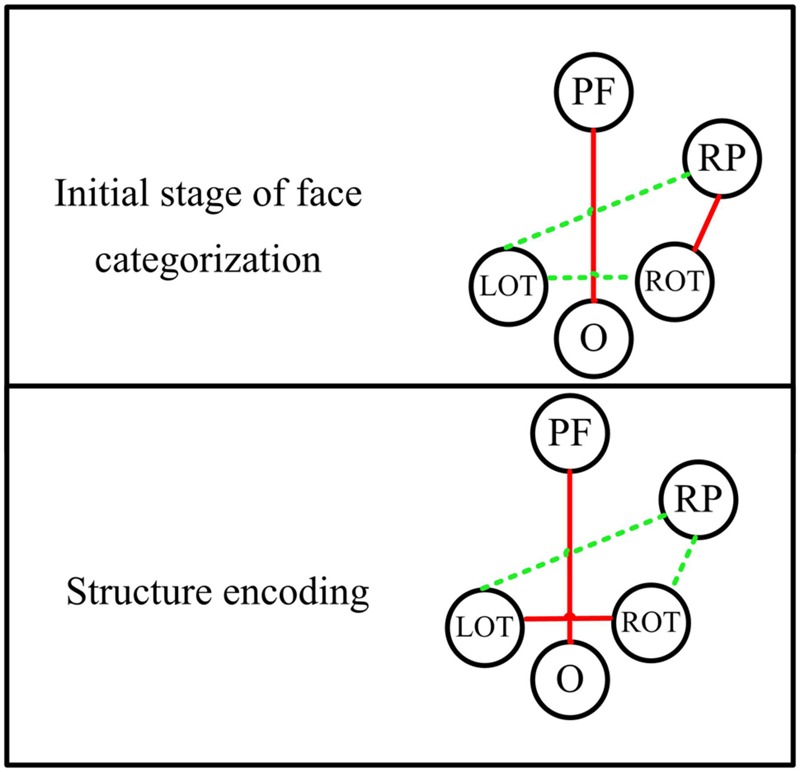
Face perception is mediated by a distributed brain network comprised of the core system at occipito-temporal areas and the extended system at other relevant brain areas involving bilateral hemispheres. In this study we explored how the brain connectivity changes over the time for face-sensitive processing. We investigated the dynamic functional connectivity in face perception by analyzing time-dependent EEG phase synchronization in four different frequency bands: theta (4-7 Hz), alpha (8-14 Hz), beta (15-24 Hz), and gamma (25-45 Hz) bands in the early stages of face processing from 30 to 300 ms. High-density EEG were recorded from subjects who were passively viewing faces, buildings, and chairs. The dynamic connectivity within the core system and between the extended system were investigated. Significant differences between faces and non-faces mainly appear in theta band connectivity: (1) at the time segment of 90-120 ms between parietal area and occipito-temporal area in the right hemisphere, and (2) at the time segment of 150-180 ms between bilateral occipito-temporal areas. These results indicate (1) the importance of theta-band connectivity in the face-sensitive processing, and (2) that different parts of network are involved for the initial stage of face categorization and the stage of face structural encoding.
Keywords: ERP; dynamic functional connectivity; face perception; high-density EEG; phase lag index.
Figures
Similar articles
-
Theta- and Gamma-Band Activity Discriminates Face, Body and Object Perception.Front Hum Neurosci. 2020 Mar 12;14:74. doi: 10.3389/fnhum.2020.00074. eCollection 2020. Front Hum Neurosci. 2020. PMID: 32226369 Free PMC article.
-
Short-range and long-range neuronal oscillatory coupling in multiple frequency bands during face perception.Int J Psychophysiol. 2020 Jun;152:26-35. doi: 10.1016/j.ijpsycho.2020.04.003. Epub 2020 Apr 8. Int J Psychophysiol. 2020. PMID: 32277957
-
Music Familiarization Elicits Functional Connectivity Between Right Frontal/Temporal and Parietal Areas in the Theta and Alpha Bands.Brain Topogr. 2024 Oct 4;38(1):2. doi: 10.1007/s10548-024-01081-z. Brain Topogr. 2024. PMID: 39367155 Free PMC article.
-
Fusing concurrent EEG-fMRI with dynamic causal modeling: application to effective connectivity during face perception.Neuroimage. 2014 Nov 15;102 Pt 1:60-70. doi: 10.1016/j.neuroimage.2013.06.083. Epub 2013 Jul 9. Neuroimage. 2014. PMID: 23850464 Review.
-
Mechanisms of face perception in humans: a magneto- and electro-encephalographic study.Neuropathology. 2005 Mar;25(1):8-20. doi: 10.1111/j.1440-1789.2004.00603.x. Neuropathology. 2005. PMID: 15822814 Review.
Cited by
-
EEG functional brain connectivity strengthens with age during attentional processing to faces in children.Front Netw Physiol. 2022 Oct 13;2:890906. doi: 10.3389/fnetp.2022.890906. eCollection 2022. Front Netw Physiol. 2022. PMID: 36926063 Free PMC article.
-
The inferior occipital gyrus is a major cortical source of the face-evoked N170: Evidence from simultaneous scalp and intracerebral human recordings.Hum Brain Mapp. 2019 Apr 1;40(5):1403-1418. doi: 10.1002/hbm.24455. Epub 2018 Nov 12. Hum Brain Mapp. 2019. PMID: 30421570 Free PMC article.
-
Psychophysical assessment of face perception deficits in adults with amblyopia through top-down and bottom-up visual processing pathways.Front Neurosci. 2025 May 21;19:1548243. doi: 10.3389/fnins.2025.1548243. eCollection 2025. Front Neurosci. 2025. PMID: 40470294 Free PMC article.
-
Multifractal Functional Connectivity Analysis of Electroencephalogram Reveals Reorganization of Brain Networks in a Visual Pattern Recognition Paradigm.Front Hum Neurosci. 2021 Oct 18;15:740225. doi: 10.3389/fnhum.2021.740225. eCollection 2021. Front Hum Neurosci. 2021. PMID: 34733145 Free PMC article.
-
Special Patterns of Dynamic Brain Networks Discriminate Between Face and Non-face Processing: A Single-Trial EEG Study.Front Neurosci. 2021 Jun 9;15:652920. doi: 10.3389/fnins.2021.652920. eCollection 2021. Front Neurosci. 2021. PMID: 34177446 Free PMC article.
References
Grants and funding
LinkOut - more resources
Full Text Sources
Other Literature Sources