

The Perivascular Niche and Self-Renewal of Stem Cells
- PMID: 26696901
- PMCID: PMC4667083
- DOI: 10.3389/fphys.2015.00367
The Perivascular Niche and Self-Renewal of Stem Cells
Abstract
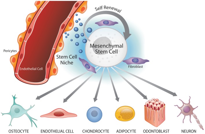
Postnatal stem cells are typically found in niches that provide signaling cues to maintain their self-renewal and multipotency. While stem cell populations may serve distinct purposes within their tissue of origin, understanding the conserved biology of stem cells and their respective niches provides insights to the behavior of these cells during homeostasis and tissue repair. Here, we discuss perivascular niches of two distinct stem cell populations (i.e., hematopoietic stem cells, mesenchymal stem cells) and explore mechanisms that sustain these stem cells postnatally. We highlight work that demonstrates the impact of cellular crosstalk to stem cell self-renewal and maintenance of functional perivascular niches. We also discuss the importance of the crosstalk within the perivascular niche to the biology of stem cells, and describe the regenerative potential of perivascular cells. We postulate that signaling events that establish and/or stabilize the perivascular niche, particularly through the modulation of self-renewing factors, are key to the long-term success of regenerated tissues.
Keywords: inflammation; perivascular niche; regenerative endodontics; tissue engineering; wound healing.
Figures
References
-
- Bianco P. (2007). Self-renewing mesenchymal progenitors in the bone marrow and in other mesodermal tissues. J. Stem Cells Regen. Med. 1, 44. - PubMed
Publication types
Grants and funding
LinkOut - more resources
Full Text Sources
Other Literature Sources