

The Right Supramarginal Gyrus Is Important for Proprioception in Healthy and Stroke-Affected Participants: A Functional MRI Study
- PMID: 26696951
- PMCID: PMC4668288
- DOI: 10.3389/fneur.2015.00248
The Right Supramarginal Gyrus Is Important for Proprioception in Healthy and Stroke-Affected Participants: A Functional MRI Study
Abstract
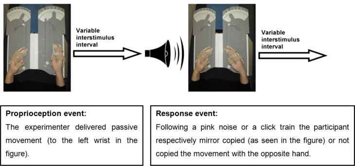
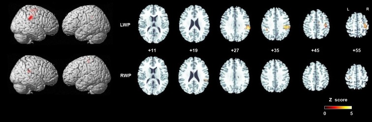
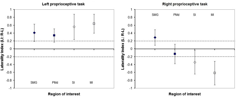
Human proprioception is essential for motor control, yet its central processing is still debated. Previous studies of passive movements and illusory vibration have reported inconsistent activation patterns related to proprioception, particularly in high-order sensorimotor cortices. We investigated brain activation specific to proprioception, its laterality, and changes following stroke. Twelve healthy and three stroke-affected individuals with proprioceptive deficits participated. Proprioception was assessed clinically with the Wrist Position Sense Test, and participants underwent functional magnetic resonance imaging scanning. An event-related study design was used, where each proprioceptive stimulus of passive wrist movement was followed by a motor response of mirror -copying with the other wrist. Left (LWP) and right (RWP) wrist proprioception were tested separately. Laterality indices (LIs) were calculated for the main cortical regions activated during proprioception. We found proprioception-related brain activation in high-order sensorimotor cortices in healthy participants especially in the supramarginal gyrus (SMG LWP z = 4.51, RWP z = 4.24) and the dorsal premotor cortex (PMd LWP z = 4.10, RWP z = 3.93). Right hemispheric dominance was observed in the SMG (LI LWP mean 0.41, SD 0.22; RWP 0.29, SD 0.20), and to a lesser degree in the PMd (LI LWP 0.34, SD 0.17; RWP 0.13, SD 0.25). In stroke-affected participants, the main difference in proprioception-related brain activation was reduced laterality in the right SMG. Our findings indicate that the SMG and PMd play a key role in proprioception probably due to their role in spatial processing and motor control, respectively. The findings from stroke--affected individuals suggest that decreased right SMG function may be associated with decreased proprioception. We recommend that clinicians pay particular attention to the assessment and rehabilitation of proprioception following right hemispheric lesions.
Keywords: cerebral cortex; functional laterality; kinesthesis; magnetic resonance imaging; proprioception; stroke; upper extremity.
Figures
Similar articles
-
Errors in proprioceptive matching post-stroke are associated with impaired recruitment of parietal, supplementary motor, and temporal cortices.Brain Imaging Behav. 2019 Dec;13(6):1635-1649. doi: 10.1007/s11682-019-00149-w. Brain Imaging Behav. 2019. PMID: 31218533
-
Investigating the neuroanatomy underlying proprioception using a stroke model.J Neurol Sci. 2021 Nov 15;430:120029. doi: 10.1016/j.jns.2021.120029. Epub 2021 Oct 12. J Neurol Sci. 2021. PMID: 34695704
-
Illusory limb movements activate different brain networks than imposed limb movements: an ALE meta-analysis.Brain Imaging Behav. 2018 Aug;12(4):919-930. doi: 10.1007/s11682-017-9756-1. Brain Imaging Behav. 2018. PMID: 28801769
-
Right hemisphere brain lateralization for knee proprioception among right-limb dominant individuals.Front Hum Neurosci. 2023 Jan 19;17:969101. doi: 10.3389/fnhum.2023.969101. eCollection 2023. Front Hum Neurosci. 2023. PMID: 36742357 Free PMC article.
-
Impaired limb position sense after stroke: a quantitative test for clinical use.Arch Phys Med Rehabil. 1996 Dec;77(12):1271-8. doi: 10.1016/s0003-9993(96)90192-6. Arch Phys Med Rehabil. 1996. PMID: 8976311 Review.
Cited by
-
Neural correlates linking trauma and physical symptoms.Psychiatry Res Neuroimaging. 2022 Dec;327:111560. doi: 10.1016/j.pscychresns.2022.111560. Epub 2022 Oct 26. Psychiatry Res Neuroimaging. 2022. PMID: 36327865 Free PMC article.
-
Localization of Impaired Kinesthetic Processing Post-stroke.Front Hum Neurosci. 2016 Oct 17;10:505. doi: 10.3389/fnhum.2016.00505. eCollection 2016. Front Hum Neurosci. 2016. PMID: 27799902 Free PMC article.
-
Frequency-Specific Changes of Amplitude of Low-Frequency Fluctuations in Patients with Acute Basal Ganglia Ischemic Stroke.Neural Plast. 2022 Jan 24;2022:4106131. doi: 10.1155/2022/4106131. eCollection 2022. Neural Plast. 2022. PMID: 35111218 Free PMC article.
-
Quantitative evaluation of shoulder proprioception 6 months following stroke.Egypt J Neurol Psychiatr Neurosurg. 2018;54(1):31. doi: 10.1186/s41983-018-0038-7. Epub 2018 Nov 6. Egypt J Neurol Psychiatr Neurosurg. 2018. PMID: 30459504 Free PMC article.
-
Beyond the Dorsal Column Medial Lemniscus in Proprioception and Stroke: A White Matter Investigation.Brain Sci. 2022 Dec 2;12(12):1651. doi: 10.3390/brainsci12121651. Brain Sci. 2022. PMID: 36552111 Free PMC article.
References
-
- Gandevia S, Burke D. Does the nervous system depend on kinesthetic information to control natural limb movements? Behav Brain Sci (1992) 15:614–32.
LinkOut - more resources
Full Text Sources
Other Literature Sources