. 2015 Dec 2:6:576.
doi: 10.3389/fimmu.2015.00576. eCollection 2015.

Augmented IFN-γ and TNF-α Induced by Probiotic Bacteria in NK Cells Mediate Differentiation of Stem-Like Tumors Leading to Inhibition of Tumor Growth and Reduction in Inflammatory Cytokine Release; Regulation by IL-10

Affiliations

Augmented IFN-γ and TNF-α Induced by Probiotic Bacteria in NK Cells Mediate Differentiation of Stem-Like Tumors Leading to Inhibition of Tumor Growth and Reduction in Inflammatory Cytokine Release; Regulation by IL-10

Vickie T Bui et al. Front Immunol. .

Abstract

Our previous reports demonstrated that the magnitude of natural killer (NK) cell-mediated cytotoxicity correlate directly with the stage and level of differentiation of tumor cells. In addition, we have shown previously that activated NK cells inhibit growth of cancer cells through induction of differentiation, resulting in the resistance of tumor cells to NK cell-mediated cytotoxicity through secreted cytokines, as well as direct NK-tumor cell contact. In this report, we show that in comparison to IL-2 + anti-CD16mAb-treated NK cells, activation of NK cells by probiotic bacteria (sAJ2) in combination with IL-2 and anti-CD16mAb substantially decreases tumor growth and induces maturation, differentiation, and resistance of oral squamous cancer stem cells, MIA PaCa-2 stem-like/poorly differentiated pancreatic tumors, and healthy stem cells of apical papillae through increased secretion of IFN-γ and TNF-α, as well as direct NK-tumor cell contact. Tumor resistance to NK cell-mediated killing induced by IL-2 + anti-CD16mAb + sAJ2-treated NK cells is induced by combination of IFN-γ and TNF-α since antibodies to both, and not each cytokine alone, were able to restore tumor sensitivity to NK cells. Increased surface expression of CD54, B7H1, and MHC-I on NK-differentiated tumors was mediated by IFN-γ since the addition of anti-IFN-γ abolished their increase and restored the ability of NK cells to trigger cytokine and chemokine release; whereas differentiated tumors inhibited cytokine release by the NK cells. Monocytes synergize with NK cells in the presence of probiotic bacteria to induce regulated differentiation of stem cells through secretion of IL-10 resulting in resistance to NK cell-mediated cytotoxicity and inhibition of cytokine release. Therefore, probiotic bacteria condition activated NK cells to provide augmented differentiation of cancer stem cells resulting in inhibition of tumor growth, and decreased inflammatory cytokine release.

Keywords: IFN-γ; NK cells; anti-IL10mAb; differentiation; monocytes; probiotic bacteria.


Figures

Figure 1
Figure 1
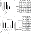
Schematic representation of steps used to differentiate cancer stem-like cells with NK cell supernatants and fixed NK cells after their activation. Highly purified NK cells were left untreated or treated with IL-2, sAJ2, monocytes, and/or anti-CD16mAb for 18 h, after which the treated NK cells were centrifuged, and the supernatants were removed and used to treat target cells. For cell–cell contact studies, treated NK cells were fixed with freshly prepared 2% paraformaldehyde for 15 min, washed three times with 1× PBS and added to target cell cultures. After the differentiation period, NK supernatants or fixed NK cells were removed from target cell cultures, and the targets were used in various assays.
Figure 2
Figure 2
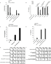
Resistance to NK cell-mediated lysis of OSCSCs treated with supernatants from IL-2 + anti-CD16mAb + sAJ2-stimulated NK cells is mediated by the combination of IFN-γ and TNF-α and not each cytokine alone. Highly purified NK cells were left untreated or treated with IL-2 (1,000 U/mL) and anti-CD16mAb (3 μg/mL) with or without sonicated AJ2 (sAJ2) at 1:3 (NK:sAJ2) ratio for 18 h before the supernatants were harvested and added to OSCSCs for 4 days. Afterwards, untreated OSCSCs and those treated with different NK cell supernatants indicated in the figure were detached from the tissue culture plates, extensively washed with 1× PBS and labeled with 51Cr. Freshly isolated NK cells were left untreated or treated with IL-2 (1,000 U/mL) for 24 h before the cells were used as effector cells in 51Cr release assay against OSCSCs treated with NK supernatants. The lytic units 30/106 cells were determined using inverse number of NK cells required to lyse 30% of the target cells ×100. Differences between untreated OSCSCs and those cultured with IL-2 + anti-CD16mAb-treated NK or those stimulated with IL-2 + anti-CD16mAb + sAJ2-treated NKs were significant at a P-value of < 0.05 (*) (A). OSCSCs were differentiated as described in (A), and after 4 days the surface expression of CD54, MHC-1, B7H1, and CD44 on untreated OSCSCs or those treated with NK cell supernatants as shown in the figure was assessed after staining with the PE-conjugated antibodies and analyzed using flow cytometry. Isotype control antibodies were used as controls. The numbers on the right hand corner are the percentages and the mean channel fluorescence intensities for each histogram (B). NK cells were treated with sAJ2 alone at 1:3 (NK:sAJ2) ratio or treated with IL-2 (1,000 U/mL) and anti-CD16mAb (3 μg/mL) and sAJ2 at 1:3 (NK:sAJ2) ratio in the presence of anti-TNF-α (1:100), anti-IFN-γ (1:100), or combination of anti-TNF-α (1:100) and anti-IFN-γ (1:100) as shown in the figure for 18 h. Afterwards, NK cell supernatants were harvested and added to OSCSCs for 4 days. Afterwards, untreated OSCSCs and those treated with NK cell supernatants were detached from the tissue culture plates, extensively washed with 1× PBS and labeled with 51Cr. Freshly isolated NK cells were left untreated or treated with IL-2 (1,000 U/mL) for 24 h before the cells were used as effector cells in 51Cr release assay against OSCSCs. The lytic units 30/106 cells were determined using inverse number of NK cells required to lyse 30% of the target cells ×100. Differences between untreated OSCSCs and those stimulated with IL-2 + anti-CD16mAb + sAJ2-treated NK with or without anti-TNF-α or anti-IFN-γ were significant at a P-value of <0.05 (*) (C). OSCSCs were treated with NK cell supernatants, as described in (C), and after 4 days the surface expression of CD54, MHC-1, and B7H1 on untreated tumor cells or those treated with NK cell supernatants was assessed after staining with the PE-conjugated antibodies and analyzed using flow cytometry. Isotype control antibodies were used as controls. The numbers on the right hand corner are the percentages and the mean channel fluorescence intensities for each histogram (D). OSCSCs were treated with NK cell supernatants as described in (C), and the percent cell death in OSCSCs was determined using PI staining followed by flow cytometric analysis. The numbers on the right hand corner are the percentages of PI-positive cells for each histogram (E). Purified NK cells were treated with a combination of IL-2 (1,000 U/mL) and anti-CD16mAb (3 μg/mL) in the presence or absence of sAJ2 at 1:3 (NK:sAJ2) ratio with or without anti-TNF-α (1:100) and/or anti-IFN-γ (1:100) for 18 h. Afterwards, supernatants were removed and used for the treatment of OSCSCs. OSCSCs (2 × 105 cells/well) were cultured overnight before they were treated with supernatants obtained from the NK cells treated as indicated in the figure for 4 days. At the end of the incubation, OSCSCs were detached, and the numbers of cells were assessed using microscopy (F). Freshly isolated NK cells were treated with IL-2 (1,000 U/mL) for 18 h. Afterwards, NK cells were added to untreated OSCSCs and those differentiated with NK cell supernatants, as described in (C), at an effector to target ratio of 0.5–1. After an overnight incubation, the supernatants were removed from the cocultures, and the levels of IFN-γ (G) and IL-8 (H) secretions were determined using specific ELISAs. Differences between untreated OSCSCs and those stimulated with IL-2 + anti-CD16mAb-treated NK or stimulated with IL-2 + anti-CD16mAb-treated NK + sAJ2 with or without anti-TNF-α were significant at a P-value of <0.05 (*).
Figure 2
Figure 2
Resistance to NK cell-mediated lysis of OSCSCs treated with supernatants from IL-2 + anti-CD16mAb + sAJ2-stimulated NK cells is mediated by the combination of IFN-γ and TNF-α and not each cytokine alone. Highly purified NK cells were left untreated or treated with IL-2 (1,000 U/mL) and anti-CD16mAb (3 μg/mL) with or without sonicated AJ2 (sAJ2) at 1:3 (NK:sAJ2) ratio for 18 h before the supernatants were harvested and added to OSCSCs for 4 days. Afterwards, untreated OSCSCs and those treated with different NK cell supernatants indicated in the figure were detached from the tissue culture plates, extensively washed with 1× PBS and labeled with 51Cr. Freshly isolated NK cells were left untreated or treated with IL-2 (1,000 U/mL) for 24 h before the cells were used as effector cells in 51Cr release assay against OSCSCs treated with NK supernatants. The lytic units 30/106 cells were determined using inverse number of NK cells required to lyse 30% of the target cells ×100. Differences between untreated OSCSCs and those cultured with IL-2 + anti-CD16mAb-treated NK or those stimulated with IL-2 + anti-CD16mAb + sAJ2-treated NKs were significant at a P-value of < 0.05 (*) (A). OSCSCs were differentiated as described in (A), and after 4 days the surface expression of CD54, MHC-1, B7H1, and CD44 on untreated OSCSCs or those treated with NK cell supernatants as shown in the figure was assessed after staining with the PE-conjugated antibodies and analyzed using flow cytometry. Isotype control antibodies were used as controls. The numbers on the right hand corner are the percentages and the mean channel fluorescence intensities for each histogram (B). NK cells were treated with sAJ2 alone at 1:3 (NK:sAJ2) ratio or treated with IL-2 (1,000 U/mL) and anti-CD16mAb (3 μg/mL) and sAJ2 at 1:3 (NK:sAJ2) ratio in the presence of anti-TNF-α (1:100), anti-IFN-γ (1:100), or combination of anti-TNF-α (1:100) and anti-IFN-γ (1:100) as shown in the figure for 18 h. Afterwards, NK cell supernatants were harvested and added to OSCSCs for 4 days. Afterwards, untreated OSCSCs and those treated with NK cell supernatants were detached from the tissue culture plates, extensively washed with 1× PBS and labeled with 51Cr. Freshly isolated NK cells were left untreated or treated with IL-2 (1,000 U/mL) for 24 h before the cells were used as effector cells in 51Cr release assay against OSCSCs. The lytic units 30/106 cells were determined using inverse number of NK cells required to lyse 30% of the target cells ×100. Differences between untreated OSCSCs and those stimulated with IL-2 + anti-CD16mAb + sAJ2-treated NK with or without anti-TNF-α or anti-IFN-γ were significant at a P-value of <0.05 (*) (C). OSCSCs were treated with NK cell supernatants, as described in (C), and after 4 days the surface expression of CD54, MHC-1, and B7H1 on untreated tumor cells or those treated with NK cell supernatants was assessed after staining with the PE-conjugated antibodies and analyzed using flow cytometry. Isotype control antibodies were used as controls. The numbers on the right hand corner are the percentages and the mean channel fluorescence intensities for each histogram (D). OSCSCs were treated with NK cell supernatants as described in (C), and the percent cell death in OSCSCs was determined using PI staining followed by flow cytometric analysis. The numbers on the right hand corner are the percentages of PI-positive cells for each histogram (E). Purified NK cells were treated with a combination of IL-2 (1,000 U/mL) and anti-CD16mAb (3 μg/mL) in the presence or absence of sAJ2 at 1:3 (NK:sAJ2) ratio with or without anti-TNF-α (1:100) and/or anti-IFN-γ (1:100) for 18 h. Afterwards, supernatants were removed and used for the treatment of OSCSCs. OSCSCs (2 × 105 cells/well) were cultured overnight before they were treated with supernatants obtained from the NK cells treated as indicated in the figure for 4 days. At the end of the incubation, OSCSCs were detached, and the numbers of cells were assessed using microscopy (F). Freshly isolated NK cells were treated with IL-2 (1,000 U/mL) for 18 h. Afterwards, NK cells were added to untreated OSCSCs and those differentiated with NK cell supernatants, as described in (C), at an effector to target ratio of 0.5–1. After an overnight incubation, the supernatants were removed from the cocultures, and the levels of IFN-γ (G) and IL-8 (H) secretions were determined using specific ELISAs. Differences between untreated OSCSCs and those stimulated with IL-2 + anti-CD16mAb-treated NK or stimulated with IL-2 + anti-CD16mAb-treated NK + sAJ2 with or without anti-TNF-α were significant at a P-value of <0.05 (*).
Figure 3
Figure 3
Paraformaldehyde fixed IL-2 + anti-CD16 mAb + sAJ2-treated NK cells mediated differentiation and resistance of OSCSCs against NK cell-mediated cytotoxicity. NK cells were left untreated or treated with a combination or IL-2 (1,000 U/mL) and anti-CD16mAb (3 μg/mL) and sAJ2 at 1:3 (NK:sAJ2) ratio in the presence of anti-TNF-α (1:100), anti-IFN-γ (1:100), or combination of anti-TNF-α (1:100) and anti-IFN-γ (1:100) for 18 h. Afterwards, supernatants were removed, and the NK cells were fixed with freshly prepared 2% paraformaldehyde for 15 min. NK cells were then washed three times with 1× PBS and added to tumor cultures. Differentiation of OSCSCs was conducted for 5 days with daily and gradual addition of increasing amounts of fixed NK cells. The complete removal of fixed NK cells from tumor cell cultures prior to the cytotoxicity assays was determined by microscopic assessment. Freshly isolated NK cells were left untreated or treated with IL-2 (1,000 U/mL) for 18 h before the cells were used as effector cells in 51Cr release assay against tumor cells cultured with the NK cells. The lytic units 30/106 cells were determined using inverse number of NK cells required to lyse 30% of the target cells ×100. Differences between untreated OSCSCs and those stimulated with fixed IL-2 + anti-CD16mAb-treated NK or IL-2 + anti-CD16mAb-treated NK + sAJ2 with or without anti-TNF-α or anti-IFN-γ but not with the combination of anti-TNF-α and anti-IFN-γ were significant at a P-value of <0.05 (*) (A). OSCSCs were treated with fixed NK cells as described in (A). The surface expressions of CD54 and MHC-1 on OSCSCs were assessed after staining with the PE-conjugated antibodies and analyzed using flow cytometry. Isotype control antibodies were used as controls. The numbers on the right hand corner are the percentages and the mean channel fluorescence intensities for each histogram (B). NK cells were prepared as described in (A) and added to OSCSCs for 5 days. Afterwards, the tumor cells were washed with 1× PBS, and their viability was determined using PI staining followed by flow cytometric analysis (C). The numbers on the right hand corner are the percentages of dead cells in each histogram.
Figure 4
Figure 4
Monocytes and probiotic bacteria synergistically induce split anergy in NK cells to increase tumor cell differentiation. Purified NK cells were left untreated, treated with IL-2 (1000U/mL) or a combination of IL-2 (1000U/mL) and anti-CD16mAb (3μg/mL) in the presence or absence of autologous monocytes (1:0.2, NK cell:monocytes) and/or sAJ2 (1:0.5, NK cell:sAJ2) ratios for 18 hours before the supernatants were harvested and added to OSCSCs. Afterwards, untreated OSCSCs and those treated with different NK cell supernatants indicated in the figure were detached from the tissue culture plates, extensively washed with 1X PBS and labeled with 51Cr. Freshly isolated NK cells were left untreated or treated with IL-2 (1000U/mL) for 24 hours before the cells were used as effector cells in 51Cr release assay against OSCSCs treated with NK supernatants. The lytic units 30/106 cells were determined using inverse number of NK cells required to lyse 30% of the target cells X100. NK cell cytotoxicity was determined using a standard 4 hours 51Cr release assay and the lytic units 30/106 were determined using inverse number of NK cells required to lyse 30% of the target cells. Differences between untreated NK cells and those cultured with monocytes and/or sAJ2 were significant at a P-value of < 0.05 (*) (A). NK cells were prepared as described in (A). After days 1, 4, 6, and 8, the supernatants were removed from the cocultures, and the levels of IFN-γ secretions were determined using specific ELISAs. Differences between untreated NK cells and those stimulated with IL-2 + anti-CD16mAb-treated NK cells or treated with IL-2 + anti-CD16mAb-treated NK cells + monocytes with or without sAJ2 were significant at a P-value of <0.05 (*) (B). Purified NK cells were left untreated or treated with a combination of IL-2 (1,000 U/mL) and anti-CD16mAb (3 μg/mL) in the presence or absence of monocytes (1:0.2, NK cell:monocytes) for 18 h. Afterwards, the supernatants were removed, and the levels of IL-10 were determined using specific ELISAs. Differences between IL-2 + anti-CD16mAb-treated NK cells and IL-2 + anti-CD16mAb-treated NK cells cultured with monocytes were significant at a P-value of <0.05 (*) (C). NK cells were prepared as described in (A). After 36 h, the supernatants were removed from the cocultures, and the levels of IL-10 secretions were determined using specific ELISAs. Differences between unstimulated NK cells and those stimulated with sAJ2 and/or monocytes were significant at a P-value of <0.05 (*) (D). Purified untreated NK cells were cultured in the presence and absence of untreated monocytes with and without sAJ2 for 18 hours. Then the supernatants were added to OSCSCs for 4 days. Afterwards, OSCSCs were removed and the surface expressions of B7H1, CD54, MHC-1, and CD44 on untreated OSCSCs and those treated with NK cells were assessed after staining with the PE conjugated antibodies and analyzed using flow cytometry (E). NK cells were left untreated or treated with IL-2 (1,000 U/mL) and anti-CD16mAb (3 μg/mL) in the presence of monocytes (1:0.5, NK cell:monocytes) and/or sAJ2 (1:0.5, NK:sAJ2) for 18 h, and then the supernatants were removed and added to OSCSCs for 4 days. Afterwards, OSCSCs were removed, and the surface expressions of B7H1, CD54, MHC-1, and CD44 on untreated OSCSCs and those treated with NK cells were assessed after staining with the PE-conjugated antibodies and analyzed using flow cytometry. Isotype control antibodies were used as controls. The numbers on the right hand corner are the percentages and the mean channel fluorescence intensities for each histogram (F).
Figure 5
Figure 5
IL-10 regulation of NK cell mediated CSC differentiation in the presence of sAJ2 and monocytes is much more pronounced in untreated NK cells when compared to those treated with IL-2 and anti-CD16mAb. Highly purified NK cells were left untreated or treated with IL-2 (1,000 U/mL) and anti-CD16mAb (3 μg/mL) with sAJ2 at 1:3 (NK:sAJ2) ratio, monocytes at 1:1 (NK:monocyte) ratio, and/or anti-IL-10 (10 μg/mL) for 18 h. Afterwards, supernatants were harvested and added to OSCSCs for a period of 3 days. Then, untreated OSCSCs and those treated with NK cell supernatants were detached from the tissue culture plates, washed with 1× PBS and labeled with 51Cr. Freshly isolated NK cells were left untreated or treated with IL-2 (1,000 U/mL) for 24 h before the cells were used as effector cells in 51Cr release assay against OSCSCs. The lytic units 30/106 cells were determined using inverse number of NK cells required to lyse 30% of the target cells ×100 (A). NK cells were prepared as described in (A), and after an overnight incubation period, the supernatants were harvested, and the levels of IFN-γ secretions were determined using specific ELISAs. Differences between untreated NK cells and those cultured with monocytes and sAJ2 were significant at a P-value of <0.05 (*) (B). Untreated NK cells were cultured with autologous monocytes at 1:1 (NK:monocyte) ratio in the presence or absence of anti-IL-10 (10 μg/mL) and/or sAJ2 at 1:3 (NK:sAJ2) ratio for 18 h. Anti-IL-10 (10 μg/mL) was added to untreated NK cells and monocytes and used as controls. After an overnight treatment period, the supernatants were removed and added to OSCSCs for 3 days (C). NK cells supernatants were prepared as described in (A) and added to OSCSCs for 3 days (D). Afterwards, untreated and treated OSCSCs were detached from the tissue culture plate and washed with 1× PBS. The surface expression of B7H1, CD54, MHC-1, and CD44 was assessed after staining with the PE-conjugated antibodies and analyzed using flow cytometry. Isotype control antibodies were used as controls. The numbers on the right-hand corner are the percentages, and the mean channel fluorescence intensities for each histogram (C) and (D). OSCSCs were differentiated with NK cell supernatants as described in (D), and at the end of the treatment period, the viability of OSCSCs was assessed with propidium iodide staining followed by flow cytometric analysis (E). The numbers on the right hand corner are the percentages of dead cells in each histogram.
Figure 5
Figure 5
IL-10 regulation of NK cell mediated CSC differentiation in the presence of sAJ2 and monocytes is much more pronounced in untreated NK cells when compared to those treated with IL-2 and anti-CD16mAb. Highly purified NK cells were left untreated or treated with IL-2 (1,000 U/mL) and anti-CD16mAb (3 μg/mL) with sAJ2 at 1:3 (NK:sAJ2) ratio, monocytes at 1:1 (NK:monocyte) ratio, and/or anti-IL-10 (10 μg/mL) for 18 h. Afterwards, supernatants were harvested and added to OSCSCs for a period of 3 days. Then, untreated OSCSCs and those treated with NK cell supernatants were detached from the tissue culture plates, washed with 1× PBS and labeled with 51Cr. Freshly isolated NK cells were left untreated or treated with IL-2 (1,000 U/mL) for 24 h before the cells were used as effector cells in 51Cr release assay against OSCSCs. The lytic units 30/106 cells were determined using inverse number of NK cells required to lyse 30% of the target cells ×100 (A). NK cells were prepared as described in (A), and after an overnight incubation period, the supernatants were harvested, and the levels of IFN-γ secretions were determined using specific ELISAs. Differences between untreated NK cells and those cultured with monocytes and sAJ2 were significant at a P-value of <0.05 (*) (B). Untreated NK cells were cultured with autologous monocytes at 1:1 (NK:monocyte) ratio in the presence or absence of anti-IL-10 (10 μg/mL) and/or sAJ2 at 1:3 (NK:sAJ2) ratio for 18 h. Anti-IL-10 (10 μg/mL) was added to untreated NK cells and monocytes and used as controls. After an overnight treatment period, the supernatants were removed and added to OSCSCs for 3 days (C). NK cells supernatants were prepared as described in (A) and added to OSCSCs for 3 days (D). Afterwards, untreated and treated OSCSCs were detached from the tissue culture plate and washed with 1× PBS. The surface expression of B7H1, CD54, MHC-1, and CD44 was assessed after staining with the PE-conjugated antibodies and analyzed using flow cytometry. Isotype control antibodies were used as controls. The numbers on the right-hand corner are the percentages, and the mean channel fluorescence intensities for each histogram (C) and (D). OSCSCs were differentiated with NK cell supernatants as described in (D), and at the end of the treatment period, the viability of OSCSCs was assessed with propidium iodide staining followed by flow cytometric analysis (E). The numbers on the right hand corner are the percentages of dead cells in each histogram.

References

    1. Jewett A, Tseng HC, Arasteh A, Saadat S, Christensen RE, Cacalano NA. Natural killer cells preferentially target cancer stem cells; role of monocytes in protection against NK cell mediated lysis of cancer stem cells. Curr Drug Deliv (2012) 9(1):5–16.10.2174/156720112798375989 - DOI - PubMed
    1. Jewett A, Man YG, Tseng HC. Dual functions of natural killer cells in selection and differentiation of stem cells; role in regulation of inflammation and regeneration of tissues. J Cancer (2013) 4(1):12–24.10.7150/jca.5045 - DOI - PMC - PubMed
    1. Jewett A, Man YG, Cacalano N, Kos J, Tseng HC. Natural killer cells as effectors of selection and differentiation of stem cells: role in resolution of inflammation. J Immunotoxicol (2014) 11(4):297–307.10.3109/1547691X.2013.877104 - DOI - PubMed
    1. Tseng HC, Bui V, Man YG, Cacalano N, Jewett A. Induction of split anergy conditions natural killer cells to promote differentiation of stem cells through cell-cell contact and secreted factors. Front Immunol (2014) 5:269.10.3389/fimmu.2014.00009 - DOI - PMC - PubMed
    1. Metchnikoff E. Essais Optimistes. Paris: A. Maloine; (1907).