. 2015 Dec 2:6:340.
doi: 10.3389/fgene.2015.00340. eCollection 2015.

Transfection of microRNA Mimics Should Be Used with Caution

Affiliations

Transfection of microRNA Mimics Should Be Used with Caution

Hyun Yong Jin et al. Front Genet. .

Abstract

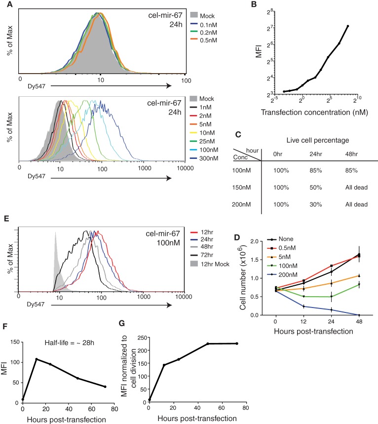
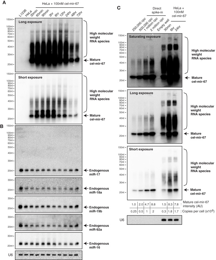
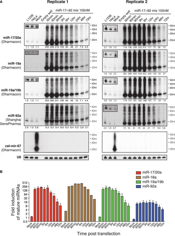
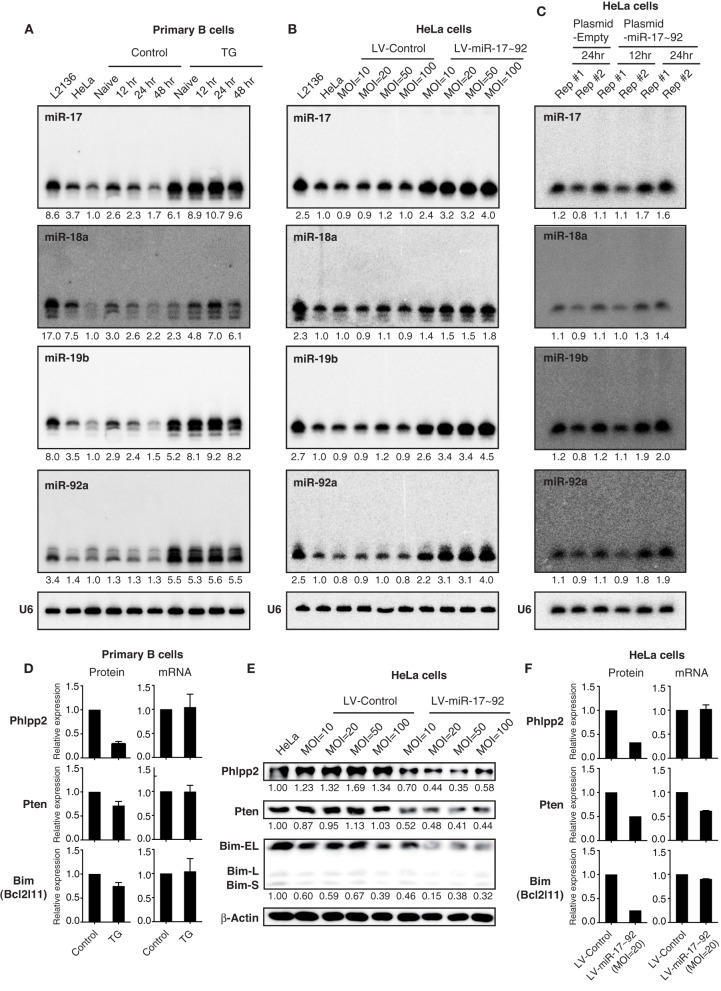
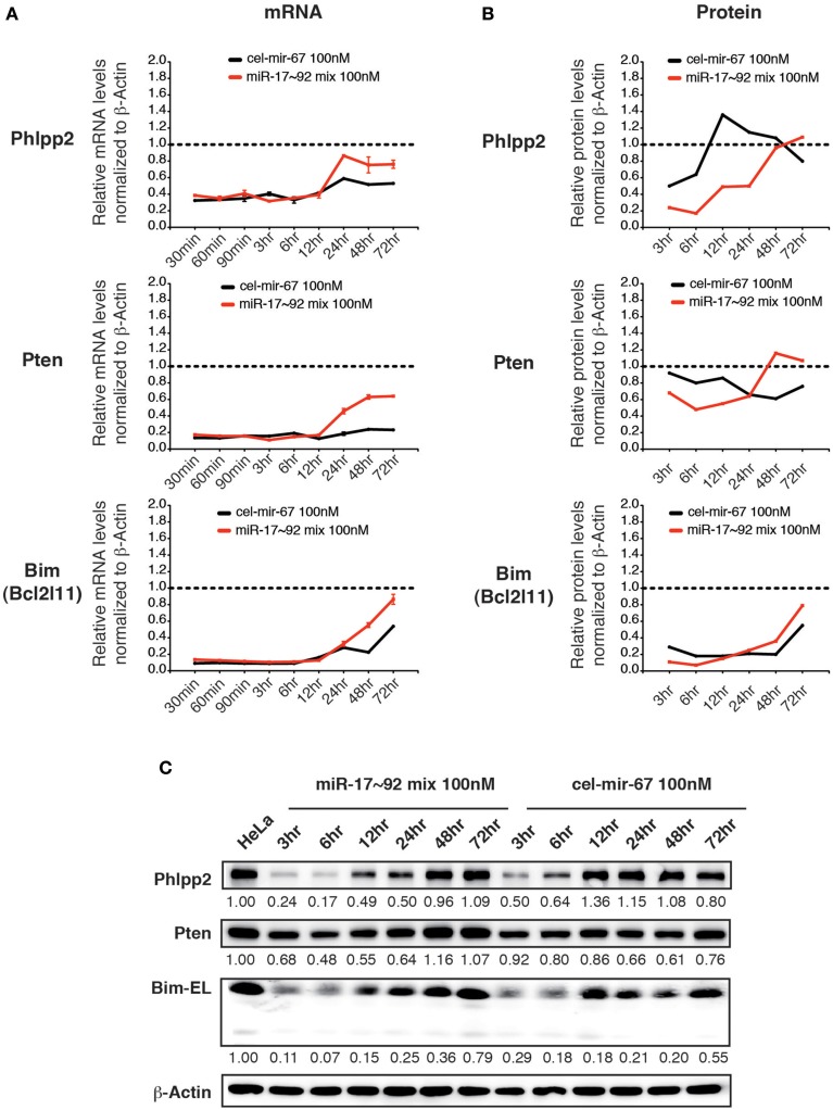
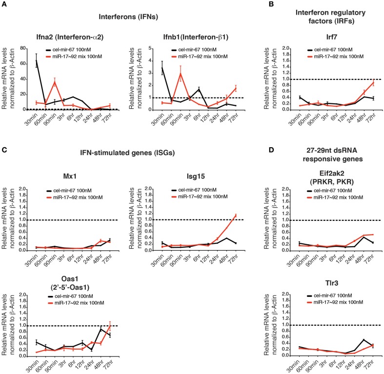
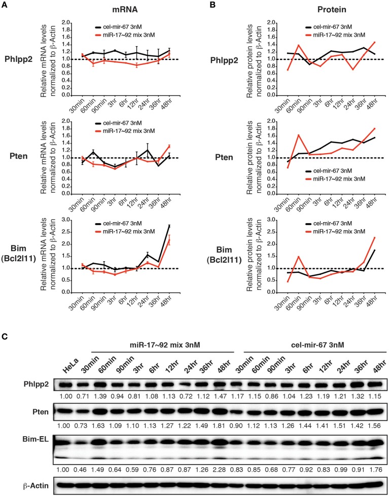
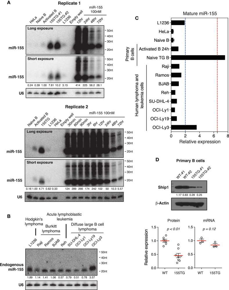
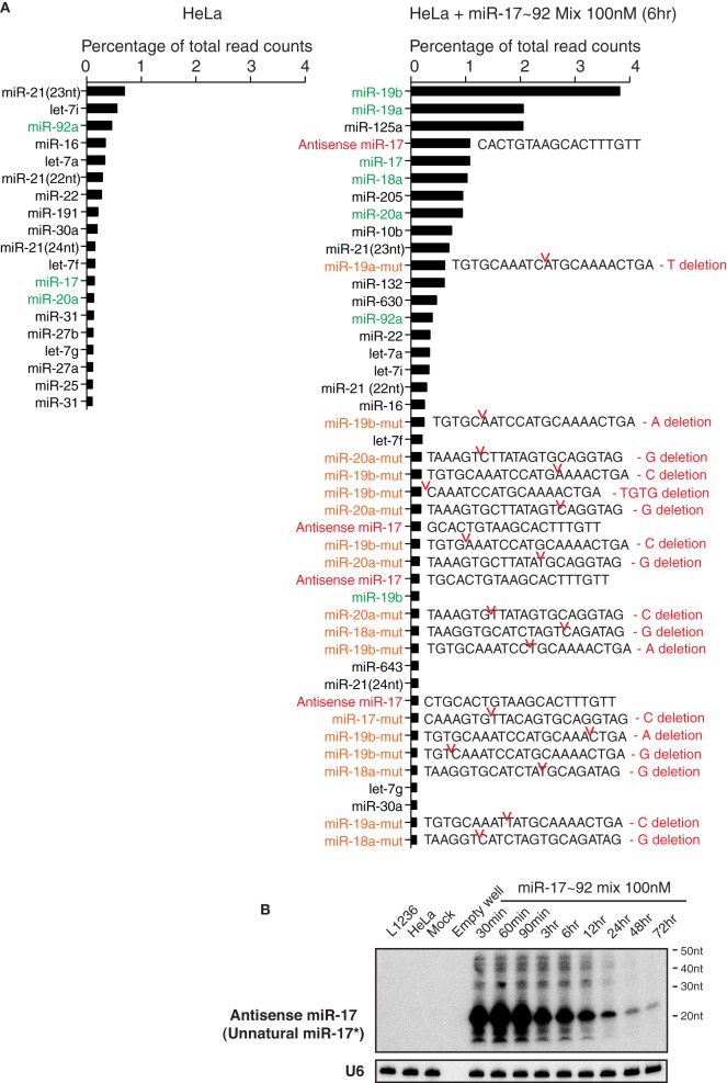
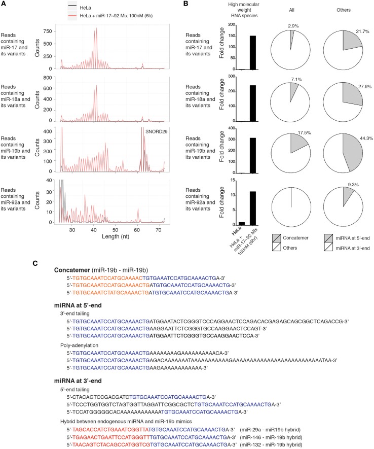
Transient transfection of chemically synthesized microRNA (miRNA) mimics is being used extensively to study the functions and mechanisms of endogenous miRNAs. However, it remains unclear whether transfected miRNAs behave similarly to endogenous miRNAs. Here we show that transient transfection of miRNA mimics into HeLa cells by a commonly used method led to the accumulation of high molecular weight RNA species and a few hundred fold increase in mature miRNA levels. In contrast, expression of the same miRNAs through lentiviral infection or plasmid transfection of HeLa cells, transgenic expression in primary lymphocytes, and endogenous overexpression in lymphoma and leukemia cell lines did not lead to the appearance of high molecular weight RNA species. The increase of mature miRNA levels in these cells was below 10-fold, which was sufficient to suppress target gene expression and to drive lymphoma development in mice. Moreover, transient transfection of miRNA mimics at high concentrations caused non-specific alterations in gene expression, while at low concentrations achieved expression levels comparable to other methods but failed to efficiently suppress target gene expression. Small RNA deep sequencing analysis revealed that the guide strands of miRNA mimics were frequently mutated, while unnatural passenger strands of some miRNA mimics accumulated to high levels. The high molecular weight RNA species were a heterogeneous mixture of several classes of RNA species generated by concatemerization, 5'- and 3'-end tailing of miRNA mimics. We speculate that the supraphysiological levels of mature miRNAs and these artifactual RNA species led to non-specific changes in gene expression. Our results have important implications for the design and interpretation of experiments primarily employing transient transfection of miRNA mimics.

Keywords: guide strand mutation; high molecular weight RNA species; miR-155; miR-17~92; microRNA mimics; transient transfection; unnatural passenger strand.


Figures

Figure 1
Figure 1
Transient transfection of cel-mir-67 into HeLa cells. (A) HeLa cells were transfected with indicated concentrations of Dy547-conjugated cel-mir-67. Dy547 signals were analyzed by flow cytometry at 24 h post-transfection. (B) Correlation summary of Dy547 mean fluorescence intensity (MFI) and transfection concentrations of cel-mir-67. (C) Live cell percentage was determined by trypan blue staining of cells transfected with indicated concentrations of Dy547-conjugated cel-mir-67 at indicated time points. (D) Growth curves of HeLa cells transfected with indicated concentrations of cel-mir-67. (E–G) HeLa cells transfected with 100 nM Dy547-conjugated cel-mir-67 and Dy547 signals were analyzed by flow cytometry at indicated time points after transfection (E). Dy547 MFI was plotted against time to calculate its half-life (F). Dy547 MFI in (F) was normalized to cell division (G). Mock, HeLa cells transfected with 100 nM un-conjugated cel-mir-67.
Figure 2
Figure 2
Northern blot analysis of cel-mir-67 and endogenous miRNAs in HeLa cells transiently transfected with 100 nM cel-mir-67. HeLa cells were transfected with 100 nM unconjugated cel-mir-67 and analyzed by Northern blot to detect cel-mir-67 (A) and endogenous miR-17, miR-18a, miR-19b, miR-92a, and miR-16 (B). Arrows indicate mature cel-mir-67 and brackets indicate high molecular weight RNA species. L1236, a human Hodgkin's lymphoma cell line that expresses 2–3-fold more miR-17~92 than HeLa cells, was used as a universal reference throughout the manuscript. (C) The copy numbers of cel-mir-67 in transfected HeLa cells were estimated by comparing to graded amounts of the same miRNA mimic loaded directly into the Northern blot gel. Three different exposures of the same blot showed that the high molecular RNA species were generated by both the manufacturing process (before transfection) and cellular processes (after transfection), cpc, copies per cell. AU, arbitrary unit. The amount of transfected mir-cel-67 was estimated by comparing the mature signals of transfected mir-cel-67 to the spiked-in cel-mir-67.
Figure 3
Figure 3
Northern blot analysis of miR-17~92 miRNAs in HeLa cells transiently transfected with miR-17~92 mimics. (A) HeLa cells were transfected with an equal molar mixture of miR-17, 18a, 19a, 20a, 19b, and 92a (16.7 nM each with a total transfection concentration of 100 nM) and analyzed by Northern blot to detect these miRNAs. Mixed probes were used for the detection of miRNAs in each subfamily. Numbers below each blot image indicate fold-induction of mature miRNAs (~22 nt). Mature miRNA expression levels in non-transfected HeLa cells were set as 1. L1236 cells and HeLa cells transfected with 100 nM cel-mir-67 (Mock) were used as controls. Small boxes in the upper left corner of Northern blots are longer exposures to show mature miRNAs in the same lanes. (B) Quantification of mature miRNA levels in (A). Endogenous miR-18a in Replicate 1 was not detectable and was excluded from quantification.
Figure 4
Figure 4
Northern blot analysis of miRNAs in HeLa cells transiently transfected with low concentrations of miR-17~92. (A) HeLa cells were transfected with an equal molar mixture of miR-17, 19a, 20a, 19b (0.5 nM each with a total transfection concentration of 2 nM) and analyzed by Northern blot to detect these miRNAs. Mixed probes were used for the detection of miRNAs in each family. Numbers below blots indicate fold increase of mature miRNA levels. Mature miRNA expression levels in non-transfected HeLa cells were set as 1. (B) Northern blot analysis of endogenous miR-17~92 levels in nine human lymphoma or leukemia cell lines. Mixed probes were used for the detection of miRNAs in each family. Numbers below blots indicate fold increase of mature miRNA levels. Mature miRNA expression levels in L1236 were set as 1. (C) Summary of miR-17 expression levels in samples used in this study. Results for primary B cells, lentivirally transduced (LV-17~92) and plasmid transfected (Plasmid-17~92) HeLa cells were from Figure 5. Red and blue dashed lines indicate the maximal and mean miR-17 expression levels in human Burkitt's lymphoma patients, respectively (Schmitz et al., 2012). Mature miR-17 expression level in naïve B cells was set as 1.
Figure 5
Figure 5
The effect of transgenic and lentiviral miR-17~92 expression on target genes. (A–C) Northern blot analysis of miR-17~92 expression. Primary B cells were purified from CD19-Cre (Control) and miR-17~92 Tg/Tg; CD19-Cre (TG) mice, stimulated in vitro for indicated amounts of time (A). HeLa cells were stably transduced with control or miR-17~92 expressing lentiviruses at indicated MOI (Multiplicity of infection) (B). HeLa cells were transfected with empty plasmid or miR-17~92-expressing plasmid (Plasmid-miR-17~92) and harvested at indicated time points (C). Numbers below blots indicate miR-17~92 expression normalized to U6. miRNA/U6 ratios in naïve control B cells (A), non-transduced HeLa cells (B), and empty plasmid transfected HeLa (C) were arbitrarily set as 1. (D) Quantification of protein and mRNA levels of target genes in primary B cells (n = 4). The representative western blot results were presented previously (Jin et al., 2013). (E) Western blot analysis of miR-17~92 target gene expression in lentivirally transduced HeLa cells. Different MOI are indicated. Numbers below blots indicate target gene expression normalized to β-Actin. The ratios in parental HeLa cells were arbitrarily set as 1. (F) Quantification of protein and mRNA levels of target genes in lentivirally transduced HeLa cells. (D–F) Target gene protein and mRNA levels were determined by Western blot and qRT-PCR, respectively.
Figure 6
Figure 6
The effect of high concentration transient transfection on target gene expression. HeLa cells were transiently transfected with miRNA mimics. Target gene mRNA and protein levels were determined by qRT-PCR and Western blot at indicated time points and normalized to β-Actin. The target gene/β-Actin ratios of non-transfected HeLa cells were arbitrarily set as 1 (dashed line). miR-17~92 mix 100 nM contains an equal molar mixture of miR-17~92 miRNAs at 16.7 nM for each miRNA with a total concentration of 100 nM. (A,B) Summary of mRNA and protein quantification results. (C) Representative Western blots.
Figure 7
Figure 7
The effect of high concentration transient transfection on innate immune responses. The mRNA levels of genes involved in innate immune response to dsRNA were determined by qRT-PCR and normalized to β-Actin. The target gene/β-Actin ratios of non-transfected HeLa cells were arbitrarily set as 1 (dashed line). Type I interferons (A), interferon regulatory factors (B), interferon stimulated genes (C) and genes specifically responding to 27–29 nt long dsRNA (D) were investigated.
Figure 8
Figure 8
The effect of low concentration transient transfection on target gene expression. HeLa cells were transfected with an equal molar mixture of miR-17~92 miRNAs at 0.5 nM for each miRNA with a total concentration of 3 nM. Target gene mRNA and protein levels were determined by qRT-PCR and Western blot at indicated time points and normalized to β-Actin. The target gene/β-Actin ratios of non-transfected HeLa cells were arbitrarily set as 1 (dashed line). (A,B) Summary of mRNA and protein quantification results. (C) Representative Western blots.
Figure 9
Figure 9
The effect of transient transfection and transgenic expression of miR-155. (A) Northern blot analysis of miR-155 expression in HeLa cells transiently transfected with a miR-155 mimic, and in primary B cells from wild type and miR-155 transgenic mice (155TG). Two different exposures of the same blot are included. (B) Northern blot analysis of endogenous miR-155 expression in nine human lymphoma or leukemia cell lines. (C) Summary of miR-155 Northern blots in (A,B). Mature miR-155 expression in in vitro activated B cells was arbitrarily set as 1 in (A–C). Red and blue dashed lines in (C) indicate the maximal and mean miR-155 expression levels in human lymphoma patients, respectively. This was estimated from absolute copy numbers of miR-155 in activated B cells (data not shown) compared to that in lymphoma patients (Eis et al., 2005). (D) Ship1 expression in miR-155 transgenic B cells. Upper panel, a representative Western blot. Lower panel, bar graphs summarizing Ship1 protein and mRNA levels in miR-155 transgenic B cells as determined by Western blot and Microarray. Each circle indicates an independent biologic sample.
Figure 10
Figure 10
Transient transfection of miRNA mimics led to the accumulation of mutated miRNAs and unnatural miRNA passenger strands. (A) Non-transfected HeLa cells and HeLa cells transfected with miR-17~92 miRNA mimics were analyzed by small RNA deep sequencing at 6 h after transfection (the same sample as in Figure 3A, replicate 1, miR-17~92 mix 100 nM). Mature miRNAs, antisense miRNAs, and their mutant forms with abundance higher than 0.1% of total reads are listed. Note that non-transfected HeLa cells do not express antisense miRNAs or mutant miRNAs at levels higher than this cut off, while HeLa cells transfected with miR-17~92 mix contain many of these species. Antisense miR-17 sequences (marked in red) are perfectly complementary to miR-17, and differ from miR-17*, the endogenous form of miR-17 passenger strand, by a few nucleotides. miR-17~92 miRNAs are marked in green, and their mutant forms are marked in orange. Sequences of the latter are included. Red notches indicate the location of deletion, with deleted nucleotides presented at the right. (B) Northern blot analysis of antisense miR-17 (as indicated in the right panel of A) in transfected HeLa cells. The blot from Figure 3A Replicate 1 was hybridized with a probe specific for antisense miR-17.
Figure 11
Figure 11
Characterization of high molecular weight RNA species. (A) Length distribution of high molecular weight RNA species identified in the small RNA deep sequencing data of Figure 10. Each peak represents the read count of high molecular weight RNA species of indicated length. SNORD29 contains sequence region highly homologous to miR-19b and is detected in both non-transfected and miR-17~92 mix-transfected HeLa cells. (B) Fold change of high molecular weight RNA species in miR-17~92 mix-transfected HeLa cells. The abundance in non-transfected HeLa cells was arbitrarily set as 1. RNA species of 30–60 nt are included in the calculation to exclude SNORD29. The relative abundance of concatemer, 5′-tailing, and 3′-tailing are presented in pie charts. (C) Representatives of high molecular weight RNA species, using miR-19-containing sequences as examples.

Similar articles

Cited by

References

    1. Agudo J., Ruzo A., Tung N., Salmon H., Leboeuf M., Hashimoto D., et al. . (2014). The miR-126-VEGFR2 axis controls the innate response to pathogen-associated nucleic acids. Nat. Immunol. 15, 54–62. 10.1038/ni.2767 - DOI - PMC - PubMed
    1. Ambros V. (2004). The functions of animal microRNAs. Nature 431, 350–355. 10.1038/nature02871 - DOI - PubMed
    1. Ameres S. L., Horwich M. D., Hung J. H., Xu J., Ghildiyal M., Weng Z., et al. . (2010). Target RNA-directed trimming and tailing of small silencing RNAs. Science 328, 1534–1539. 10.1126/science.1187058 - DOI - PMC - PubMed
    1. Ameres S. L., Zamore P. D. (2013). Diversifying microRNA sequence and function. Nat. Rev. Mol. Cell Biol. 14, 475–488. 10.1038/nrm3611 - DOI - PubMed
    1. Baek D., Villén J., Shin C., Camargo F. D., Gygi S. P., Bartel D. P. (2008). The impact of microRNAs on protein output. Nature 455, 64–71. 10.1038/nature07242 - DOI - PMC - PubMed