. 2016:2016:3710836.
doi: 10.1155/2016/3710836. Epub 2015 Nov 30.

Three-Dimensional Gastrointestinal Organoid Culture in Combination with Nerves or Fibroblasts: A Method to Characterize the Gastrointestinal Stem Cell Niche

Affiliations

Three-Dimensional Gastrointestinal Organoid Culture in Combination with Nerves or Fibroblasts: A Method to Characterize the Gastrointestinal Stem Cell Niche

Agnieszka Pastuła et al. Stem Cells Int. 2016.

Abstract

The gastrointestinal epithelium is characterized by a high turnover of cells and intestinal stem cells predominantly reside at the bottom of crypts and their progeny serve to maintain normal intestinal homeostasis. Accumulating evidence demonstrates the pivotal role of a niche surrounding intestinal stem cells in crypts, which consists of cellular and soluble components and creates an environment constantly influencing the fate of stem cells. Here we describe different 3D culture systems to culture gastrointestinal epithelium that should enable us to study the stem cell niche in vitro in the future: organoid culture and multilayered systems such as organotypic cell culture and culture of intestinal tissue fragments ex vivo. These methods mimic the in vivo situation in vitro by creating 3D culture conditions that reflect the physiological situation of intestinal crypts. Modifications of the composition of the culture media as well as coculturing epithelial organoids with previously described cellular components such as myofibroblasts, collagen, and neurons show the impact of the methods applied to investigate niche interactions in vitro. We further present a novel method to isolate labeled nerves from the enteric nervous system using Dclk1-CreGFP mice.


Figures

Figure 1
Figure 1
Summary of three-dimensional methods to culture intestinal epithelium. (a) Tissue culture ex vivo. Arrowhead indicates stromal cells. (b) Organotypic cell culture. (c) Organoid culture. PAS staining. R-Spo, R-Spondin.
Figure 2
Figure 2
Characterization of small intestinal organoids and small intestinal myofibroblasts used to reconstruct intestinal stem cell niche ex vivo. (a) H&E staining, PAS staining, and Ki-67 staining of small intestine organoids. Scale bar, 50 μm. (b) Lgr5-GFP-positive epithelial cells in cultured conditions (marked with arrowheads). Scale bar, 100 μm. (c) Morphology of small intestine myofibroblasts, phase contrast microscopy. (d) Confirmation of the purity of small intestine organoids and small intestine myofibroblasts by Reverse-Transcription PCR. RT- (reaction without the addition of reverse transcriptase) served as the negative control.
Figure 3
Figure 3
Fibroblasts improve organoid growth and organoid culture survival. (a) Macroscopic image of the crypt monoculture and coculture in an organotypic cell culture system at day 7. (b) Characterization of the crypt monoculture (left panel) and coculture (right panel) in an organotypic cell culture system at day 7 by immunohistochemistry staining: H&E (scale bar 100 μm), PAS (scale bar 100 μm), Ki-67 (scale bar 50 μm), and α-SMA staining (scale bar 100 μm), with adjacent magnification. (c) Organoid diameter measured on H&E slides from crypt monoculture and coculture in organotypic cell culture system after 2 and 7 days of culture. Each dot represents a single organoid. P < 0.0001 (one-way ANOVA with Bonferroni comparison). (d) Lower, example of a nonviable crypt in organotypic cell culture system, H&E staining. Scale bar, 25 μm. Upper, survival of crypts in an organotypic cell culture system, for the monoculture 169 crypts were quantified; for the coculture 213 crypts were quantified. P < 0.0001, two-tailed t-test.
Figure 4
Figure 4
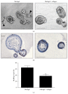
Collagen modulates the phenotype of small intestine organoids. (a) Morphology of small intestine organoids cultured in the presence of EGF, Noggin, and R-Spondin. Arrowheads indicate buds. Scale bar, 100 μm. (b) Ki-67 staining of small intestine organoids, scale bar, 50 μm. (c) Collagen reduces budding of small intestine organoids. In total 1700–1800 crypts were quantified per condition. P = 0.0045, two-tailed t-test.
Figure 5
Figure 5
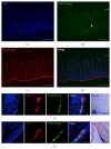
Positive Dclk1 lineage shown in the enteric nervous system with GFP labeled neurons. Immunofluorescence of small intestine from Dclk1.Cre.GFP-Tom fl/fl GFP mice. (a) DAPI for cell nuclei. (b) Positive GFP-fluorescence in neurons (Dclk1.Cre.GFP-Tom fl/fl GFP label recombined cells of the Dclk1 lineage from birth on) invading organoids and single Dclk1-positive cells in epithelium, arrowhead indicating lineage of Dclk1-positive neurons, (c) Tom-Red ubiquitously expressed as marker protein complex. (d) Merge of (a)–(c), scale bars 100 μm. (e) Cultured neurons identified by positive GFP-fluorescence and overlapping with PGP9.5 staining as proof of lineage, last panel demonstrating lacZ stained neuronal structures next to smooth musculature within the GI tract, scale bar, 50 μm. (f) Overlapping fluorescence with GFAP as proof of lineage of glia within myenteric plexus, last panel showing lacZ stained glia within myenteric plexus, scale bar, 50 μm.
Figure 6
Figure 6
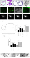
Cardia organoid cultures and cocultures with neurons as a model to investigate the gastrointestinal stem cell niche. (a) IHC for H&E, PAS, Ki-67, and α-SMA for further characterization of the method, arrowheads indicating positive stained cells, scale bars, 100 μm. (b) Light microscopy of Lgr5-GFP-positive organoid, scale bar, 50 μm. (c) Immunofluorescence of Dclk1-labeled neurons in cultured conditions (NBM). (d) Cultured neurons (NBM) stained with beta-III Tubulin, scale bar, 100 μm. (e) Cocultured neuron with cardia organoid, light microscopy and immunofluorescence,  Dclk1-labeled neuron (green fluorescence) neighboring crypt wall, scale bar, 100 μm. (f) Ca-imaging of neuron in cocultured conditions (NBM), different points in time beginning from application of nicotinic acid to Fluo-4-labeled neurons, brightening of neuron indicating Ca-influx. Scale bar, 50 μm. (g) Analysis of the RLI of stimulated neuron, ROI I, ROI II, and ROI III measuring different regions of the neuron as indicated in (f), picture 2 (regions labeled 1, 2, and 3). ((h), (i)) Analysis of organoids and cocultured neurons in different conditions, distribution of mean diameters and circumferences per group and point in time and standard error of the mean (SEM) (CM = crypt medium, CaM = Wnt-conditioned cardia medium, NBM = cocultured neurons in neurobasal complete medium, groups compared at 7 d and 10 d), measured in μm; comparison of average organoid growth between groups applying two-tailed t-test ( level of statistical significance, ∗∗∗∗ P < 0.0001), n = 3 experiments per group and point in time. (j) Representative light microscopic image of each group and point in time (arrowheads indicating neurons), scale bar, 100 μm.
Figure 7
Figure 7
Reconstruction of stem cell niche in the intestine in vitro and proposed workflow. Intestinal crypts and stromal cells (neurons and/or myofibroblasts) are isolated and combined together in three-dimensional system such as organoid culture. Role of stromal cells as stem cell niche can be analyzed, for example, by the measurement of organoid diameter, cell viability assay, clonogenicity assay, gene expression analysis, and different types of staining. Identification of stem cell markers and individual cell types can be analyzed by FACS. In addition, genetic manipulations in organoids can be performed by lentiviral transfection and CRISPR-CASP9 system.

Similar articles

Cited by

References

    1. Voog J., Jones D. L. Stem cells and the niche: a dynamic duo. Cell Stem Cell. 2010;6(2):103–115. doi: 10.1016/j.stem.2010.01.011. - DOI - PMC - PubMed
    1. Walker M. R., Patel K. K., Stappenbeck T. S. The stem cell niche. Journal of Pathology. 2009;217(2):169–180. doi: 10.1002/path.2474. - DOI - PubMed
    1. Fuchs E., Tumbar T., Guasch G. Socializing with the neighbors: stem cells and their niche. Cell. 2004;116(6):769–778. doi: 10.1016/s0092-8674(04)00255-7. - DOI - PubMed
    1. Zhang J., Niu C., Ye L., et al. Identification of the haematopoietic stem cell niche and control of the niche size. Nature. 2003;425(6960):836–841. doi: 10.1038/nature02041. - DOI - PubMed
    1. Brittan M., Wright N. A. Gastrointestinal stem cells. The Journal of Pathology. 2002;197(4):492–509. doi: 10.1002/path.1155. - DOI - PubMed