. 2015:2015:852986.
doi: 10.1155/2015/852986. Epub 2015 Nov 30.

Expression of HGF and c-Met Proteins in Human Keratoconus Corneas

Affiliations

Expression of HGF and c-Met Proteins in Human Keratoconus Corneas

Jingjing You et al. J Ophthalmol. 2015.

Erratum in

Abstract

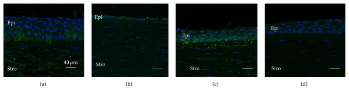
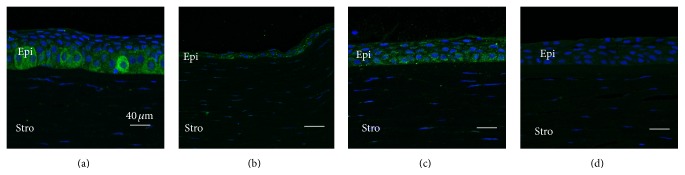
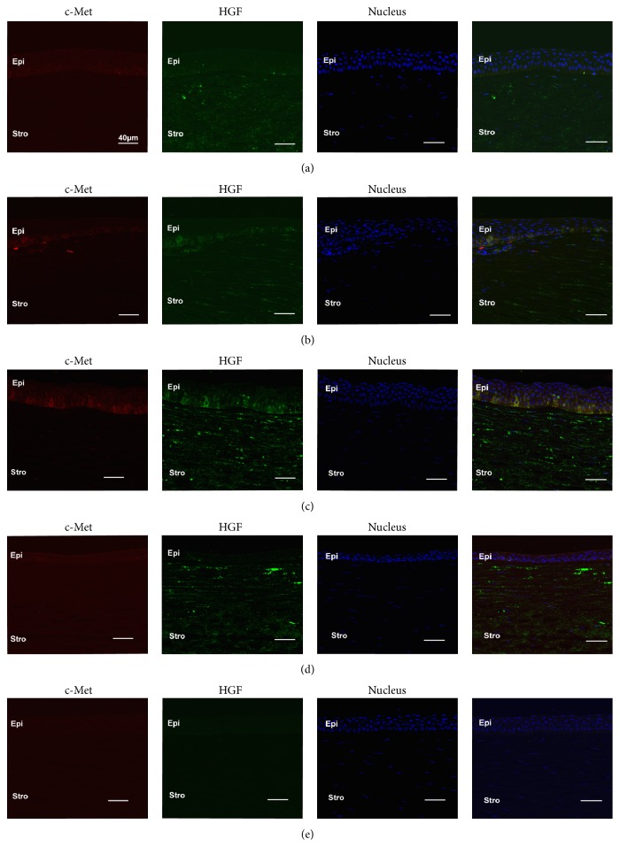
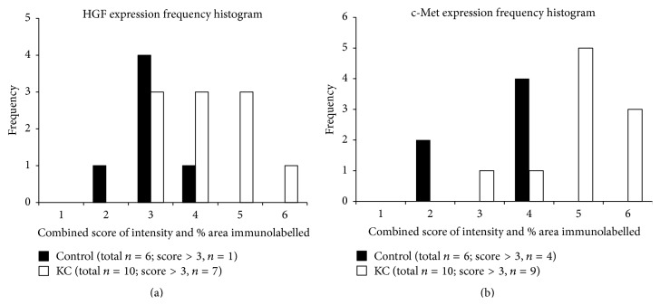
Keratoconus (KC) is a progressive degenerative inflammatory-related disease of the human cornea leading to decreased visual function. The pathogenesis of KC remains to be understood. Recent genetic studies indicate that gene variants of an inflammation-related molecule, hepatocyte growth factor (HGF), are associated with an increased susceptibility for developing KC. However HGF protein expression in KC has not been explored. In this initial study, we investigated late-stage KC and control corneas for the expression of HGF and its receptor mesenchymal-epithelial transition factor (c-Met/Met). KC buttons (~8 mm diameter) (n = 10) and whole control corneas (n = 6) were fixed in 10% formalin or 2% paraformaldehyde, paraffin embedded and sectioned. Sections were immunolabelled with HGF and c-Met antibodies, visualised using immunofluorescence, and examined with scanning laser confocal microscopy. Semiquantitative grading was used to compare HGF and c-Met immunostaining in KC and control corneas. Overall, KC corneas showed increased HGF and c-Met immunostaining compared to controls. KC corneal epithelium displayed heterogeneous moderate-to-strong immunoreactivity for HGF and c-Met, particularly in the basal epithelium adjacent to the cone area. Taken together with the recent genetic studies, our results further support a possible role for HGF/c-Met in the pathogenesis of KC.


Figures

Figure 1
Figure 1
Representative images showing increased HGF expression in basal epithelial cells adjacent to the cone region in KC (a), compared to the relatively more uniform expression seen in all layers of control cornea epithelium at a similar location (c). Strong HGF stromal staining (keratocyte and extracellular matrix) is also observed in KC and control corneas (a to c). No immunostaining is apparent in the Ig negative control (d). (a) Region adjacent to the cone in KC cornea, (b) cone region in KC cornea, (c) central region of control cornea, and (d) Ig negative control cornea. Epi: epithelium, Stro: stromal. Green: HGF; blue: nuclei.
Figure 2
Figure 2
Representative images showing increased c-Met expression in basal epithelial cells adjacent to the cone region in KC (a), compared to the relatively more uniform expression seen in all layers of control cornea epithelium at a similar location (c). Weak c-Met staining is detected in the stroma in both KC and control corneas (a to c). No immunostaining is apparent in the Ig negative control (d). (a) Region adjacent to the cone in KC cornea, (b) cone region in KC cornea, (c) central region of control cornea, and (d) Ig negative control. Epi: epithelium; Stro: stromal. Green: HGF; blue: nuclei.
Figure 3
Figure 3
Representative images of central region of corneas with weak, uniform HGF (green), and c-Met (red) expression in all corneal layers (a). Patchy staining of HGF and c-Met is colocalised in the basal epithelium of both limbal regions (b) and adjacent to cone region of KC (c), with KC showing stronger staining than the limbal region. Strong HGF expression is seen within the stroma of both KC (c and d) and control corneas (a and b). (a) Central region of control cornea, (b) limbal region of control cornea, (c) region adjacent to the cone in KC cornea, (d) cone region in KC cornea, and (e) Ms and Rb Ig control.
Figure 4
Figure 4
Frequency histograms for HGF (a) and c-Met (b) expression in KC and control corneas (combined score of staining intensity and % area coverage; range = 0 to 6). Note the right skewed distribution of HGF and c-Met immunostaining for KC corneas compared to controls. HGF staining had a combined score >3 in 7/10 (70%) KC corneas, compared to 1/6 (16%) control corneas. c-Met staining with a combined score >3 was seen in 9/10 (90%) KC corneas compared to 4/6 (66%) control corneas.

Similar articles

Cited by

References

    1. Rabinowitz Y. S. Keratoconus. Survey of Ophthalmology. 1998;42(4):297–319. doi: 10.1016/S0039-6257(97)00119-7. - DOI - PubMed
    1. Mcghee C. N. J. 2008 Sir Norman McAlister gregg lecture: 150 years of practical observations on the conical cornea—what have we learned? Clinical and Experimental Ophthalmology. 2009;37(2):160–176. doi: 10.1111/j.1442-9071.2009.02009.x. - DOI - PubMed
    1. Wagner H., Barr J. T., Zadnik K. Collaborative Longitudinal Evaluation of Keratoconus (CLEK) Study: methods and findings to date. Contact Lens and Anterior Eye. 2007;30(4):223–232. doi: 10.1016/j.clae.2007.03.001. - DOI - PMC - PubMed
    1. Williams K. A., Lowe M. T., Keane M. C., et al. The Australian Corneal Graft Registry 2012 Report. Adelaide, Australia: Flinders University Press; 2012.
    1. Snibson G. R. Collagen cross-linking: a new treatment paradigm in corneal disease—a review. Clinical and Experimental Ophthalmology. 2010;38(2):141–153. doi: 10.1111/j.1442-9071.2010.02228.x. - DOI - PubMed

LinkOut - more resources