. 2015 Sep 14;4(5):e429-33.
doi: 10.1016/j.eats.2015.04.002. eCollection 2015 Oct.

Diagnosis of Knee Osteochondral Lesions With Ultrasound Imaging

Affiliations

Diagnosis of Knee Osteochondral Lesions With Ultrasound Imaging

Pekko Penttilä et al. Arthrosc Tech. .

Abstract

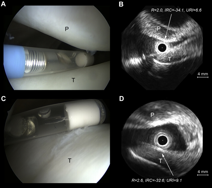
Evaluation of articular cartilage and subchondral bone is essential in the diagnosis of joint diseases and injuries. Interobserver and intraobserver reproducibilities of arthroscopic grading are only poor to moderate. Thus, for quantitative and objective evaluation of cartilage and subchondral bone, ultrasound arthroscopy (UA) has been introduced to clarify this dilemma. Assessment of the clinical feasibility of high-frequency ultrasonography (US) during 6 knee arthroscopies was conducted, and the surgical technique is presented. US imaging was conducted with a flexible 9-MHz US catheter inserted into the joint through conventional portals. US and arthroscopy videos were synchronously recorded, and US parameters for cartilage and subchondral bone characteristics were measured. Arthroscopy and US imaging were combined to perform cartilage grading. UA produced quantitative data on lesion size, as well as cartilage quality, and showed subchondral bone changes. Visualization of an osteochondritis dissecans lesion not detected by conventional arthroscopy and US-guided retrograde drilling were possible with UA. To conclude, UA proved to be clinically feasible and aided in the diagnosis when assessing knee osteochondral lesions.


Figures

Fig 1
Fig 1
(A, B) The characteristic subchondral separation (oval) encountered in osteochondritis dissecans lesions can be visualized with ultrasound arthroscopy as gaps of otherwise intact subchondral bone signal. (C, D) Furthermore, if ultrasound arthroscopy shows fluid beneath the cartilage surface, an unstable osteochondritis dissecans can be suspected. (f, fluid; F, femur; IRC, integrated reflection coefficient [decibels]; m, meniscus; R, ultrasound reflection coefficient [percent]; s, arthroscope artifact; sb, subchondral bone; Ti, tibia; URI, ultrasound roughness index [micrometers]).
Fig 2
Fig 2
Still ultrasound arthroscopy images showing (A, B) normal International Cartilage Repair Society (ICRS) grade 0 cartilage and (C, D) nearly normal ICRS grade 1 cartilage. Both surfaces produce similar ultrasound reflection (ultrasound reflection coefficient [R], as a percent). However, the ultrasound roughness index (URI) (in micrometers) is elevated for ICRS grade 1 compared with ICRS grade 0 articular cartilage, as clearly seen in the (A, C) corresponding arthroscopy images. (IRC, integrated reflection coefficient [decibels]; P, patella; T, trochlea.)
Fig 3
Fig 3
Still ultrasound arthroscopy images showing a decrease in ultrasound reflection (ultrasound reflection coefficient [R], as a percent, and integrated reflection coefficient [IRC], in decibels) and a respective increase in ultrasound roughness index (URI) (in micrometers) with progressive cartilage degradation, as seen in (A, B) abnormal patellar International Cartilage Repair Society grade 2 and (C, D) severely abnormal femoral International Cartilage Repair Society grade 3 lesions compared with normal cartilage (Fig 2). (D) Concomitant meniscus pathology (encircled in red) can be visualized by ultrasound. (F, femur; m, meniscus; P, patella; T, trochlea; Ti, tibia.)
Fig 4
Fig 4
(A, B) Ultrasound arthroscopy shows high reflection and intermediate roughness values for exposed sclerotic subchondral bone. (C, D) Other subchondral bone changes such as microfracture picking holes (mf) can also be visualized by ultrasound arthroscopy. (F, femur; IRC, integrated reflection coefficient [decibels]; R, ultrasound reflection coefficient [percent], sb, subchondral bone; Ti, tibia; URI, ultrasound roughness index [micrometers].)

References

    1. Reed M.E., Villacis D.C., Hatch G.F., III 3.0-Tesla MRI and arthroscopy for assessment of knee articular cartilage lesions. Orthopedics. 2013;36:e1060–e1064. - PubMed
    1. Harris J.D., Brophy R.H., Siston R.A., Flanigan D.C. Treatment of chondral defects in the athlete’s knee. Arthroscopy. 2010;26:841–852. - PubMed
    1. Sugita T., Aizawa T., Uozumi H. Can the fragment stability of osteochondritis dissecans be interpreted by arthroscopic findings alone? Arthroscopy. 2011;27:1171–1172. - PubMed
    1. Spahn G., Klinger H.M., Baums M., Pinkepank U., Hofmann G.O. Reliability in arthroscopic grading of cartilage lesions: Results of a prospective blinded study for evaluation of inter-observer reliability. Arch Orthop Trauma Surg. 2011;131:377–381. - PubMed
    1. Spahn G., Klinger H.M., Hofmann G.O. How valid is the arthroscopic diagnosis of cartilage lesions? Results of an opinion survey among highly experienced arthroscopic surgeons. Arch Orthop Trauma Surg. 2009;129:1117–1121. - PMC - PubMed

LinkOut - more resources