. 2015:2015:917156.
doi: 10.1155/2015/917156. Epub 2015 Nov 30.

Inhibition of ACE Retards Tau Hyperphosphorylation and Signs of Neuronal Degeneration in Aged Rats Subjected to Chronic Mild Stress

Affiliations

Inhibition of ACE Retards Tau Hyperphosphorylation and Signs of Neuronal Degeneration in Aged Rats Subjected to Chronic Mild Stress

Said AbdAlla et al. Biomed Res Int. 2015.

Abstract

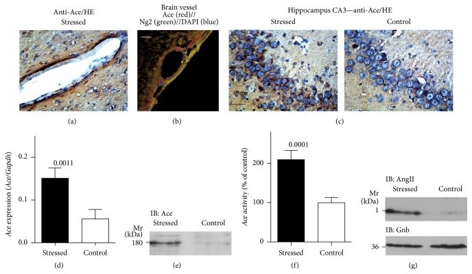
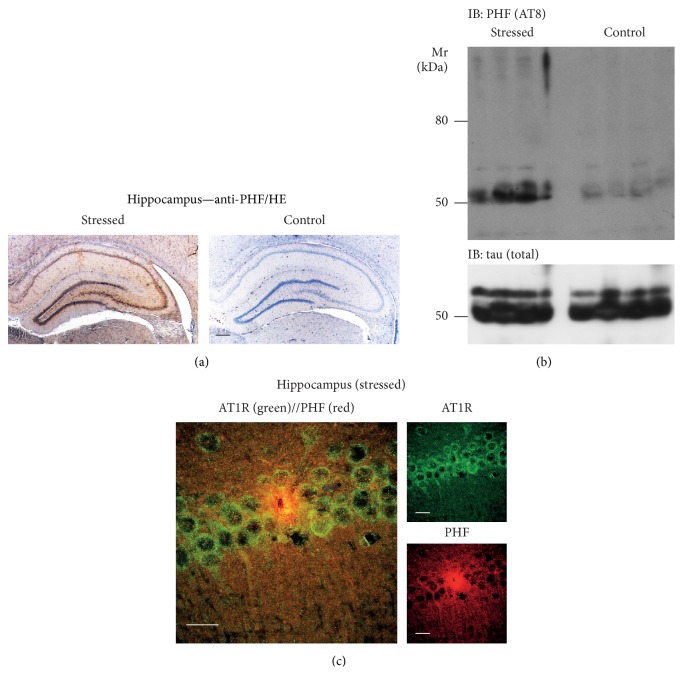
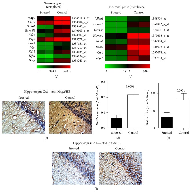
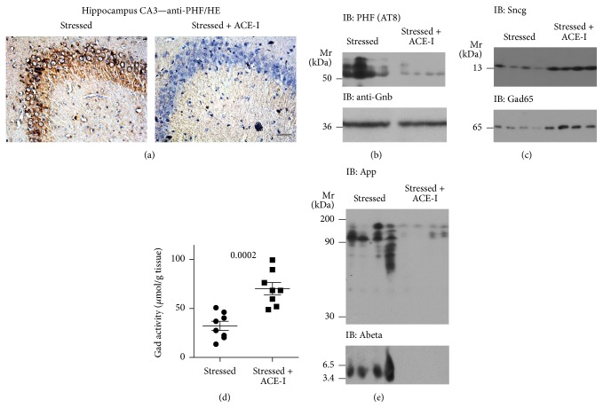
With increasing life expectancy, Alzheimer's disease (AD) and other types of age-associated dementia are on the rise worldwide. Treatment approaches for dementia are insufficient and novel therapies are not readily available. In this context repurposing of established drugs appears attractive. A well-established class of cardiovascular drugs, which targets the angiotensin II system, is such a candidate, which currently undergoes a paradigm shift with regard to the potential benefit for treatment of neurodegenerative symptoms. In search for additional evidence, we subjected aged rats to chronic unpredictable mild stress, which is known to enhance the development of AD-related neuropathological features. We report here that four weeks of chronic mild stress induced a strong upregulation of the hippocampal angiotensin-converting enzyme (Ace) at gene expression and protein level. Concomitantly, tau protein hyperphosphorylation developed. Signs of neurodegeneration were detected by the significant downregulation of neuronal structure proteins such as microtubule-associated protein 2 (Map2) and synuclein-gamma (Sncg). Ace was involved in neurodegenerative symptoms because treatment with the brain-penetrating ACE inhibitor, captopril, retarded tau hyperphosphorylation and signs of neurodegeneration. Moreover, ACE inhibitor treatment could counteract glutamate neurotoxicity by preventing the downregulation of glutamate decarboxylase 2 (Gad2). Taken together, ACE inhibition targets neurodegeneration triggered by environmental stress.


Figures

Figure 1
Figure 1
Chronic unpredictable mild stress induced upregulation of hippocampal Ace protein and gene expression. (a) Immunohistological localization of Ace in a brain vessel of a stressed rat. Nuclei were stained with hematoxylin (HE); bar: 20 μm. (b) Immunofluorescence localization of Ace (red) and the pericyte marker Ng2 (green) in a brain vessel of a stressed rat. Ace was detected with affinity-purified rabbit anti-Ace antibodies followed by F(ab)2 fragments of Alexa Fluor 546-labeled (red) secondary antibodies, and Ng2 was detected with affinity-purified murine anti-NG2 antibody followed by F(ab)2 fragments of Alexa Fluor 488-labeled (green) secondary antibodies. Nuclei were stained with DAPI (bar: 20 μm). (c) Immunohistological localization of Ace in hippocampal CA3 neurons of a stressed rat (left) relative to a nonstressed control (right). Nuclei were stained with hematoxylin (HE); bar: 20 μm. Histological experiments are representative of 4 rats/group (a–c). (d) Hippocampal Ace gene expression was determined by qRT-PCR and is presented as the ratio of Ace/Gapdh expression (±s.d.; n = 4; P = 0.0011). (e) Immunoblot detection of the hippocampal Ace protein with anti-Ace antibodies in stressed rats relative to nonstressed controls (n = 4/group). (f) Hippocampal Ace activity of stressed rats relative to nonstressed controls (i.e., 100%; ±s.d., n = 8; P = 0.0001). (g) The hippocampal angiotensin II content was determined by immunoblot in stressed rats relative to nonstressed controls (upper panel). The lower panel shows a control immunoblot, which detects Gnb (n = 4/group).
Figure 2
Figure 2
Tau hyperphosphorylation in the hippocampus of stressed rats. (a) Immunohistological detection of the hyperphosphorylated tau protein (anti-PHF) in the hippocampus of a stressed rat (left panel) relative to a nonstressed control (right panel). Nuclei were counterstained with hematoxylin (HE; bar: 200 μm). (b) Immunoblot detection of hyperphosphorylated tau in hippocampal extracts of stressed rats relative to nonstressed controls was performed with anti-PHF antibody (IB: PHF (AT8)). The lower panel is a control immunoblot detecting total tau protein (n = 4/group). (c) Immunofluorescence localization of AT1R (green) and PHF (red) in hippocampal CA1 neurons of a stressed rat (bar: 20 μm). The AT1R was detected with affinity-purified rabbit anti-AT1R antibodies followed by F(ab)2 fragments of Alexa Fluor 488-labeled (green) secondary antibodies, and hyperphosphorylated tau was visualized with affinity-purified mouse anti-PHF antibody followed by F(ab)2 fragments of Alexa Fluor 546-labeled secondary antibodies (red). Histological experiments are representative of 4 rats/group ((a) and (c)).
Figure 3
Figure 3
Whole genome microarray gene expression profiling detected stress-induced signs of hippocampal neurodegeneration. (a) and (b) Whole genome microarray gene expression profiling of hippocampal gene expression documents that stress promoted the significant downregulation of probe sets detecting neuron-specific genes with cytosolic localization (a) or membrane localization (b) according to GO analysis. All probe sets were significantly downregulated in the hippocampus of stressed rats relative to nonstressed controls (P < 0.05 and ≥2-fold difference). The heat map visualizes signal intensities (centered to the median value). (c) Immunohistological detection of Map2 in hippocampal CA1 neurons of a stressed rat (left panel) relative to a nonstressed control (right panel; bar: 20 μm). (d) Hippocampal Map2 expression level was determined by qRT-PCR and is presented as the ratio of Map2/Gapdh expression (±s.d.; n = 4; P = 0.0004). (e) Hippocampal Gad activity of stressed rats relative to nonstressed controls (±s.d.; n = 8; P = 0.0001). (f) The decreased hippocampal Grin3a protein level of a stressed rat (left panel) relative to that of a nonstressed control (right panel) was detected by immunohistology in hippocampal CA1 neurons; bar: 20 μm. Immunohistology data are representative of 4 rats/group ((c) and (f)).
Figure 4
Figure 4
Treatment with the brain-penetrating ACE inhibitor captopril retarded signs of neurodegeneration induced by chronic mild stress. (a) Immunohistological detection of hyperphosphorylated tau protein with anti-PHF antibody in hippocampal CA3 neurons of a stressed rat without treatment (left panel) relative to a stressed rat treated with the ACE inhibitor captopril (Stressed + ACE-I) during the stress protocol (right panel); bar: 40 μm. Immunohistological experiments are representative of 4 rats/group. (b) Immunoblot detection of hyperphosphorylated tau protein (IB: PHF (AT8)) in hippocampal extracts of stressed rats without treatment relative to stressed rats treated with the ACE inhibitor captopril (Stressed + ACE-I) during the stress protocol (n = 4/group). (c) Immunoblot detection of Sncg (upper panel) and Gad65 (lower panel) in hippocampal extracts of stressed rats without treatment relative to stressed rats with ACE inhibitor (Stressed + ACE-I) treatment (n = 4/group). (d) Hippocampal Gad activity of stressed rats without treatment relative to stressed rats treated with the ACE inhibitor (Stressed + ACE-I) captopril (n = 8/group; P = 0.0002). (e) Immunoblot detection of the amyloid beta (A4) precursor protein, App (IB: App; upper panel), and β-amyloid (IB: Abeta; lower panel) in hippocampal extracts of stressed rats without treatment relative to stressed rats treated with the ACE inhibitor captopril (Stressed + ACE-I) during the stress protocol (n = 4/group).

References

    1. Scully T. Demography: to the limit. Nature. 2012;492(7427):S2–S3. - PubMed
    1. Abbott A. Dementia: a problem for our age. Nature. 2011;475(7355):S2–S4. doi: 10.1038/475s2a. - DOI - PubMed
    1. Marcello E., Gardoni F., Di Luca M. Alzheimer's disease and modern lifestyle: what is the role of stress? Journal of Neurochemistry. 2015;134(5):795–798. doi: 10.1111/jnc.13210. - DOI - PubMed
    1. Willner P., Towell A., Sampson D., Sophokleous S., Muscat R. Reduction of sucrose preference by chronic unpredictable mild stress, and its restoration by a tricyclic antidepressant. Psychopharmacology. 1987;93(3):358–364. doi: 10.1007/bf00187257. - DOI - PubMed
    1. Cuadrado-Tejedor M., Ricobaraza A., Del Río J., et al. Chronic mild stress in mice promotes cognitive impairment and CDK5-dependent tau hyperphosphorylation. Behavioural Brain Research. 2011;220(2):338–343. doi: 10.1016/j.bbr.2011.01.005. - DOI - PubMed

Publication types

MeSH terms