Rev7/Mad2B plays a critical role in the assembly of a functional mitotic spindle
- PMID: 26697843
- PMCID: PMC4825701
- DOI: 10.1080/15384101.2015.1120922
Rev7/Mad2B plays a critical role in the assembly of a functional mitotic spindle
Abstract
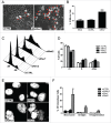
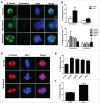
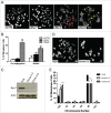
The spindle assembly checkpoint (SAC) acts as a guardian against cellular threats that may lead to chromosomal missegregation and aneuploidy. Mad2, an anaphase-promoting complex/cyclosome-Cdc20 (APC/C(Cdc20)) inhibitor, has an additional homolog in mammals known as Mad2B, Mad2L2 or Rev7. Apart from its role in Polζ-mediated translesion DNA synthesis and double-strand break repair, Rev7 is also believed to inhibit APC/C by negatively regulating Cdh1. Here we report yet another function of Rev7 in cultured human cells. Rev7, as predicted earlier, is involved in the formation of a functional spindle and maintenance of chromosome segregation. In the absence of Rev7, cells tend to arrest in G2/M-phase and display increased monoastral and abnormal spindles with misaligned chromosomes. Furthermore, Rev7-depleted cells show Mad2 localization at the kinetochores of metaphase cells, an indicator of activated SAC, coupled with increased levels of Cyclin B1, an APC(Cdc20) substrate. Surprisingly unlike Mad2, depletion of Rev7 in several cultured human cell lines did not compromise SAC activity. Our data therefore suggest that besides its role in APC/C(Cdh1) inhibition, Rev7 is also required for mitotic spindle organization and faithful chromosome segregation most probably through its physical interaction with RAN.
Keywords: Mad2; RAN; Rev7/Mad2B; aneuploidy; mitosis; spindle assembly checkpoint.
Figures


Similar articles
-
Mitotic slippage is determined by p31comet and the weakening of the spindle-assembly checkpoint.Oncogene. 2020 Mar;39(13):2819-2834. doi: 10.1038/s41388-020-1187-6. Epub 2020 Feb 6. Oncogene. 2020. PMID: 32029899 Free PMC article.
-
Active transport can greatly enhance Cdc20:Mad2 formation.Int J Mol Sci. 2014 Oct 21;15(10):19074-91. doi: 10.3390/ijms151019074. Int J Mol Sci. 2014. PMID: 25338047 Free PMC article.
-
Role of ubiquitylation of components of mitotic checkpoint complex in their dissociation from anaphase-promoting complex/cyclosome.Proc Natl Acad Sci U S A. 2018 Feb 20;115(8):1777-1782. doi: 10.1073/pnas.1720312115. Epub 2018 Feb 5. Proc Natl Acad Sci U S A. 2018. PMID: 29432156 Free PMC article.
-
The spindle checkpoint: how do cells delay anaphase onset?SEB Exp Biol Ser. 2008;59:243-56. SEB Exp Biol Ser. 2008. PMID: 18368927 Review.
-
Dual inhibition of Cdc20 by the spindle checkpoint.J Biomed Sci. 2007 Jul;14(4):475-9. doi: 10.1007/s11373-007-9157-3. Epub 2007 Mar 17. J Biomed Sci. 2007. PMID: 17370142 Review.
Cited by
-
Translesion Synthesis in Plants: Ultraviolet Resistance and Beyond.Front Plant Sci. 2019 Oct 9;10:1208. doi: 10.3389/fpls.2019.01208. eCollection 2019. Front Plant Sci. 2019. PMID: 31649692 Free PMC article. Review.
-
Mitotic Arrest-Deficient Protein 2B Overexpressed in Lung Cancer Promotes Proliferation, EMT, and Metastasis.Oncol Res. 2019 Aug 8;27(8):859-869. doi: 10.3727/096504017X15049209129277. Epub 2017 Sep 11. Oncol Res. 2019. PMID: 28899455 Free PMC article.
-
REV7 in Cancer Biology and Management.Cancers (Basel). 2023 Mar 11;15(6):1721. doi: 10.3390/cancers15061721. Cancers (Basel). 2023. PMID: 36980607 Free PMC article. Review.
-
Repair of programmed DNA lesions in antibody class switch recombination: common and unique features.Genome Instab Dis. 2021;2(2):115-125. doi: 10.1007/s42764-021-00035-0. Epub 2021 Mar 26. Genome Instab Dis. 2021. PMID: 33817557 Free PMC article. Review.
-
SHLD2/FAM35A co-operates with REV7 to coordinate DNA double-strand break repair pathway choice.EMBO J. 2018 Sep 14;37(18):e100158. doi: 10.15252/embj.2018100158. Epub 2018 Aug 28. EMBO J. 2018. PMID: 30154076 Free PMC article.
References
-
- Rajagopalan H, Lengauer C. Aneuploidy and cancer. Nature 2004; 432:338-41; PMID:15549096; http://dx.doi.org/10.1038/nature03099 - DOI - PubMed
-
- Farr KA, Cohen-Fix O. The metaphase to anaphase transition: a case of productive destruction. European J Biochem / FEBS 1999; 263:14-19; http://dx.doi.org/10.1046/j.1432-1327.1999.00510.x - DOI - PubMed
-
- Musacchio A, Salmon ED. The spindle-assembly checkpoint in space and time. Nat Rev Mol Cell Biol 2007; 8:379-93; PMID:17426725; http://dx.doi.org/10.1038/nrm2163 - DOI - PubMed
-
- Jeganathan KB, Baker DJ, van Deursen JM. Securin associates with APCCdh1 in prometaphase but its destruction is delayed by Rae1 and Nup98 until the metaphase/anaphase transition. Cell Cycle 2006; 5:366-70; PMID:16479161; http://dx.doi.org/10.4161/cc.5.4.2483 - DOI - PubMed
-
- Jeganathan KB, Malureanu L, van Deursen JM. The Rae1-Nup98 complex prevents aneuploidy by inhibiting securin degradation. Nature 2005; 438:1036-9; PMID:16355229; http://dx.doi.org/10.1038/nature04221 - DOI - PubMed
Publication types
MeSH terms
Substances
LinkOut - more resources
Full Text Sources
Other Literature Sources
Miscellaneous
