. 2016 Feb 19;291(8):3860-70.
doi: 10.1074/jbc.M115.680801. Epub 2015 Dec 23.

Characterization and Structural Insights into Selective E1-E2 Interactions in the Human and Plasmodium falciparum SUMO Conjugation Systems

Affiliations

Characterization and Structural Insights into Selective E1-E2 Interactions in the Human and Plasmodium falciparum SUMO Conjugation Systems

Katherine H Reiter et al. J Biol Chem. .

Abstract

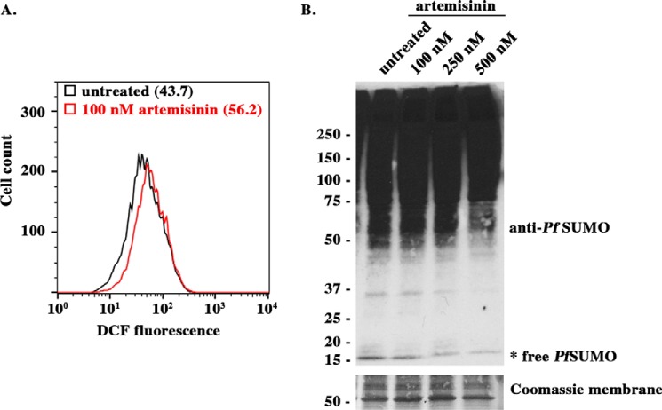
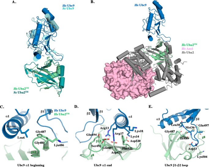
Protein modification by small ubiquitin-related modifiers (SUMOs) is essential and conserved in the malaria parasite, Plasmodium falciparum. We have previously shown that interactions between the SUMO E1-activating and E2-conjugating enzyme in P. falciparum are distinct compared with human, suggesting a potential target for development of parasite-specific inhibitors of SUMOylation. The parasite asexual trophozoite stage is susceptible to iron-induced oxidative stress and is subsequently a target for many of the current anti-malarial drugs. Here, we provide evidence that SUMOylation plays a role in the parasite response to oxidative stress during red blood cell stages, indicative of a protective role seen in other organisms. Using x-ray crystallography, we solved the structure of the human SUMO E1 ubiquitin fold domain in complex with the E2, Ubc9. The interface defined in this structure guided in silico modeling, mutagenesis, and in vitro biochemical studies of the P. falciparum SUMO E1 and E2 enzymes, resulting in the identification of surface residues that explain species-specific interactions. Our findings suggest that parasite-specific inhibitors of SUMOylation could be developed and used in combination therapies with drugs that induce oxidative stress.

Keywords: crystallography; malaria; oxidative stress; plasmodium; small ubiquitin-like modifier (SUMO); ubiquitin fold domain (Ufd); ubiquitin-conjugating enzyme (E2 enzyme).


Figures

FIGURE 1.
FIGURE 1.
SUMO levels are modulated by oxidative stress. A, trophozoites were incubated with the oxidative stress reporter dye H2DCFDA and treated with the pro-oxidant artemisinin. Fluorescent DCF signal was analyzed by flow cytometry. A representative histogram is shown for the gated infected red blood cells, representing the trophozoite population based on SYTO-61 signal. Median DCF fluorescence is indicated in parentheses. B, trophozoites treated with artemisinin were lysed and diluted in SDS sample buffer. The samples were analyzed by SDS-PAGE and immunoblotting with PfSUMO antibodies. Both low and high exposures are shown for comparison. Equivalent lysate loading is demonstrated by a Coomassie-stained membrane.
FIGURE 2.
FIGURE 2.
Structural conservation of a human Uba2Ufd-Ubc9 complex. A, ribbon representation of the human Uba2Ufd-Ubc9 complex (light green and dark blue) superimposed with the previously solved S. cerevisiae Uba2Ufd-Ubc9 structure (teal and light blue) (Protein Data Bank code 3ONG) (25). B, superposition of the human complex onto the previously solved Ufd domain of a human Aos1/Uba2-SUMO1 adenylate structure (pink and gray) (Protein Data Bank code 3KYC) (18). C–E, residues involved in the human Uba2Ufd-Ubc9 interaction are highlighted, organized by binding region: beginning and end of HsUbc9 α1 helix and β1-β2 loop.
FIGURE 3.
FIGURE 3.
P. falciparum has a divergent Uba2Ufd-Ubc9 interface. A, multiple sequence alignment of Ubc9 α1 helix (positions 1–21) from human (Hs), S. cerevisiae (Sc), S. pombe (Sp), P. falciparum (Pf), P. vivax (Pv), P.œnowlesi (Pk), and P. berghei (Pb) ubiquitin E2s, Ubc1, Ubc2, and Ubc4, respectively. Ubc9 α1 helix residues important for human and Sc Ubc9-Ufd binding are denoted by Δ. Additional residues mediating Ubc2-Ufd interactions are denoted by •. Conserved basic N-terminal residues are highlighted with red shading. B, ribbon representation of the PfUbc9 (yellow) (4JUE) docked with the predicted PfUba2Ufd structure (orange) (iTasser), superimposed on the Hs Uba2Ufd-Ubc9 complex by alignment of the Ufd domains. C–E, residues involved in the PfUba2Ufd-Ubc9 interaction based on the docked structure are highlighted, organized by binding region: beginning and end of Ubc9 α1 helix and β1-β2 loop. Interactions denoted by • and Δ from A are specified above the residue label.
FIGURE 4.
FIGURE 4.
Single mutations throughout the PfUba2Ufd-Ubc9 interface are sufficient to disrupt binding. Noncovalent complexes between 33 μg of wild type GST-PfUba2Ufd and 9.9 μg of mutant PfUbc9 (A) and mutant GST-PfUba2Ufd and wild type PfUbc9 (B) were formed at room temperature for 2 h and resuspended in SDS sample buffer. The samples were analyzed by SDS-PAGE and Coomassie staining. Mutants are organized by Ubc9 binding regions.
FIGURE 5.
FIGURE 5.
Mutations in PfUbc9 α1 helix and β1-β2 loop disrupt RanGAP1 conjugation. 1 μm GST-RanGAP1 was incubated with 50 nm PfE1, 5 μm PfSUMO, 5 mm ATP, and 100 nm of the indicated PfUbc9 mutant. The reactions were stopped with the addition of SDS sample buffer at the indicated time points. The samples were separated by SDS-PAGE and analyzed by immunoblotting with GST antibodies. Panels are organized by binding region: beginning and end of Ubc9 α1 helix and β1-β2 loop. R1 and R1S denote unmodified and SUMO-modified RanGAP1, respectively.
FIGURE 6.
FIGURE 6.
PfUbc9 charge reversal mutants are properly folded. CD spectra of wild type PfUbc9 and charge reversal mutants (K5E, K6E, R12E, R16E, K26E, D32K, and K34E) show characteristic bands representative of structured proteins. The averages of three scans are reported, and values are given in terms of ellipticity, measured in millidegrees (mdeg).
FIGURE 7.
FIGURE 7.
Select residues mediate Plasmodium specific Uba2Ufd-Ubc9 binding and conjugation activity. A, ribbon representation of the PfUba2Ufd-Ubc9 docked structure with open book rotation of the interface. Surface residues important for binding are colored light blue to dark blue based on increasing importance. E2 residues important for conjugation activity are scored by asterisks (ranging from least important (*) to most important (****)). B, surface residue conservation map of the Uba2Ufd-Ubc9 interface between P. falciparum and human, colored from gray (identical amino acids) to light pink (similar amino acids) to dark pink (nonconservative substitution). Residues were clustered by similar chemical characteristics (GAVLI, FYW, CM, ST, KRH, DENQ, and P), with nonconservative substitutions deviating from these groups (46).

References

    1. World Health Organization (2014) World Malaria Report 2014, World Health Organization, Geneva, Switzerland
    1. Miller L. H., Ackerman H. C., Su X. Z., and Wellems T. E. (2013) Malaria biology and disease pathogenesis: insights for new treatments. Nat. Med. 19, 156–167 - PMC - PubMed
    1. Issar N., Roux E., Mattei D., and Scherf A. (2008) Identification of a novel post-translational modification in Plasmodium falciparum: protein sumoylation in different cellular compartments. Cell Microbiol. 10, 1999–2011 - PMC - PubMed
    1. Artavanis-Tsakonas K., Misaghi S., Comeaux C. A., Catic A., Spooner E., Duraisingh M. T., and Ploegh H. L. (2006) Identification by functional proteomics of a deubiquitinating/deNeddylating enzyme in Plasmodium falciparum. Mol. Microbiol. 61, 1187–1195 - PMC - PubMed
    1. Chung D. W., Ponts N., Cervantes S., and Le Roch K. G. (2009) Post-translational modifications in Plasmodium: more than you think!. Mol. Biochem. Parasitol. 168, 123–134 - PubMed

Publication types

MeSH terms

Substances

LinkOut - more resources