. 2016 Apr 1;310(7):F607-F620.
doi: 10.1152/ajprenal.00169.2015. Epub 2015 Dec 23.

G protein α12 (Gα12) is a negative regulator of kidney injury molecule-1-mediated efferocytosis

Affiliations

G protein α12 (Gα12) is a negative regulator of kidney injury molecule-1-mediated efferocytosis

Ola Z Ismail et al. Am J Physiol Renal Physiol. .

Abstract

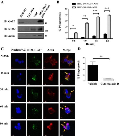
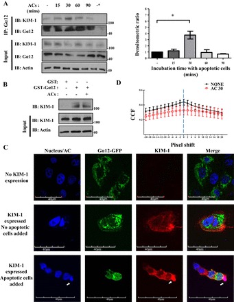
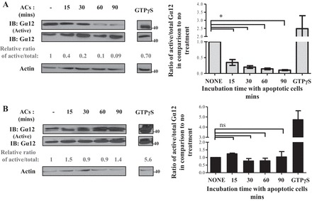
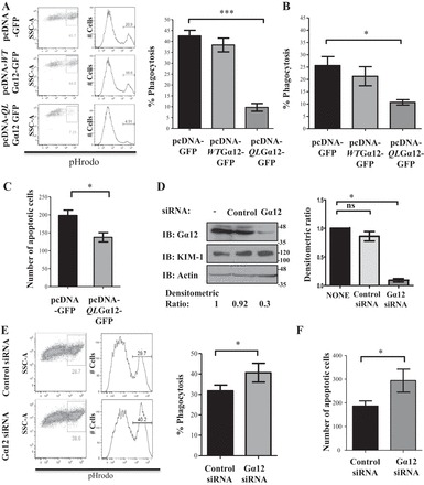
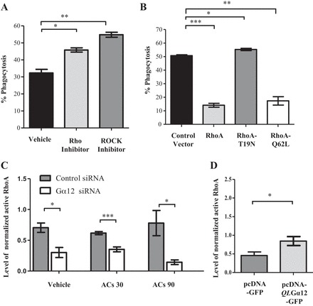
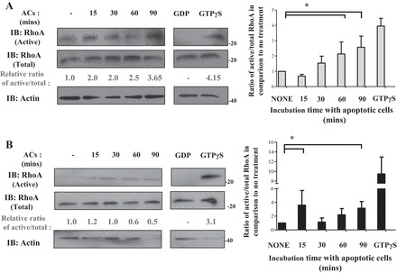
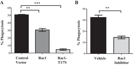
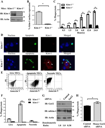
Kidney injury molecule-1 (KIM-1) is a receptor for the "eat me" signal, phosphatidylserine, on apoptotic cells. The specific upregulation of KIM-1 by injured tubular epithelial cells (TECs) enables them to clear apoptotic cells (also known as efferocytosis), thereby protecting from acute kidney injury. Recently, we uncovered that KIM-1 binds directly to the α-subunit of heterotrimeric G12 protein (Gα12) and inhibits its activation by reactive oxygen species during renal ischemia-reperfusion injury (Ismail OZ, Zhang X, Wei J, Haig A, Denker BM, Suri RS, Sener A, Gunaratnam L. Am J Pathol 185: 1207-1215, 2015). Here, we investigated the role that Gα12 plays in KIM-1-mediated efferocytosis by TECs. We showed that KIM-1 remains bound to Gα12 and suppresses its activity during phagocytosis. When we silenced Gα12 expression using small interefering RNA, KIM-1-mediated engulfment of apoptotic cells was increased significantly; in contrast overexpression of constitutively active Gα12 (QL12) resulted in inhibition of efferocytosis. Inhibition of RhoA, a key effector of Gα12, using a chemical inhibitor or expression of dominant-negative RhoA, had the same effect as inhibition of Gα12 on efferocytosis. Consistent with this, silencing Gα12 suppressed active RhoA in KIM-1-expressing cells. Finally, using primary TECs from Kim-1+/+ and Kim-1-/- mice, we confirmed that engulfment of apoptotic cells requires KIM-1 expression and that silencing Gα12 enhanced efferocytosis by primary TECs. Our data reveal a previously unknown role for Gα12 in regulating efferocytosis and that renal TECs require KIM-1 to mediate this process. These results may have therapeutic implications given the known harmful role of Gα12 in acute kidney injury.

Keywords: G protein; Gα12; kidney; kidney injury molecule-1 (KIM-1); phagocytosis.


Figures

Fig. 1.
Fig. 1.
Kidney injury molecule-1 (KIM-1) mediates the uptake of apoptotic cells (AC). A: various cell lines were screened for level of KIM-1 and Gα12 expression via Western blotting. These cells included human embryonic kidney-293 (HEK-293), porcine proximal tubular epithelial cells (LLC-PK1), and human renal adenocarcinoma cell lines (769P). HEK-293 transfected with human KIM-1 and Gα12 served as a positive control. Lysates were obtained and run on Western blots and immunoblotted (IB) for KIM-1, Gα12, and actin. B: HEK-293 stably expressing either a control vector (pcDNA-GFP) or KIM-1-green fluorescent protein (GFP) were fed pHrodo-labeled (Red) AC for various time points (in hours) as indicated. The percentage of phagocytosis was determined by flow cytometry measurement of GFP- and high pHrodo Red double-positive cells (n = 3). *P < 0.05, **P < 0.005, ***P < 0.01 by unpaired t-test. C: HEK-293 cells stably expressing KIM-1-GFP were fed pHrodo-labeled AC (red) for various time points. Cells were fixed and stained with rhodamine-phalloidin to visualize F-actin (red) and 4,6-diamidino-2-phenylindole (DAPI) to visualize the nucleus and apoptotic cells (blue; ×600; bars = 5 μm). AC labeled with both pHrodo Red and DAPI (blue) appear as purple/pink in the merged image. Arrows indicates the AC in the vicinity of KIM-1. D: HEK-293 stably expressing KIM-1-GFP were treated with vehicle (DMSO) or the actin polymerization inhibitor cytochalasin D (1 μM) for 30 min and were then fed fluorescently labeled AC for 90 min. The percentage of phagocytosis represents the percentage of KIM-1-GFP-positive cells with high fluorescence of pHrodo Red of engulfed AC as measured by flow cytometry (n = 3). **P < 0.005 by unpaired t-test.
Fig. 2.
Fig. 2.
KIM-1 interacts with Gα12 during efferocytosis. A: HEK-293 cells stably expressing human KIM-1-GFP with a high level of endogenous Gα12 were stimulated with AC for indicated time points and used for coimmunoprecipitation (IP) using antibody against Gα12. Densitometric ratio of the coimmunoprecipitated KIM-1 during AC stimulation compared with unstimulated sample was quantified (n = 3). *P < 0.05 1-way ANOVA. B: glutathione-S-transferase (GST) and GST-Gα12 pull down was performed on HEK-293-KIM-1-GFP cells that were left unstimulated or stimulated with AC for 90 min. All samples were analyzed by SDS-PAGE followed by IB with antibodies against KIM-1, Gα12, and actin. The input lane represents 5% of the lysate. The data represent 3 independent experiments. C: HEK-293 cells were transfected with KIM-1-wild-type (WT) and a GFP-tagged Gα12 construct and fed pHrodo Red fluorescently labeled AC (blue/red) for 30 min before fixation, and detection of surface KIM-1 using anti-KIM-1 antibody against mucin domain (AKG) and Alexa 555-conjugated secondary antibody (×600; bars = 40 μm). Arrows indicate where phagocytic cup formed. D: images of colocalization of both KIM-1 and Gα12 were analyzed using the Van Steensel approach, where the cross correlation function (CCFs) was calculated with a pixel shift ± 20. The results shown are representative of 4 images of 3 independent experiments (1-way ANOVA).
Fig. 3.
Fig. 3.
KIM-1 downregulates Gα12 activity during KIM-1-mediated efferocytosis. HEK-293 stably expressing KIM-1-GFP (A) or pcDNA (control vector; B) were either left untreated (none) or fed AC for various time points (15, 30, 60, and 90 min). Cell lysate were subjected to a GST-tetratricopeptide repeat (TPR) pull-down assay to measure active Gα12 as described in experimental procedures. Samples were analyzed by SDS-PAGE followed by IB with antibodies against Gα12 and actin. The total Gα12 lanes represent 5% of the lysate were used for the GST-TPR pull down. Densitometric analysis of the relative ratio of the active Gα12 to total Gα12 compared with nontreated samples is shown as numbers below blots. The average of the ratio of the active to total Gα12 compared with no treatment was blotted as a representation of 3 independent experiments (n = 3). *P < 0.05 by unpaired t-test.
Fig. 4.
Fig. 4.
12 negatively regulates KIM-1-mediated efferocytosis. A: HEK-293 cells stably expressing wild-type KIM-1 were transfected with either a control vector encoding GFP (pcDNA-GFP), wild-type GFP-tagged Gα12 (pcDNA-WT12-GFP), or constitutively active GFP-tagged Gα12 (pcDNA-QL12-GFP). B: LLC-PK1 cells stably expressing KIM-1 were transfected with the same constructs as in A. Cells in A and B were fed AC that are fluorescently labeled with pHrodo Red for 90 min. The percentage of uptake of AC was determined by flow cytometry as described in experimental procedures. The ratio of phagocytosis compared with the control vector was determined by flow cytometry after gating on GFP-positive cells (n = 3). *P < 0.05, ***P < 0.001 by unpaired t-test. C: HEK-293 cells stably expressing wild-type KIM-1 were grown on coverslips and transfected with either pcDNA-GFP or pcDNA-QL12-GFP and fed fluorescently labeled (pHrodo Red) AC for 90 min. Confocal microscopy images were taken to quantify the number of AC bound to the phagocytic cells. The results were plotted as a representation of 8 sections from 4 independent experiments (n = 4; ×400; bars = 5 μm). *P < 0.05. D: HEK-293 cells stably expressing KIM-1 were transfected with 50 pmol of control small interfering (si) RNA or siRNA against Gα12 for 24 h, and level of Gα12, KIM-1, and actin was determined by IB with indicated antibodies. A graph of the densitometric ratio of the Gα12 in siRNA-treated samples to nontransfected samples represents 4 independent experiments (n = 4). *P < 0.05 by unpaired t-test. E: HEK-293-KIM-1-GFP-expressing cells transfected with either control or Gα12 siRNA were fed fluorescently labeled (pHrodo Red) AC to measure phagocytosis as in A and B. F: cells in E were visualized with confocal microscopy to count the number of AC. The number of AC bound to the cells that were seen in each field was plotted based on analysis of 8 representative sections from each of 4 independent experiments with ∼120 GFP-positive (KIM-1-GFP) cells counted for each section (n = 4). *P < 0.05 by unpaired t-test.
Fig. 5.
Fig. 5.
RhoA is a downstream mediator of Gα12 in KIM-1-mediated phagocytosis. A: HEK-293 cells stably expressing KIM-1 were treated for 4 h with vehicle (DMSO), Rho inhibitor (cell-permeable C3 transferase; 0.5 μg/ml), or Rho kinase (ROCK) inhibitor Y27632 (10 μM). B: cells were transfected with cyan fluorescent protein (CFP)-tagged control vector, CFP-tagged wild-type RhoA, CFP-tagged dominant negative RhoA (T19N), or CFP-tagged constitutively active RhoA (Q62L). These cells (A and B) were incubated with pHrodo Red-labeled AC for 90 min, and the percentage of phagocytosis by CFP-positive cells was measured by flow cytometry (n = 3). *P < 0.05, **P < 0.01, ***P < 0.001 by unpaired t-test. C: HEK-293 cells stably expressing KIM-1 were transfected with either control siRNA or siRNA against Gα12 for 24 h and then stimulated with AC for 30 or 90 min. D: HEK-293-KIM-1 cells were transfected with a control vector or constitutively active Gα12 (QL12). Levels of active RhoA (in C and D) were measured using a G-LISA RhoA activation assay kit. Total RhoA levels in lysates were measured using an RhoA ELISA. The levels of normalized active RhoA were calculated and graphed (n = 3). *P < 0.05, ***P < 0.001 by unpaired t-test.
Fig. 6.
Fig. 6.
RhoA activity is increased during the course of KIM-1-mediated efferocytosis. HEK-293 stably expressing KIM-1-GFP (A) or pcDNA (control vector; B) were fed AC for various time points (15, 30, 60, and 90 min). Cell lysate were loaded with nonhydrolyzable GTP (GTPyS) as a positive control or GDP as a negative control. The level of active RhoA was measured using an active RhoA (RhoA-GTP) pull-down assay as described in experimental procedures. Samples were analyzed by SDS-PAGE followed by IB with antibodies against RhoA and actin. Densitometric analysis of the relative ratio of active RhoA to the respective total fractions compared with the untreated sample is shown as numbers below blots. The average ratio of active to total RhoA compared with no treatment was graphed as a representation of 3 independent experiments (n = 3). *P < 0.05 by unpaired t-test.
Fig. 7.
Fig. 7.
Rac1 is involved in KIM-1-mediated efferocytosis. A: HEK-293 cells were cotransfected with a construct encoding wild-type KIM-1 and either a CFP-tagged control vector (control) or CFP-tagged dominant negative Rac1 (Rac1-T17N). B: HEK-293-KIM-1 cells were treated for 4 h with vehicle (DMSO) or a Rac1 inhibitor (NSC23766; 50 μM). Cells in A and B were fed to pHrodo-labeled AC for 90 min, and the percentage of uptake was measured by flow cytometry. In A, the percentage of pHrodo Red high fluorescent cells was determined for CFP-positive cells (n = 3). **P < 0.01, ***P < 0.001 by unpaired t-test.
Fig. 8.
Fig. 8.
KIM-1 is a major phagocytic receptor in proximal tubular epithelial cells (TECs), and Gα12 is a negative regulator. A: proximal TECs isolated from C57BL/6 wild-type (Kim-1+/+) and Kim-1 knockout (Kim-1−/−) mice were cultured, and the level of mouse Kim-1 expressed was measured by Western blotting. B: surface expression of mouse Kim-1 on TECs was determined by flow cytometry using PE-conjugated anti-mouse Tim-1/Kim-1 antibody (R&D Systems; n = 3). *P < 0.05 by unpaired t-test. C: TECs were fed pHrodo red-labeled AC for various time points (in hour), and the percentage of phagocytosis of fluorescently labeled AC was measured by flow cytometry (n = 3). *P < 0.05 by 1-way ANOVA. D: TECs were cultured on glass coverslips and fed green-labeled AC for 3 h. Cells were fixed and surface stained for mouse Kim-1 using an anti-mouse Kim-1 antibody and Cy5-conjugated secondary antibody (×600; bars = 10 μm). E: TECs isolated from wild-type C57BL/6 mice were left untreated (live), subjected to UV for 3 min, followed by incubation overnight (to stimulate apoptosis), or subjected to heat shock at 65°C for 15 min (to stimulate necrosis). The different type of cell death was confirmed using PI and annexin V staining followed by flow cytometry. Data represent 3 independent experiments. F: live, apoptotic, or necrotic cells labeled with pHrodo Red were fed to either Kim-1+/+ or Kim-1−/− TECs, and the percentage of phagocytosis was measured using flow cytometry (n = 3) **P < 0.005 by unpaired t-test. G: TECs isolated from wild-type C57BL/6 mice were transfected with 50 pmol of control siRNA or siRNA against mouse Gα12 for 24 h, and the level of Gα12, mouse Kim-1, and actin was determined by Western blotting. H: Gα12 siRNA-treated cells were subjected to phagocytosis assay, and the percentage of phagocytosis of pHrodo-labeled AC was measured by flow cytometry (n = 4). *P < 0.5 by unpaired t-test.

Similar articles

Cited by

References

    1. Ajay AK, Kim TM, Ramirez-Gonzalez V, Park PJ, Frank DA, Vaidya VS. A bioinformatics approach identifies signal transducer and activator of transcription-3 and checkpoint kinase 1 as upstream regulators of kidney injury molecule-1 after kidney injury. J Am Soc Nephrol 25: 105–118, 2014. - PMC - PubMed
    1. Arandjelovic S, Ravichandran KS. Phagocytosis of apoptotic cells in homeostasis. Nat Immunol 16: 907–917, 2015. - PMC - PubMed
    1. Bailly V, Zhang Z, Meier W, Cate R, Sanicola M, Bonventre JV. Shedding of kidney injury molecule-1, a putative adhesion protein involved in renal regeneration. J Biol Chem 277: 39739–39748, 2002. - PubMed
    1. Balasubramanian S, Kota SK, Kuchroo VK, Humphreys BD, Strom TB. TIM family proteins promote the lysosomal degradation of the nuclear receptor NUR77. Sci Signal 5: ra90, 2012. - PMC - PubMed
    1. Binne LL, Scott ML, Rennert PD. Human TIM-1 associates with the TCR complex and up-regulates T cell activation signals. J Immunol 178: 4342–4350, 2007. - PubMed

Publication types

Substances

LinkOut - more resources