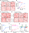
Drosophila p120-catenin is crucial for endocytosis of the dynamic E-cadherin-Bazooka complex
- PMID: 26698216
- PMCID: PMC4760304
- DOI: 10.1242/jcs.177527
Drosophila p120-catenin is crucial for endocytosis of the dynamic E-cadherin-Bazooka complex
Abstract
The intracellular functions of classical cadherins are mediated through the direct binding of two catenins: β-catenin and p120-catenin (also known as CTNND1 in vertebrates, and p120ctn in Drosophila). Whereas β-catenin is crucial for cadherin function, the role of p120-catenin is less clear and appears to vary between organisms. We show here that p120-catenin has a conserved role in regulating the endocytosis of cadherins, but that its ancestral role might have been to promote endocytosis, followed by the acquisition of a new inhibitory role in vertebrates. In Drosophila, p120-catenin facilitates endocytosis of the dynamic E-cadherin-Bazooka subcomplex, which is followed by its recycling. The absence of p120-catenin stabilises this subcomplex at the membrane, reducing the ability of cells to exchange neighbours in embryos and expanding cell-cell contacts in imaginal discs.
Keywords: Cell adhesion; E-cadherin trafficking; Epithelial morphogenesis.
© 2016. Published by The Company of Biologists Ltd.
Conflict of interest statement
The authors declare no competing or financial interests.
Figures


Similar articles
-
Binding site for p120/delta-catenin is not required for Drosophila E-cadherin function in vivo.J Cell Biol. 2003 Feb 3;160(3):313-9. doi: 10.1083/jcb.200207160. Epub 2003 Jan 27. J Cell Biol. 2003. PMID: 12551956 Free PMC article.
-
p120-catenin binding masks an endocytic signal conserved in classical cadherins.J Cell Biol. 2012 Oct 15;199(2):365-80. doi: 10.1083/jcb.201205029. J Cell Biol. 2012. PMID: 23071156 Free PMC article.
-
Numb controls E-cadherin endocytosis through p120 catenin with aPKC.Mol Biol Cell. 2011 Sep;22(17):3103-19. doi: 10.1091/mbc.E11-03-0274. Epub 2011 Jul 20. Mol Biol Cell. 2011. PMID: 21775625 Free PMC article.
-
Evolution and diversity of cadherins and catenins.Exp Cell Res. 2017 Sep 1;358(1):3-9. doi: 10.1016/j.yexcr.2017.03.001. Epub 2017 Mar 6. Exp Cell Res. 2017. PMID: 28268172 Review.
-
Role of p120-catenin in cadherin trafficking.Biochim Biophys Acta. 2007 Jan;1773(1):8-16. doi: 10.1016/j.bbamcr.2006.07.005. Epub 2006 Jul 21. Biochim Biophys Acta. 2007. PMID: 16949165 Review.
Cited by
-
Interactions and Feedbacks in E-Cadherin Transcriptional Regulation.Front Cell Dev Biol. 2021 Jun 28;9:701175. doi: 10.3389/fcell.2021.701175. eCollection 2021. Front Cell Dev Biol. 2021. PMID: 34262912 Free PMC article.
-
Multifaceted control of E-cadherin dynamics by Adaptor Protein Complex 1 during epithelial morphogenesis.Mol Biol Cell. 2022 Aug 1;33(9):ar80. doi: 10.1091/mbc.E21-12-0598. Epub 2022 May 24. Mol Biol Cell. 2022. PMID: 35609212 Free PMC article.
-
Adherens Junction and E-Cadherin complex regulation by epithelial polarity.Cell Mol Life Sci. 2016 Sep;73(18):3535-53. doi: 10.1007/s00018-016-2260-8. Epub 2016 May 5. Cell Mol Life Sci. 2016. PMID: 27151512 Free PMC article. Review.
-
NDRG2 regulates adherens junction integrity to restrict colitis and tumourigenesis.EBioMedicine. 2020 Nov;61:103068. doi: 10.1016/j.ebiom.2020.103068. Epub 2020 Oct 21. EBioMedicine. 2020. PMID: 33099085 Free PMC article.
-
The homophilic receptor PTPRK selectively dephosphorylates multiple junctional regulators to promote cell-cell adhesion.Elife. 2019 Mar 29;8:e44597. doi: 10.7554/eLife.44597. Elife. 2019. PMID: 30924770 Free PMC article.
References
-
- Ciesiolka M., Delvaeye M., Van Imschoot G., Verschuere V., McCrea P., van Roy F. and Vleminckx K. (2004). p120 catenin is required for morphogenetic movements involved in the formation of the eyes and the craniofacial skeleton in Xenopus. J. Cell Sci. 117, 4325-4339. 10.1242/jcs.01298 - DOI - PubMed
Publication types
MeSH terms
Substances
Grants and funding
LinkOut - more resources
Full Text Sources
Other Literature Sources
Molecular Biology Databases
Miscellaneous

