. 2016 Feb 19;291(8):3705-24.
doi: 10.1074/jbc.M115.701698. Epub 2015 Dec 23.

Structural Basis for Toughness and Flexibility in the C-terminal Passenger Domain of an Acinetobacter Trimeric Autotransporter Adhesin

Affiliations

Structural Basis for Toughness and Flexibility in the C-terminal Passenger Domain of an Acinetobacter Trimeric Autotransporter Adhesin

Kotaro Koiwai et al. J Biol Chem. .

Abstract

Trimeric autotransporter adhesins (TAAs) on the cell surface of Gram-negative pathogens mediate bacterial adhesion to host cells and extracellular matrix proteins. However, AtaA, a TAA in the nonpathogenic Acinetobacter sp. strain Tol 5, shows nonspecific high adhesiveness to abiotic material surfaces as well as to biotic surfaces. It consists of a passenger domain secreted by the C-terminal transmembrane anchor domain (TM), and the passenger domain contains an N-terminal head, N-terminal stalk, C-terminal head (Chead), and C-terminal stalk (Cstalk). The Chead-Cstalk-TM fragment, which is conserved in many Acinetobacter TAAs, has by itself the head-stalk-anchor architecture of a complete TAA. Here, we show the crystal structure of the Chead-Cstalk fragment, AtaA_C-terminal passenger domain (CPSD), providing the first view of several conserved TAA domains. The YadA-like head (Ylhead) of the fragment is capped by a unique structure (headCap), composed of three β-hairpins and a connector motif; it also contains a head insert motif (HIM1) before its last inner β-strand. The headCap, Ylhead, and HIM1 integrally form a stable Chead structure. Some of the major domains of the CPSD fragment are inherently flexible and provide bending sites for the fiber between segments whose toughness is ensured by topological chain exchange and hydrophobic core formation inside the trimer. Thus, although adherence assays using in-frame deletion mutants revealed that the characteristic adhesive sites of AtaA reside in its N-terminal part, the flexibility and toughness of the CPSD part provide the resilience that enables the adhesive properties of the full-length fiber across a wide range of conditions.

Keywords: Acinetobacter; Gram-negative bacteria; adhesin; autotransporter; bacterial adhesion; crystal structure; membrane protein; protein domain; protein structure; structure-function.


Figures

FIGURE 1.
FIGURE 1.
Structure of the C-terminal part of the AtaA passenger domain. A, schematic representations of AtaA and of recombinant constructs from its C-terminal part (AtaA_CPSD): CheadCstalk (2905–3561 aa), Chead1 (2905–3168 aa), Chead2 (2777–3168 aa), CstalkFL (3170–3561 aa), CstalkN (3170–3332 aa), CstalkC1 (3334–3474 aa), and CstalkC2 (3334–3561 aa). All constructs were connected to GCN4 tags (white boxes). The Ylhead, FGG, GANG, Trp ring, DALL1, and GIN domains are labeled and numbered from the N terminus of full-length AtaA. Neck domains are not labeled. Pro-3061 of the CheadCstalk and Chead1 was mutated to Gly. The numbers above the structures indicate amino acid residues. Signal peptide, YadA-like head, and transmembrane anchor, which are annotated by the daTAA program, are abbreviated as SP, Ylhead, and TM, respectively. The headCap was newly annotated from the solved crystal structure in this study and consists of β-hairpins, an N-terminally truncated FGG motif (ΔN-FGG), and a HANS motif. B, SDS-PAGE analyses of recombinant CheadCstalk proteins. CheadCstalk samples (WT and P3061G) purified by nickel-affinity chromatography were fractionated by SDS-PAGE and detected by Coomassie Blue staining (CBB). Only WT was sensitive to proteolysis, being degraded into 55- and 23-kDa fragments (WT fragment 55 and WT fragment 23, respectively), whereas the P3061G mutant was protease-resistant. C, Western blotting analyses of purified CheadCstalk samples using an anti-His tag antibody (IB). IB, immunoblot. The recombinant proteins carried the His tag at the C terminus, and only the 55-kDa fragment of the WT protein was detected. This confirms that cleavage had occurred in the region of Pro-3061 and that proline at this position represents a folding obstacle, presumably due to a necessary cis-trans isomerization step that overloads the endogenous prolyl isomerase during overexpression. D, three-dimensional structure of the AtaA_CPSD. The structures of Chead1, CstalkFL, CstalkN, and CstalkC1 were determined experimentally. CheadCstalk is a model constructed from these crystal structures. Of the three CstalkC1 structures (CstalkC1i (PDB code 3WPO); CstalkC1ii (PDB code 3WQA); and CstalkC1iii (PDB code 3WPP)), CstalkC1i is shown. In each structure, the three polypeptide chains forming a homotrimer are colored brown, green, and yellow, respectively; GCN4 tags are colored gray. Ions in CstalkC1i, CstalkFL, and CheadCstalk are shown as spheres: blue, chloride, and black, unidentified. Neck domains occur following the HIM1, GIN_15, YDD, and DALL3 domains. PDB code are indicated below the construct names.
FIGURE 2.
FIGURE 2.
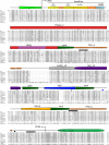
Domain structure of the C-terminal region of AtaA (AtaA_CPSD) marked on a sequence alignment with the same region of other Acinetobacter TAAs. Well characterized Acinetobacter TAAs with the Chead and A. baumannii Ata were aligned by ClustalW2. A. baumannii Ata has Trp ring and DALL1 domains instead of GANG and YDD, respectively. Tol 5 AtaA is highly similar to a hypothetical TAA of A. bereziniae, and the amino acids differing between these TAAs are marked by asterisks. Pro-3061 is marked by @. Coiled coils with hendecad periodicity are indicated by bold lines. The domains of AtaA are numbered from the N terminus. Circles colored in blue and black indicate residues capturing chloride and unknown ions, respectively. Hydrophobic residues are shaded gray.
FIGURE 3.
FIGURE 3.
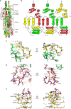
C-terminal head domain contains a unique headCap structure. A, overall structure of the AtaA Chead1 construct, consisting of headCap, Ylhead, HIM1, and neck domains. The headCap is composed of a helical core with three peripheral β-hairpin insertions, the third of which is a truncated FGG domain; it ends with a HANS motif that makes the transition to the Ylhead. Colors are as in Fig. 1. The characteristic residues Asn-2930 of the second β-hairpin and Gln-2950 of the third β-hairpin are shown in stick representation. P3061G in each chain is indicated by an asterisk. B, topological diagram of the headCap, with loops as lines, α-helices as cylinders, and β-strands as arrows. The hairpins are colored as in A. A β-strand with HANS motif is indicated by HANS_β. The residues Asn-2930 of the second β-hairpin and Gln-2950 of the third β-hairpin are shown in stick representation. C–E, enlarged views of the headCap β-hairpins, as marked by squares in A, highlighting the extensive stabilizing interactions that each chain makes in this region with the chain preceding it by 120° counter-clockwise around the trimer axis, as viewed from the N terminus. All views are in stick representation; dotted lines indicate hydrogen bonds. Colors are as in A. Side chains that do not contribute specific interactions are not shown. Orange and blue atoms indicate oxygen and nitrogen, respectively. Gray spheres indicate captured water molecules. C, interchain interactions between the first and second β-hairpins via a shared hydrophobic core and a hydrogen bond network mediated by a structural water molecule. D, intrachain interactions between the second and third β-hairpins. E, interchain interactions among the third-hairpin, a C-terminal extension of the HANS motif, and the following N-terminal strand of the Ylhead domain.
FIGURE 4.
FIGURE 4.
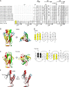
HIM1 of AtaA_CPSD. A, sequence alignment of HIMs. Amino acid sequences of AtaA HIM1, BpaA HIM2, UspA1 HIM2, and SadA HIM3 were aligned with ClustalW2. Of the three head insert motif variants, only HIM1 contains an α-helix (α). Hydrophobic residues are shaded gray. B, superimposition of HIM structures. AtaA_HIM1, BpaA_HIM2, UspA1_HIM2, and SadA_HIM3 are colored in brown, green, yellow, and blue, respectively. The regions adjacent to HIMs, including a part of Ylhead, the neck, and the following α-helix, are colored gray. HIMs are superimposed using the following neck.
FIGURE 5.
FIGURE 5.
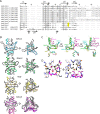
FGG domains in AtaA_CPSD. A, sequence alignment of FGG domains. The two FGG domains of AtaA_CPSD, as well as the headCap, were aligned to the FGG domains of SadA and BpaA. For the headCap, only the helix leading to the insertion and the third β-hairpin are structurally equivalent to the corresponding regions of the FGG motif; the remaining sequence of the headCap is shown in bold italics. Hydrophobic residues are shaded gray. The conserved hydrophobic Gly-Gly sequence of FGG motifs is indicated by asterisks. A conserved aromatic residue, tyrosine or phenylalanine, is indicated by a caret (). The β-strands of the domains are indicated by straight arrows. N- and C-terminal α-helices in FGG domains are indicated by wavy arrows labeled αN and αC, respectively. αN has a hendecad periodicity. B, comparison of FGG structures. The structures were superimposed using the C-terminal helix (residues 2958–2968 of the AtaA headCap, brown; residues 3214–3224 of AtaA FGG_4, green; residues 3514–3524 of AtaA FGG_5, yellow; residues 292–302 of SadA, gray; residues 2335–2345 of BpaA, black). The conserved aromatic residue indicated by a caret in A is shown in stick representation. C, hydrophobic interaction between the β-hairpins of FGG_5 and the cognate coiled coil with hendecad periodicity. Side chains of residues contributing to the hydrophobic interaction between the coiled coil and β-hairpins are shown in stick representation. The residues on the β-hairpins are shown by bold italics. Colors are as in Fig. 1. The tyrosine residue (Tyr-3504) indicated by a caret is the conserved residue in A. D, contact between HIM1 and FGG_4. Colors are as in Fig. 1. Both HIM1 and β-hairpins of FGG_4 project to outside at positions rotated 60° around the trimer axis. Both HIM1 and FGG_4 have high B-factors of Cα in the crystal structures of Chead1 and CstalkN, suggesting that HIM1 and FGG_4 can form different conformations to contact each other. E, contact between the neck following DALL3 and FGG_5. Colors are as in Fig. 1. The neck following the DALL3 and β-hairpins of FGG_5 project to the outside at 120 and 60° around the trimer axis, respectively. F, surface of models of AtaA_CheadCstalk, FGG (+), and a truncation of β-hairpins in FGG_4 and FGG_5 of CheadCstalk, FGG (−). The surface is colored by distance from the trimer axis in a gradient from red to white and from white to green as shown by a scale bar.
FIGURE 6.
FIGURE 6.
GANG domain is a truncated variant of the Trp ring domain. A, sequence alignment of the 10 GANG domains of AtaA with two Trp ring domains, one from A. baumannii Ata, which substitutes in that protein for GANG_10, and the other from B. henselae BadA. Asterisks (bold) and carets (bold) indicate the conserved GANG and Tyr-polar-Val motifs, respectively. The characteristic tryptophan of Trp ring domains is highlighted in yellow. Residues forming the hydrophobic core are shaded gray. The β-strands of the domains are indicated by arrows, above the alignment for GANG and below for Trp ring. B, overall structure of GANG_10. Colors are as in Fig. 1. The GANG motif (NADG in this domain) is colored blue and shown in stick representation, as is the conserved tyrosine, Tyr-3256. Water molecules are shown as spheres; a water molecule at the trimer axis is colored red and the others gray. C, overall structure of the Trp ring domain, exemplified by the domain of B. henselae BadA (PDB code 3D9X). The conserved tryptophan and phenylalanine residues of the Trp ring domain are shown in stick representation. The phenylalanine is at the equivalent position to the conserved tyrosine of GANG domains. D, topological diagram of GANG and Trp ring domains, with loops as lines and β-strands as arrows. One chain of the trimer is colored, yellow for the part common between the GANG and Trp ring domains and black for the two additional β-strands of Trp ring domains (β′1 and β′2). E, superimposition of single monomers from AtaA GANG_10 (brown) and BadA Trp ring (gray) obtained by structural alignment of the following GIN domains.
FIGURE 7.
FIGURE 7.
GIN domain of AtaA_CPSD. A, sequence alignment of the GIN domains of AtaA and BadA. Asterisks indicate the conserved GIN motif. Residues forming the hydrophobic core are shaded gray. The β-strands of the domains are indicated by arrows, above the alignment for the GIN domain. B, superimposition of the GANG-GIN and Trp ring-GIN structures. The structures of GANG_10-GIN_15 from CstalkFL (colored in red (helix), yellow (strand), and green (loop)) and Trp ring-GIN from the BadA head (PDB code 3D9X, blue) are superimposed using their C-terminal neck and helix. C and D, hydrophobic residues of the GIN domain, colored gray and shown in stick representation. The monomer (C) and trimer (D) of CstalkFL are shown, colored as in Fig. 1. Ile-3315, Val-3318, and Ala-3321 are in a neck domain.
FIGURE 8.
FIGURE 8.
Structure of DALL domain variants. A, sequence alignment of DALL domain variants from AtaA and SadA. AtaA has eight DALL1 domains in its Nstalk region (see Fig. 1) and single YDD and DALL3 domains in the Cstalk. A histidine residue highly conserved in DALL2 domains is highlighted in yellow. A key aromatic residue of DALL1, DALL2, and YDD is indicated by an asterisk. DALL1 differs from DALL2/YDD by an extended β1–β2 hairpin; in β2, the extension is separated from the rest of the β-strand by a β-bulge at a conserved tyrosine residue corresponding to Tyr-1325 in SadA, indicated by a caret and marked as β″ in the figure. The N-terminal half of DALL3, shown in lowercase letters, is topologically different from DALL1 and DALL2/YDD, containing two additional β-strands (β1′ and β2′) and orienting β1 in the opposite direction. Residues forming the hydrophobic core are shaded gray. B, structures of DALL domain variants and the following necks. DALL1 and DALL2 are from SadA ((PDB code 2YO3 and 2YNZ, respectively); YDD and DALL3 are from CstalkFL. In each structure, the main chain of one subunit forming the DALL domain is shown in color and stick representation. Water molecules interacting with this subunit are indicated by spheres of the same color. In DALL3, water molecules interacting with the other subunits are colored black. Water molecules along the 3-fold axes of DALL1, DALL2, and DALL3 are colored red. YDD alone does not contain an axial water molecule, allowing the central water molecule of the following neck to be visible (colored in white). C, superimposition of DALL domain monomers, obtained by structural alignment of the following neck domains. The residues of the two additional β-strands in DALL3 are labeled. D, hydrogen bond network of water molecules in DALL3, showing the 13 water molecules of a trimer in the same colors as in B. Hydrogen bonds are indicated by dotted lines.
FIGURE 9.
FIGURE 9.
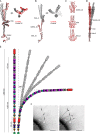
Bending of the AtaA fiber at GANG, YDD, and DALL3. A, superimposition of monomers from CstalkC1ii (gray) and CstalkFL (red), obtained by structural alignment of the neck-coiled coil tandem following DALL3. All three monomers of the asymmetric CstalkC1ii structure are shown. B, superimposition of monomers from CstalkN (gray) and CstalkFL (red), obtained by structural alignment of the GIN_15-neck-coiled coil segment. All three monomers of the asymmetric CstalkN structure are shown. The N terminus of CstalkFL was not traceable in the electron density. C, gallery of the domains causing bending in the AtaA fiber. Extended (red) and bent (gray) structures for GANG, YDD, and DALL3, respectively, were superimposed using their C-terminally adjacent domains. D, model of bending in the CheadCstalk structure. The bent conformation (gray) was modeled from the structures of the entire Chead1 and of CstalkN for FGG_4 and GANG_10; CstalkFL for GIN_15, coiled coil, and FGG_5; and CstalkC1ii for YDD and DALL3. The extended conformation (red) is as in Fig. 1. The two models were superimposed using FGG_5 and the C-terminal coiled coil. E, schematic representation of bending in the full-length AtaA fiber. Models colored in gray differ by the number of DALL1 domains assumed to be bent (none, two, and four). F, bent AtaA fibers on the cell surface. Tol 5 4140/pAtaA cells were imaged by transmission electron microscopy using negative staining. Arrows indicate bent fibers. The right panel is the same image as the left panel, with the bent AtaA fibers traced in black. The scale bar, 100 nm.
FIGURE 10.
FIGURE 10.
Construction, cell surface display, and functional analyses of in-frame deletion mutants of AtaA. A, schematic representation of IFD mutants of AtaA. WT, IFD-ΔCheadCstalk (truncation of 2906–3475 aa), IFD-ΔChead (truncation of 2902–3167 aa), IFD-ΔCstalk1 (truncation of 3170–3475 aa), IFD-ΔCstalk2 (truncation of 3177–3475 aa), and IFD-CPSD (truncation of 108–2996 aa) were inserted into pARP3 plasmids for expression in Tol 5 4140 (ΔataA). Details of these IFD mutants are shown in Fig. 11. B, models of the wild-type and the new domain connections arising from the deletions in IFD-ΔCheadCstalk and IFD-ΔChead, obtained by homology modeling from the equivalent domains of CstalkN, Chead, and CstalkFL. Colors are as in Fig. 1. C, models of the wild type and the new domain connections arising from the deletions in IFD-ΔCstalk1 and IFD-ΔCstalk2, obtained by homology modeling from the equivalent domains of Chead, CstalkN, and CstalkFL. Colors are as in Fig. 1. D, immunoelectron microscopy of Tol 5 cells displaying IFD mutants, imaged with an anti-Nhead antibody and colloidal gold-conjugated anti-rabbit IgG antibody. Scale bars, 200 nm. E, confirmation of cell surface-displayed IFD-AtaA constructs by flow cytometry and CLSM (insets). Scale bars, 2 μm. F, transmission electron microscope image of negatively stained IFD-CPSD fibers on Tol 5 cells. Scale bar, 200 nm. G, adherence assays of Tol 5 IFD mutants. Bacterial cells adhering to bare PS (blank bar) and to collagen-coated PS plates (filled bar) were quantified by a crystal violet staining method. Assays were performed in triplicate. Error bars indicate S.E. H, autoagglutination assay of Tol 5 IFD mutants. The autoagglutination was quantified by a decrease in OD660 in tube-settling assays. Assays were performed in triplicate. Error bars indicate S.E. I, adherence assay of the Tol 5 IFD-CPSD mutant. Bacterial cells adhering to bare PS and to fibronectin-, laminin-, and collagen-coated PS plates were quantified by a crystal violet staining method. Assays were performed in triplicate. Error bars indicate S.E. J, far-Western blotting analysis of the recombinant proteins forming AtaA_CPSD (Chead, CstalkN, and CstalkC2), compared with the YadA_head protein (26–210 aa) fused with a C-terminal GCN4-His tag. Each of these His-tagged proteins was applied as input to membranes, along with three ECMs (fibronectin, collagen type I, and laminin); for the negative control, the input was His-tagged Chead1. The membranes were then subjected to a reaction with His-tagged proteins as listed on the right of the panels. After the reaction, the proteins were detected immunologically using an anti-His-tag antibody. IB, immunoblot.
FIGURE 11.
FIGURE 11.
Domain configuration and amino acid sequence of AtaA_CPSD and in-frame deletion constructs. Domain colors are the same in the schematic figure and the amino acid sequence, except for Ylhead_2 where only the inner β-strands are colored red. Trimeric coiled coils are underlined. In-frame deletion constructs of AtaA were truncated as indicated; IFD-ΔChead (red line), IFD-ΔCheadCstalk (narrow black line), IFD-ΔCstalk1 (dotted line), and IFD-ΔCstalk2 (bold black line). IFD-ΔChead and IFD-ΔCheadCstalk were designed to connect the neck to a coiled-coil segment. To avoid steric hindrance between HIM1 and FGG_5, IFD-ΔCstalk2 was designed to include seven additional residues (KAVGNQV) in the coiled coil separating the two motifs.
FIGURE 12.
FIGURE 12.
Domain architecture of Acinetobacter TAAs. Domains were predicted by daTAA and manually refined. Incomplete sequences are only shown for TAAs containing a Chead and were marked by dotted lines. The scale bar above the sequences indicates residue numbers. The domains are shown schematically as in Fig. 1. Sequences are labeled with the species and strain name of the source organism, and the sequence ID in the NCBI nr database. AcTAA, Acinetobacter TAA.

References

    1. Leo J. C., Grin I., and Linke D. (2012) Type V secretion: mechanism(s) of autotransport through the bacterial outer membrane. Philos. Trans. R. Soc. Lond. B. Biol. Sci. 367, 1088–1101 - PMC - PubMed
    1. Bernstein H. D. (2015) Looks can be deceiving: recent insights into the mechanism of protein secretion by the autotransporter pathway. Mol. Microbiol. 97, 205–215 - PMC - PubMed
    1. Linke D., Riess T., Autenrieth I. B., Lupas A., and Kempf V. A. (2006) Trimeric autotransporter adhesins: variable structure, common function. Trends Microbiol. 14, 264–270 - PubMed
    1. Bassler J., Hernandez Alvarez B., Hartmann M. D., and Lupas A. N. (2015) A domain dictionary of trimeric autotransporter adhesins. Int. J. Med. Microbiol. 305, 265–275 - PubMed
    1. Hoiczyk E., Roggenkamp A., Reichenbecher M., Lupas A., and Heesemann J. (2000) Structure and sequence analysis of Yersinia YadA and Moraxella UspAs reveal a novel class of adhesins. EMBO J. 19, 5989–5999 - PMC - PubMed

Publication types

MeSH terms

Substances