. 2015 Nov-Dec;18(6):504-11.
doi: 10.2350/15-10-1732-OA.1. Epub 2015 Dec 23.

Pleuropulmonary Blastoma: Evolution of an Entity as an Entry into a Familial Tumor Predisposition Syndrome

Affiliations

Pleuropulmonary Blastoma: Evolution of an Entity as an Entry into a Familial Tumor Predisposition Syndrome

Louis P Dehner et al. Pediatr Dev Pathol. 2015 Nov-Dec.

Abstract

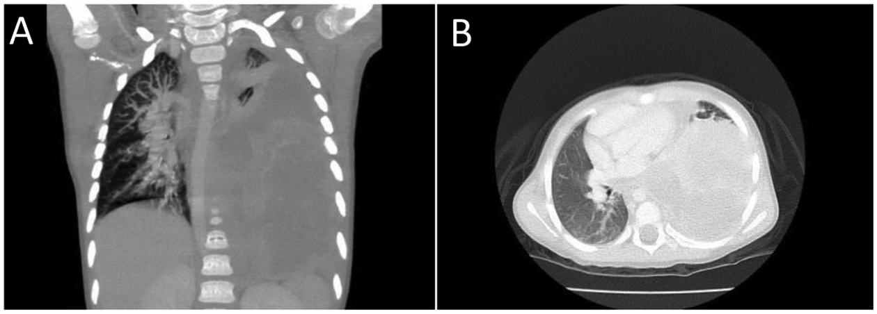
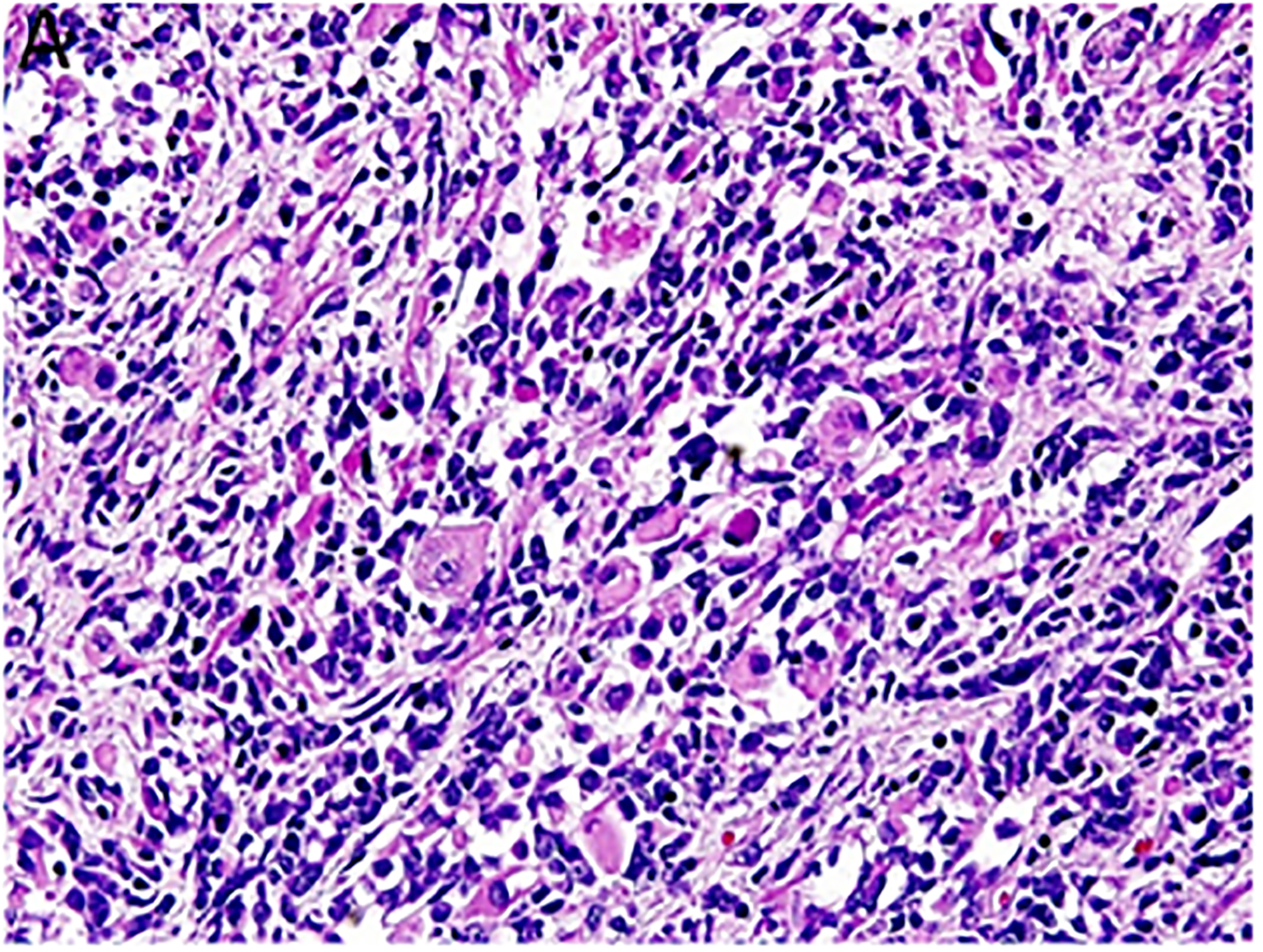
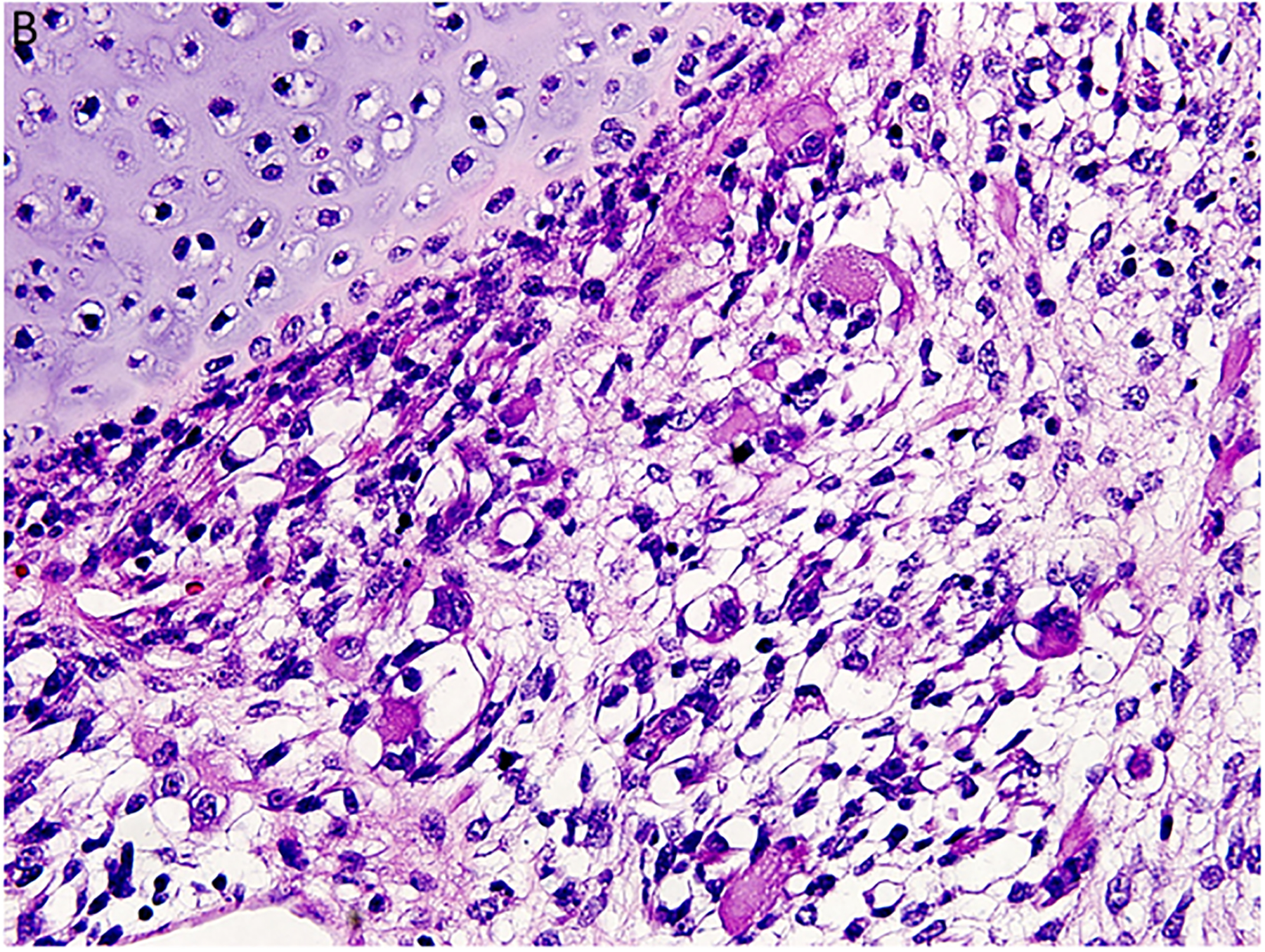
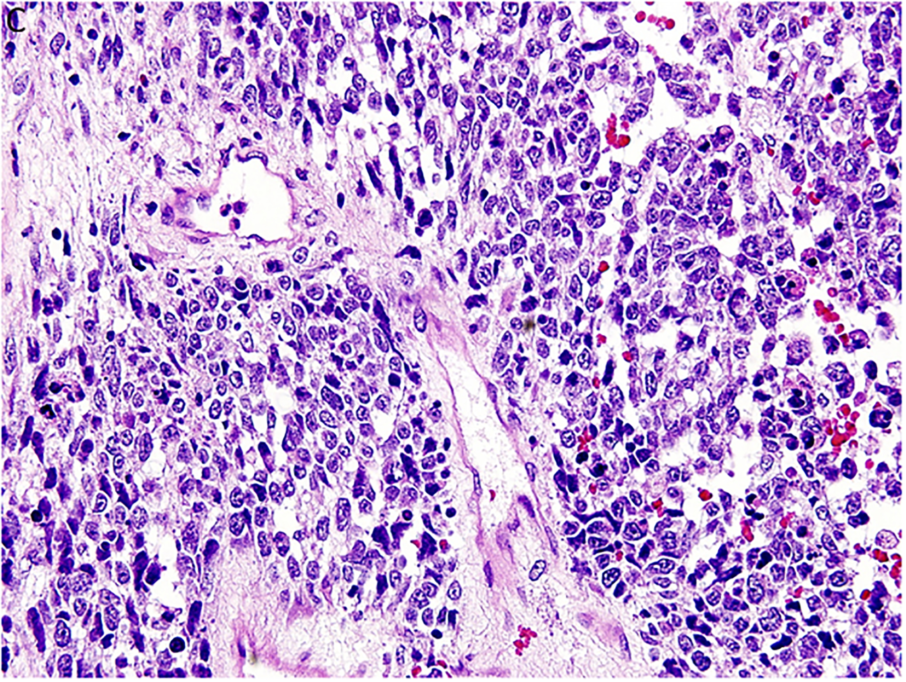
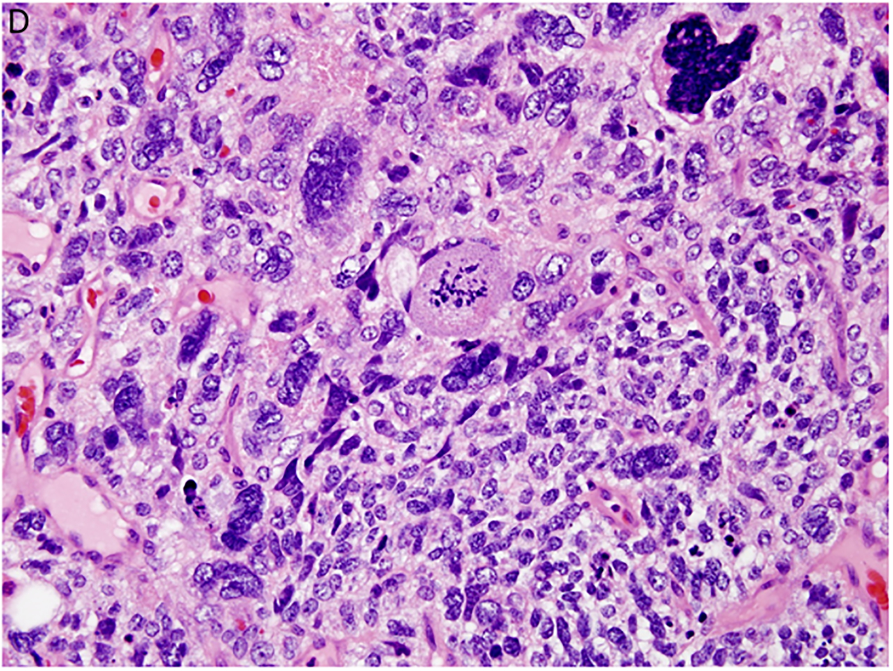
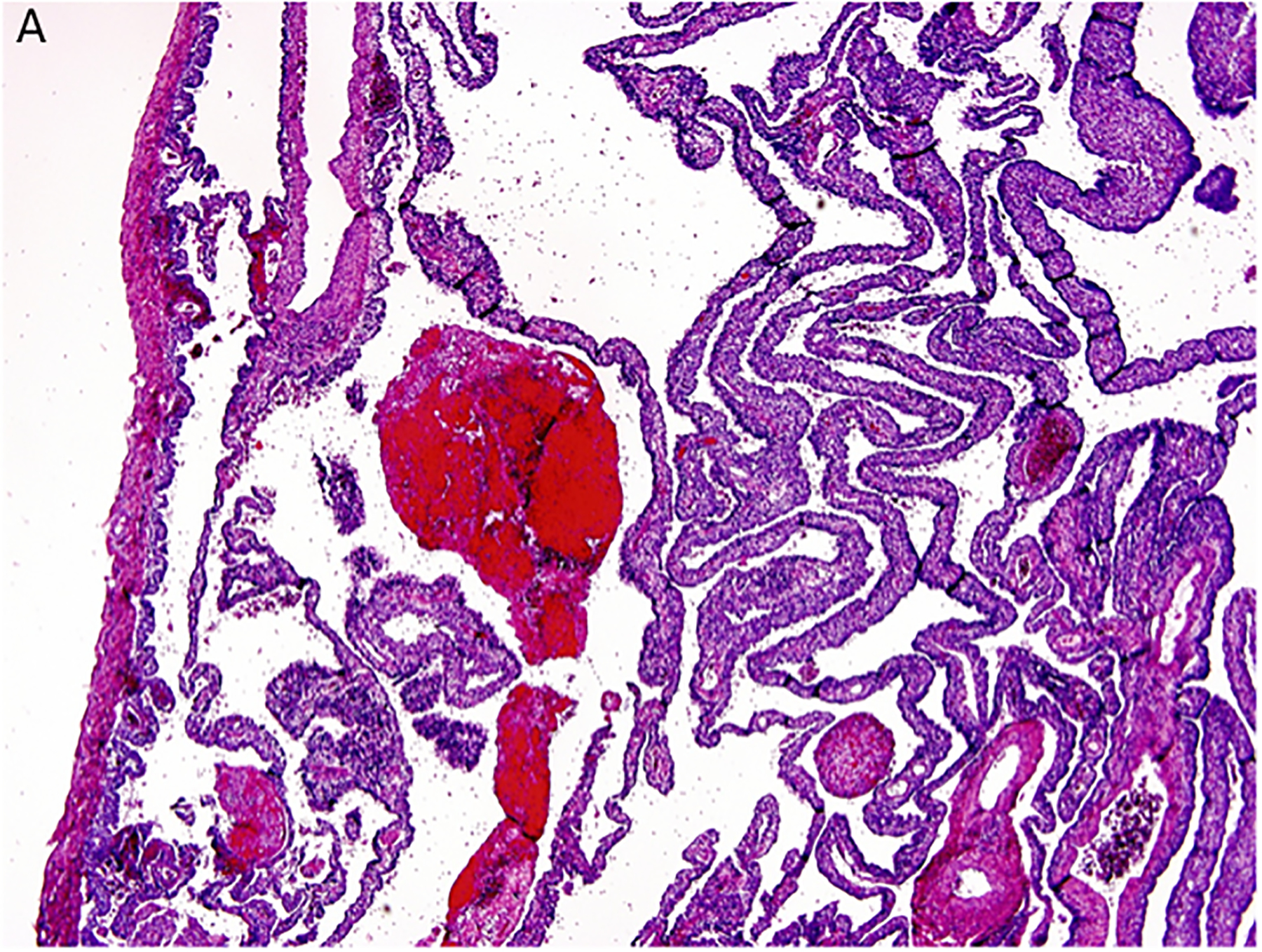
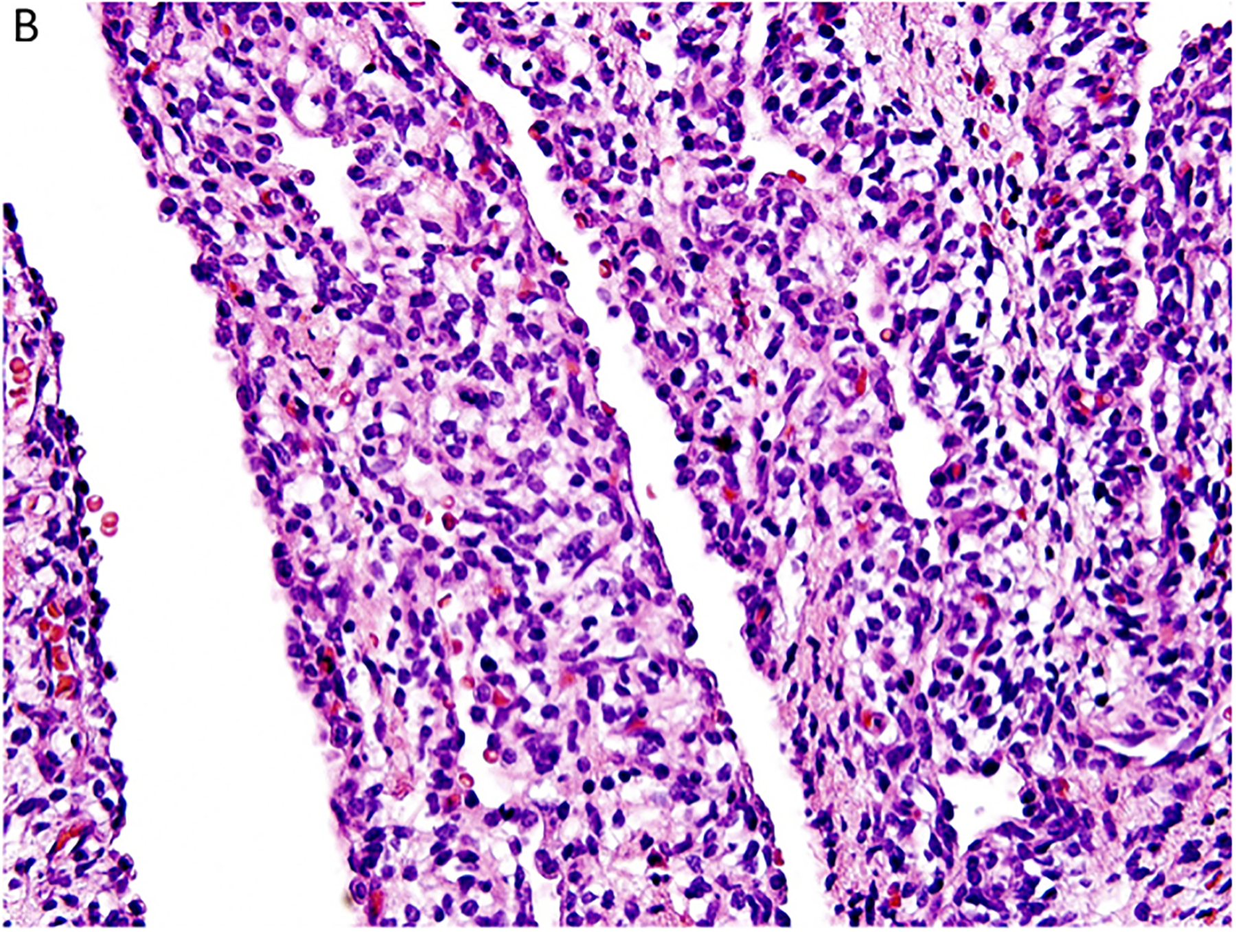
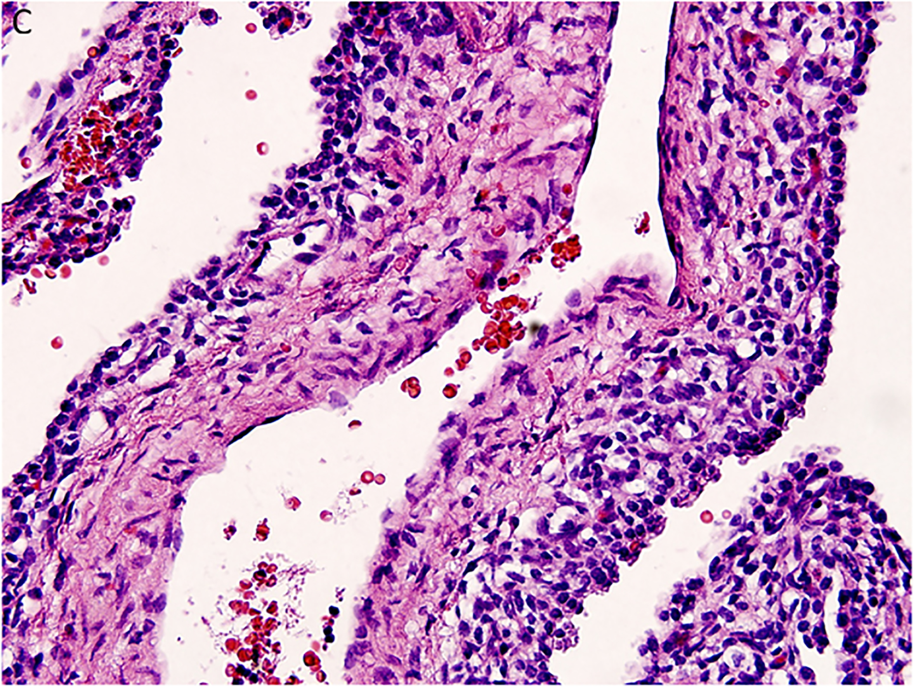
Pleuropulmonary blastoma (PPB) is the most common primary malignant neoplasm of the lung in children. Like other solid dysontogenic neoplasms, this tumor typically presents before 7 years of age. The earliest manifestation is the presence of a lung cyst(s), which is usually recognized in the first year of life and is difficult to differentiate on the basis of imaging studies from non-neoplastic cysts of early childhood. From a multilocular cyst, PPB has the potential to progress to a high-grade multipatterned primitive sarcoma. More than 65% of all affected children have a heterozygous germline mutation in DICER1. The DICER1 PPB familial tumor predisposition syndrome is initially recognized in most cases on the basis of PPB alone but also by several other unique and characteristic extrapulmonary tumors, including pediatric cystic nephroma, nasal chondromesenchymal hamartoma, nodular lesions of the thyroid, embryonal rhabdomyosarcoma of the cervix, and ciliary body medulloepithelioma.

Keywords: DICER1; cystic nephroma; lung; pleuropulmonary blastoma; rhabdomyosarcoma.


Figures

Figure 1.
Figure 1.
Pleuropulmonary blastoma presented in a 4-year-old girl with respiratory symptoms. A,B. The entire left thoracic cavity is occupied by a 15 × 9.5 × 9.7 cm. heterogeneous enhancing mass which has crossed the midline. Mass effect on the descending intrathoracic aorta, heart and left hemidiaphragm has occurred. This appearance is typical of type II or type III PPB. Both rhabdomyoblastic and chondroid elements were seen in the biopsy.
Figure 2.
Figure 2.
Pleuropulmonary blastoma has a heterogeneous appearance within the solid areas of either type II or type III PPB. A. Rhabdomyosarcomatous pattern includes large rhabdomyoblasts with intense eosinophilic cytoplasm surrounded by smaller, more primitive rhabdomyoblasts (x400). B. The periphery of a nodule of fetal appearing cartilage is adjacent to a focus of rhabdomyosarcoma (x400). C. Nests of primitive malignant small cells within poorly formed nests representing the blastemal pattern (x400). D. A focus of primitive sarcoma with enlarged, hyperchromatic tumor cells and bizarre mitotic figures representing anaplasia (x400).
Figure 2.
Figure 2.
Pleuropulmonary blastoma has a heterogeneous appearance within the solid areas of either type II or type III PPB. A. Rhabdomyosarcomatous pattern includes large rhabdomyoblasts with intense eosinophilic cytoplasm surrounded by smaller, more primitive rhabdomyoblasts (x400). B. The periphery of a nodule of fetal appearing cartilage is adjacent to a focus of rhabdomyosarcoma (x400). C. Nests of primitive malignant small cells within poorly formed nests representing the blastemal pattern (x400). D. A focus of primitive sarcoma with enlarged, hyperchromatic tumor cells and bizarre mitotic figures representing anaplasia (x400).
Figure 3.
Figure 3.
Pleuropulmonary blastoma type I presented with spontaneous pneumothorax in a 14-month-old girl. A cystic lesion was detected in the right lung. A. This low magnification demonstrates the typical multicystic pattern. Note that the cyst is covered with pleura along one edge (x40). B. The septal stroma is occupied by a uniform population of small primitive cells (x400). C. The septa demonstrates a cambium layer of small primitive cells and a fibrous stroma. Some septa only had the fibrous stroma (x400). D. Desmin immunostain shows the presence of individual positive cells in the septa. A similar pattern of immunostaining was obtained with myogenin (x400).
Figure 3.
Figure 3.
Pleuropulmonary blastoma type I presented with spontaneous pneumothorax in a 14-month-old girl. A cystic lesion was detected in the right lung. A. This low magnification demonstrates the typical multicystic pattern. Note that the cyst is covered with pleura along one edge (x40). B. The septal stroma is occupied by a uniform population of small primitive cells (x400). C. The septa demonstrates a cambium layer of small primitive cells and a fibrous stroma. Some septa only had the fibrous stroma (x400). D. Desmin immunostain shows the presence of individual positive cells in the septa. A similar pattern of immunostaining was obtained with myogenin (x400).
Figure 4.
Figure 4.
Pleuropulmonary blastoma type II presented in this 3-year-old female as a solid and cystic mass in the right chest. A. The only epithelium in a PPB is the remnant of the cyst lining (x400). B. This field shows the presence of a continuous epithelium of a residual cyst and an expanded stroma composed of uniform malignant small cells (x200). C. Focus of anaplastic cells are identified among the primitive malignant cells (x400). D. p53 immunostaining is especially intense in the nuclei of the anaplastic cells (x400).
Figure 4.
Figure 4.
Pleuropulmonary blastoma type II presented in this 3-year-old female as a solid and cystic mass in the right chest. A. The only epithelium in a PPB is the remnant of the cyst lining (x400). B. This field shows the presence of a continuous epithelium of a residual cyst and an expanded stroma composed of uniform malignant small cells (x200). C. Focus of anaplastic cells are identified among the primitive malignant cells (x400). D. p53 immunostaining is especially intense in the nuclei of the anaplastic cells (x400).

References

    1. Frances D, Jacobsen M. Pulmonary blastoma. Curr Top Pathol 1983; 73:265–294. - PubMed
    1. Spencer H Pulmonary blastoma. J Pathol Bacteriol 1961; 82:161–165.
    1. Travis WD, Brambilla E, Burke AP, Mars A, Nicholson AG. (Eds): World Health Organization Classification of Tumours of the Lung, Pleura, Thymus and Heart. Fourth edition. IARC Press: Lyon: 2015. - PubMed
    1. Mechoulan A, Leclair MD, Yvinec M, Philippe HJ, Winer N. [Pleuropulmonary blastoma: a case of early neonatal diagnosis through antenatal scan screening]. Gynecol Obstet Fertil, 2007; 35:437–441. - PubMed
    1. Miniati DN, Chintagumpala M, Langston C, et al. Prenatal presentation and outcome of children with pleuropulmonary blastoma. J Pediatr Surg 2006; 41:66–71. - PubMed

Publication types

MeSH terms

Supplementary concepts