Review
. 2016 Feb;49(2):73-80.
doi: 10.5483/bmbrep.2016.49.2.265.

Structure biology of selective autophagy receptors

Affiliations
Review

Structure biology of selective autophagy receptors

Byeong-Won Kim et al. BMB Rep. 2016 Feb.

Abstract

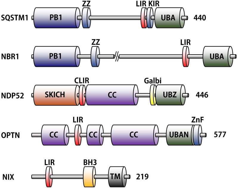
Autophagy is a process tightly regulated by various autophagy-related proteins. It is generally classified into non-selective and selective autophagy. Whereas non-selective autophagy is triggered when the cell is under starvation, selective autophagy is involved in eliminating dysfunctional organelles, misfolded and/or ubiquitylated proteins, and intracellular pathogens. These components are recognized by autophagy receptors and delivered to phagophores. Several selective autophagy receptors have been identified and characterized. They usually have some common domains, such as LC3-interacting- region (LIR) motif, a specific cargo interacting (ubiquitin-dependent or ubiquitin-independent) domain. Recently, structural data of these autophagy receptors has been described, which provides an insight of their function in the selective autophagic process. In this review, we summarize the most up-to-date findings about the structure-function of autophagy receptors that regulates selective autophagy.


Figures

Fig. 1.
Fig. 1.. Domain structures of autophagy receptor proteins. LC3-interacting motifs (LIR) and ubiquitin binding domains (UBA, UBZ, and UBAN) are distinguished by red and green color, respectively. The other unique domains represent different colors. The following abbreviations are used for each domain: PB1, Phox and Bem1p; ZZ, ZZ-type zinc finger; LIR, LC3-interacting region; KIR, Keap1-interacting region, UBA, ubiquitin-associated; SKICH, skeletal muscle and kidney-enriched inositol phosphatase carboxyl homology; CLIR, non-canonical LIR; CC, coiled-coil; Galbi, galectin-8 binding region; UBZ, ubiquitin-binding zinc finger; UBAN, ubiquitin-binding in ABIN and NEMO; ZnF, Zn-finger; BH3, Bcl-2 homology region 3; TM, transmembrane.
Fig. 2.
Fig. 2.. Structures of LC3-LIR complex. (A) LC3B/SQSTM1-LIR peptide complex [2ZJD], (B) NIX-LIR fused LC3B [4WAA], (C) GABARAPL1/NBR1-LIR peptide complex [2L8J], (D) LC3C/NDP52 SKICHLIR region complex [3VVW], and (E) LC3B/OPTN-phosphoLIR peptide complex [2LUE]. Surface of LC3 or GABARAP protein is shown in dark gray color, and the ribbon of LIR motifs in green. The side chain of LC3 interacting residues is also shown in stick model, colored green, red, blue, and orange for carbon, oxygen, nitrogen, and phosphorous, respectively.
Fig. 3.
Fig. 3.. Overall structures of ubiquitin binding domain (UBD) and ubiquitin complex. (A) Ubiquitin (model) with SQSTM1 UBA domain [1Q02] and (B) Ubiquitin with NBR1 UBA domain [2MJ5] are shown in ribbon. The ubiquitin interacting Gln434 and Tyr435 of SQSTM1 UBA, and Glu926, Phe929, and Leu954 of NBR1 UBA are shown in stick model. (C) Ubiquitin with NDP52 ZF2 domain [4XKL]. A bound zinc atom is shown as a gray ball. UBD and ubiquitin are colored green and cyan, respectively.
Fig. 4.
Fig. 4.. Structures of ubiquitin-independent binding domains in autophagy receptors. (A) PB1 domains of SQSTM1 (magenta) and NBR1 (yellow) are superposed. Basic side chains of Lys7, Arg21, Arg22, and Arg94 residues and acidic side chains of Asp67, Glu68, Asp69, Asp71, and Glu80 residues involved in the oligomerization are shown in stick model. (B) Complex of Keap1 (orange) and SQSTM1-KIR peptide (green). The β-propeller structure of Keap1 recognizes KIR peptide containing phosphorylated Ser351. (C) NDP52 SKICH domain (slate) [3VVV], and (D) Complex between galectin-8 (salmon) and NDP52 Galbi peptide (green) [4HAN]. N-terminal carbohydrate binding domain (N-CRD) and C-terminal carbohydrate binding domain (C-CRD) are labeled. Ribbon diagram of overall structure and stick model of bound peptides are shown.

References

    1. Klionsky DJ. Autophagy revisited: a conversation with Christian de Duve. Autophagy. (2008);4:740–743. doi: 10.4161/auto.6398. - DOI - PubMed
    1. Ohsumi Y. Historical landmarks of autophagy research. Cell Res. (2014);24:9–23. doi: 10.1038/cr.2013.169. - DOI - PMC - PubMed
    1. Mizushima N, Noda T, Yoshimori T, et al. A protein conjugation system essential for autophagy. Nature. (1998);395:395–398. doi: 10.1038/26506. - DOI - PubMed
    1. Nakatogawa H, Suzuki K, Kamada Y, Ohsumi Y. Dynamics and diversity in autophagy mechanisms: lessons from yeast. Nat Rev Mol Cell Biol. (2009);10:458–467. doi: 10.1038/nrm2708. - DOI - PubMed
    1. Ohsumi Y. Molecular dissection of autophagy: two ubiquitin-like systems. Nat Rev Mol Cell Biol. (2001);2:211–216. doi: 10.1038/35056522. - DOI - PubMed

Publication types