. 2016 Feb 2;113(5):E509-18.
doi: 10.1073/pnas.1512952113. Epub 2015 Dec 22.

Nicastrin functions to sterically hinder γ-secretase-substrate interactions driven by substrate transmembrane domain

Affiliations

Nicastrin functions to sterically hinder γ-secretase-substrate interactions driven by substrate transmembrane domain

David M Bolduc et al. Proc Natl Acad Sci U S A. .

Abstract

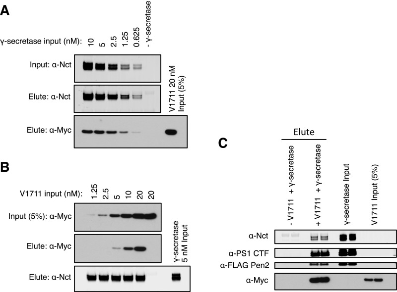
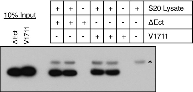
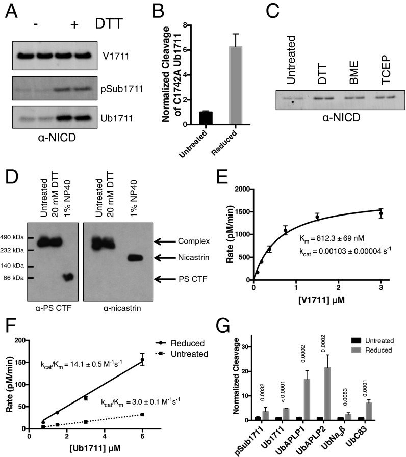
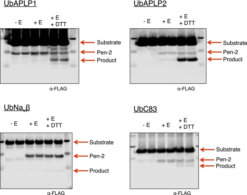
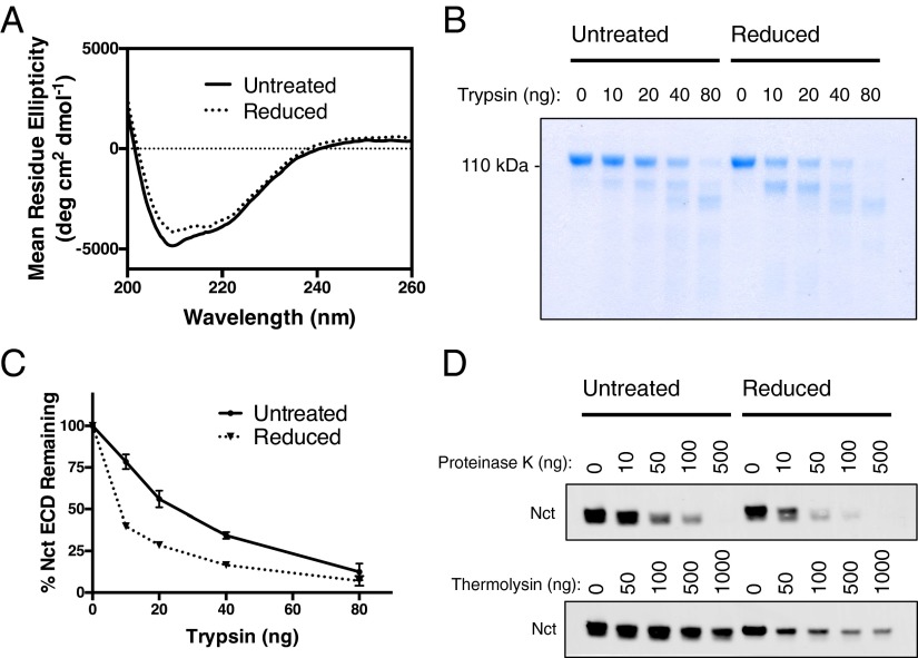
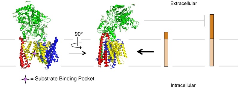
γ-Secretase is an intramembrane-cleaving protease that processes many type-I integral membrane proteins within the lipid bilayer, an event preceded by shedding of most of the substrate's ectodomain by α- or β-secretases. The mechanism by which γ-secretase selectively recognizes and recruits ectodomain-shed substrates for catalysis remains unclear. In contrast to previous reports that substrate is actively recruited for catalysis when its remaining short ectodomain interacts with the nicastrin component of γ-secretase, we find that substrate ectodomain is entirely dispensable for cleavage. Instead, γ-secretase-substrate binding is driven by an apparent tight-binding interaction derived from substrate transmembrane domain, a mechanism in stark contrast to rhomboid--another family of intramembrane-cleaving proteases. Disruption of the nicastrin fold allows for more efficient cleavage of substrates retaining longer ectodomains, indicating that nicastrin actively excludes larger substrates through steric hindrance, thus serving as a molecular gatekeeper for substrate binding and catalysis.

Keywords: Azheimer’s disease; intramembrane-cleaving protease; nicastrin; notch; γ-secretase.


Conflict of interest statement

The authors declare no conflict of interest.

Figures

Fig. S1.
Fig. S1.
Establishment of a detergent-solubilized γ-secretase assay with a notch-based substrate. (A) Cleavage of V1711 with γ-secretase was monitored by Western blot to cleaved NICD. Cleavage was linear with respect to time for the first 45 min. (B) The reaction rate doubled with doubling enzyme concentration (mean ± SD). (C) The amount of active γ-secretase enzyme used in the detergent-soluble assay was titrated with the highly potent inhibitor LY411,575.
Fig. 1.
Fig. 1.
γ-secretase activity toward N-terminally manipulated notch. (A) Summary of kinetic data from γ-secretase cleavage of notch substrates containing varying N-terminal amino acids. Cleavage was monitored by Western blot using a cleavage-specific antibody to NICD (mean ± SD, n = 3). (B) Coomassie-stained gel of semisynthetic native V1711 and N-terminally acetylated AcV1711 generated by native chemical ligation. (C) Electrospray ionization time-of-flight mass spectrometry of V1711 and AcV1711. Intact masses were obtained after deconvolution of the multiply charged states of each protein. (D) γ-secretase activity toward semisynthetic V1711 and AcV1711 (mean ± SD, n = 2). (E) In trans peptide inhibition of recombinant V1711 with notch ectodomain and Aβ (1–12) peptides. Data points are normalized to untreated V1711 cleavage (mean ± SD, n = 2).
Fig. 2.
Fig. 2.
The effects of notch ectodomain length on its interaction with γ-secretase. (A) Schematic diagram of notch-based substrates. N43 is a synthetic peptide, whereas the other substrates were produced recombinantly. (B) Kinetic analysis of notch with varying ectodomain length (mean ± SD, n = 3) and (C) of N43 substrate (mean ± SD, n = 3). (D) Comparing the ability of γ-secretase to immunoprecipitate in complex with V1711 and Ub1711. γ-secretase was immunoprecipitated via its HA tag on Aph-1. Substrates were visualized by their C-terminal myc tag. (E) Summary of kinetic data from B and C (mean ± SD, n = 3; ND, not determined).
Fig. S2.
Fig. S2.
Representative Western blot images from kinetic assays in Fig. 2.
Fig. S3.
Fig. S3.
Estimation of the binding affinity between γ-secretase and substrate. (A) γ-secretase concentrations ranging from 0.625–10 nM were incubated with 20 nM V1711 before co-IP via the HA tag on Aph-1. (B) Myc-tagged V1711 concentrations ranging from 1.25–20 nM were incubated with 5 nM γ-secretase before co-IP via the HA tag on Aph-1. (C) A total of 5 nM γ-secretase was incubated with 20 nM V1711 before pull-down of the complex via the myc tag of V1711 with anti-myc affinity resin. Quantification reveals apparent Kd values averaging in the mid-nM range.
Fig. S4.
Fig. S4.
Immunoprecipitation of ΔEct and V1711 with γ-secretase. Lysate from S20 cells was incubated with notch substrate before co-IP via the HA tag on γ-secretase. ΔEct and V1711 were visualized by Western blot (anti-myc).
Fig. 3.
Fig. 3.
Intramembrane kinetics of γ-secretase within a proteoliposome. (A) Fluorescence quenching of N43 when incorporated into vesicles is alleviated after dissolving the proteoliposome with 0.25% Nonidet P-40 detergent. (B) Circular dichroism spectrum of N43 incorporated into a proteoliposome reveals an α-helical structure. (C) Labeling of a notch peptide with a Cys on its N terminus with a membrane impermeable dye in the presence and absence of melittin to allow dye passage into the interior of the proteoliposome (mean ± SD, n = 2). (D) After reconstitution of γ-secretase and N43 substrate into proteoliposomes, the reaction was initiated by resuspending the proteoliposomes in 50 mM Hepes pH 7.0 and 150 mM NaCl. Product was formed linearly with time. (E) Kinetics of N43 cleavage by γ-secretase within a proteoliposome (mean ± SD, n = 4).
Fig. S5.
Fig. S5.
Kinetics of Ub-N41 cleavage in the proteoliposome assay. (A) Ligation of ubiquitin–thioester to N41 notch peptide. (B) Purified Ub-N41 and N43 visualized via their C-terminal fluorescein tag. (C) After 18 h, Ub-N41 cleavage product was monitored by running proteoliposome reactions on a 16.5% Tris–tricine gel and imaging with a fluorescence scanner. Cleavage was greatly reduced compared with N43 (compare with Fig. 3E), and saturating concentrations were not achieved (n = 2).
Fig. 4.
Fig. 4.
Activity of γ-secretase after treatment with reducing reagents. (A) Western blot of NICD product after the cleavage of V1711, pSub1711, and Ub1711 with γ-secretase in the presence and absence of DTT. (B) Cleavage of cysteine-free Ub1711 with untreated and reduced γ-secretase (mean ± SD, n = 2). (C) Western blot of NICD product after γ-secretase cleavage of pSub1711 untreated and in the presence of DTT, BME, or TCEP. (D) Native blue gel electrophoresis of γ-secretase untreated or in the presence of DTT or Nonidet P-40 detergent. Nicastrin and presenilin CTFs were detected by Western blot. (E) Kinetic analysis of V1711 cleavage in the presence of DTT (mean ± SD, n = 3). (F) Kinetic analysis of Ub1711 cleavage in the presence and absence of DTT (mean ± SD, n = 3). (G) Cleavage of pSub1711 (n = 6) and N-terminally ubiquitin (Ub)-tagged notch (n = 3), APLP1 (n = 2), APLP2 (n = 2), Navβ (n = 2), and C83 (n = 2) (mean ± SD, t test; P values are shown above each substrate).
Fig. S6.
Fig. S6.
Representative Western blot images of cleavage of N-terminally ubiquitin-tagged γ-secretase substrates APLP1, APLP2, Navβ, and C83 in the presence and absence of reducing reagents from Fig. 4H.
Fig. 5.
Fig. 5.
The effects of chemical reduction of disulfides on nicastrin structure. (A) Circular dichroism spectrum of purified nicastrin ectodomain with and without TCEP present. (B) Limited proteolysis of pure nicastrin ectodomain with varying amounts of trypsin in the presence and absence of DTT. (C) Quantification of intact nicastrin ectodomain from B (mean ± SD, n = 2). (D) Limited proteolysis of nicastrin in the γ-secretase complex with varying amounts of proteinase K and thermolysin proteases.
Fig. S7.
Fig. S7.
Purification of human nicastrin ectodomain. The Fc tag on nicastrin–Fc was removed with factor Xa protease. Pure nicastrin ectodomain used in circular dichroism and limited proteolysis studies was obtained after anion exchange and size exclusion chromatography.
Fig. 6.
Fig. 6.
Model of the nicastrin-mediated substrate-gatekeeping mechanism of γ-secretase. The ectodomain of nicastrin sits on top of the extracellular side of the γ-secretase TMDs with its large lobe extending over the presenilin active site. This prevents substrates with large ectodomains from entering the active site and subsequently being cleaved. Substrates with small ectodomains are able to pass under the nicastrin ectodomain gatekeeper and be cleaved by presenilin. Front and side views of the atomic resolution structure of γ-secretase are depicted (Protein Data Bank ID code 5A63).

Comment in

  • Nicastrin guards Alzheimer's gate.
    Urban S. Urban S. Proc Natl Acad Sci U S A. 2016 Feb 2;113(5):1112-4. doi: 10.1073/pnas.1524151113. Epub 2016 Jan 25. Proc Natl Acad Sci U S A. 2016. PMID: 26811479 Free PMC article. No abstract available.

References

    1. Urban S. Mechanisms and cellular functions of intramembrane proteases. Biochim Biophys Acta. 2013;1828(12):2797–2800. - PubMed
    1. Huppert SS, et al. Embryonic lethality in mice homozygous for a processing-deficient allele of Notch1. Nature. 2000;405(6789):966–970. - PubMed
    1. Rawson RB, et al. Complementation cloning of S2P, a gene encoding a putative metalloprotease required for intramembrane cleavage of SREBPs. Mol Cell. 1997;1(1):47–57. - PubMed
    1. Ye J, et al. ER stress induces cleavage of membrane-bound ATF6 by the same proteases that process SREBPs. Mol Cell. 2000;6(6):1355–1364. - PubMed
    1. Stevenson LG, et al. Rhomboid protease AarA mediates quorum-sensing in Providencia stuartii by activating TatA of the twin-arginine translocase. Proc Natl Acad Sci USA. 2007;104(3):1003–1008. - PMC - PubMed

Publication types

MeSH terms

LinkOut - more resources