. 2016 Jan 5;113(1):206-11.
doi: 10.1073/pnas.1508249112. Epub 2015 Dec 22.

Effects of aging on circadian patterns of gene expression in the human prefrontal cortex

Affiliations

Effects of aging on circadian patterns of gene expression in the human prefrontal cortex

Cho-Yi Chen et al. Proc Natl Acad Sci U S A. .

Abstract

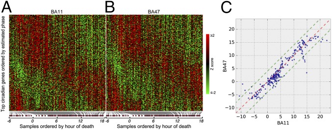
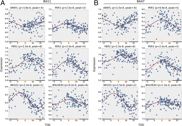
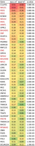
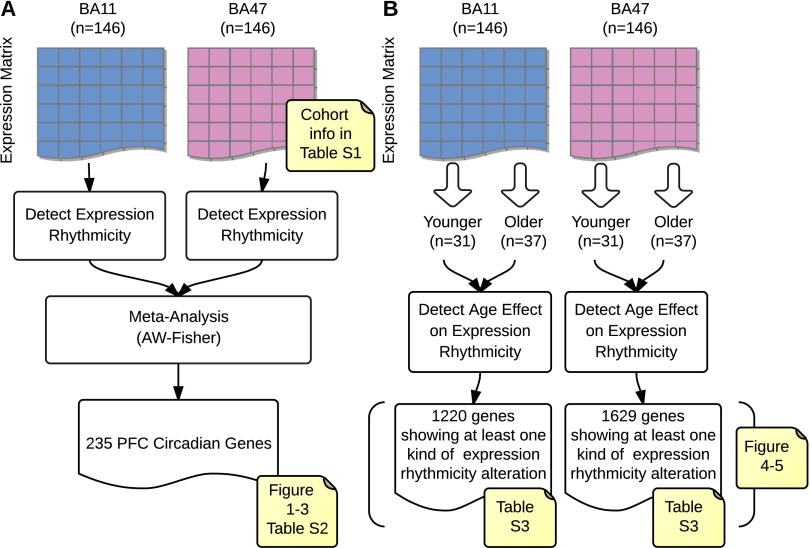
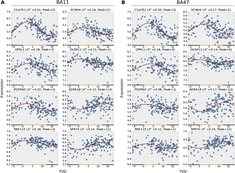
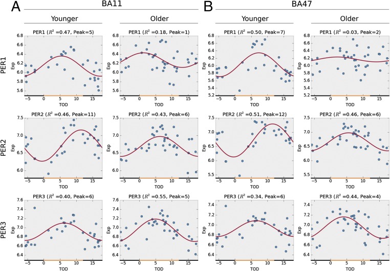
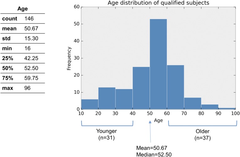
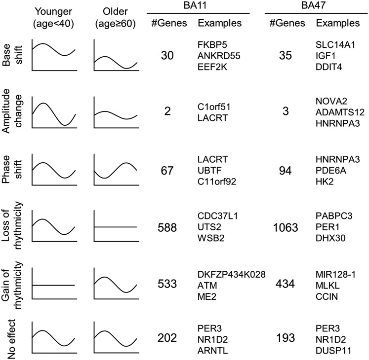
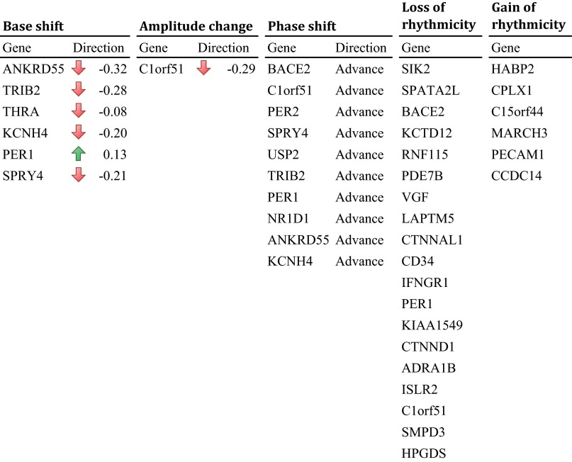
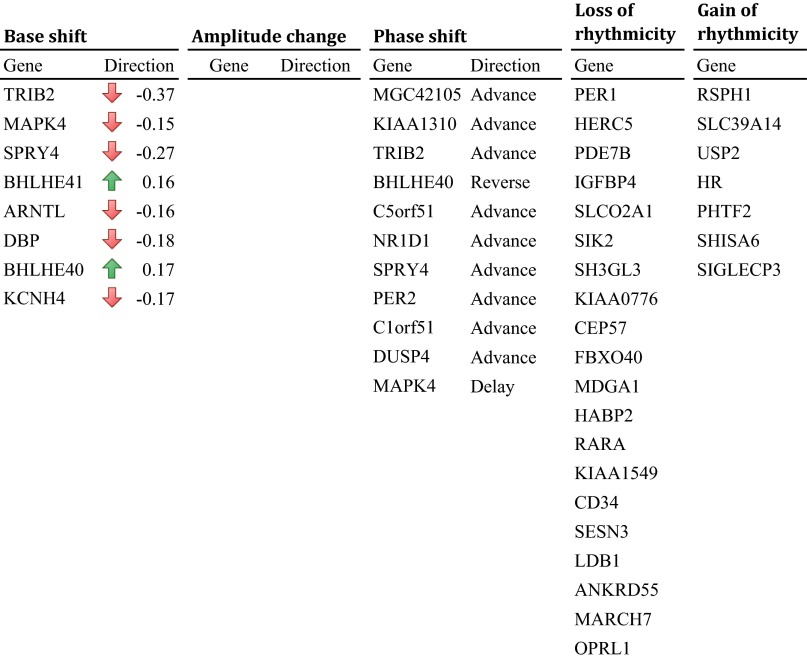
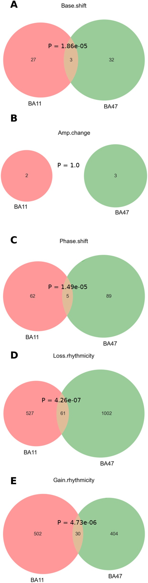
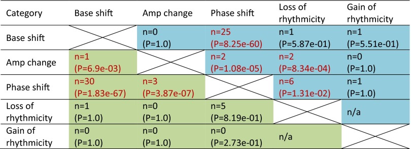
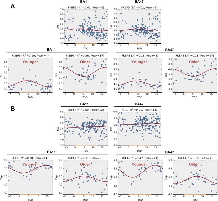
With aging, significant changes in circadian rhythms occur, including a shift in phase toward a "morning" chronotype and a loss of rhythmicity in circulating hormones. However, the effects of aging on molecular rhythms in the human brain have remained elusive. Here, we used a previously described time-of-death analysis to identify transcripts throughout the genome that have a significant circadian rhythm in expression in the human prefrontal cortex [Brodmann's area 11 (BA11) and BA47]. Expression levels were determined by microarray analysis in 146 individuals. Rhythmicity in expression was found in ∼ 10% of detected transcripts (P < 0.05). Using a metaanalysis across the two brain areas, we identified a core set of 235 genes (q < 0.05) with significant circadian rhythms of expression. These 235 genes showed 92% concordance in the phase of expression between the two areas. In addition to the canonical core circadian genes, a number of other genes were found to exhibit rhythmic expression in the brain. Notably, we identified more than 1,000 genes (1,186 in BA11; 1,591 in BA47) that exhibited age-dependent rhythmicity or alterations in rhythmicity patterns with aging. Interestingly, a set of transcripts gained rhythmicity in older individuals, which may represent a compensatory mechanism due to a loss of canonical clock function. Thus, we confirm that rhythmic gene expression can be reliably measured in human brain and identified for the first time (to our knowledge) significant changes in molecular rhythms with aging that may contribute to altered cognition, sleep, and mood in later life.

Keywords: aging; circadian rhythms; gene expression; postmortem; prefrontal cortex.


Conflict of interest statement

The authors declare no conflict of interest.

Figures

Fig. 1.
Fig. 1.
Heat map of expression levels and comparison of circadian acrophase for the top circadian genes (n = 235, q < 0.05) in BA11 and BA47. (A and B) Expression levels were Z-transformed for each gene. Red indicates higher expression level; green indicates lower expression levels. (C) The circadian phase (peak hours) of 235 circa genes derived from metaanalysis are plotted on TOD axes for BA11 (x axis) and BA47 (y axis). Red dashed line: 1:1 diagonal line. Green dashed lines: ±4-h phase concordance boundaries. Ninety-two percent of circa genes (217 of 235) are located within the concordance interval.
Fig. 2.
Fig. 2.
Circadian gene expression patterns of six canonical circadian genes in BA11 (A) and BA47 (B). The x axis denotes the time of death (TOD) on ZT scale (−6–18 h). Approximate day–night interval within a pseudoday is represented by yellow (day) and black (night) bands. Data points from 146 subjects are plotted in blue. The best-fitted sinusoidal curves are depicted in red. The empirical P value and the estimated peak hour are also reported above each panel.
Fig. 3.
Fig. 3.
Top 50 clock-regulated genes across two brain regions. Gene list is sorted by q value after adaptive weighted (AW)-Fisher procedure. Genes in red are known circadian-related genes according to their records in GeneCards database (59, 60).
Fig. S1.
Fig. S1.
Overview of analysis flows. (A) Detection of clock-regulated genes in human prefrontal cortex (PFC) via metaanalysis. We first estimated the P values of expression rhythmicity for all of the genes in brain regions BA11 and BA47 independently. Then we combined these P values to meta-P values via AW-Fisher and corrected them into q values. A total of 235 cross-region PFC circadian genes were detected at q < 0.05. (B) Detection of age effect on circadian expression rhythmicity. The work flow was independent from A; i.e., the analysis in B did not rely on any a priori results yielded in A. It is possible that a gene shows expression rhythmicity only in younger individuals but not in older individuals (or could be vice versa). Therefore, if younger subjects were mixed with older subjects (e.g., when all 146 subjects were included as how we did in A), we might be unable to detect the expression rhythmicity of certain genes (see Fig. S4 for such examples).
Fig. S2.
Fig. S2.
Top eight rhythmic genes in BA11 (A) and BA47 (B). The x axis denotes the time of death (TOD) on ZT scale (−6–18 h). Approximate day–night interval within a pseudoday is represented by yellow (day) and black (night) bands. Data points from 146 subjects are plotted in blue. The best-fitted sinusoidal curves are depicted in red. The empirical P value and the estimated peak hour are also reported above each panel.
Fig. 4.
Fig. 4.
Aging has unique effects on the circadian gene expression patterns of period genes (PER family) in both BA11 (A) and BA47 (B). Older people have disrupted PER1 circadian expression patterns (P = 0.027 in BA11; P = 0.0005 in BA47), together with a phase advance of PER2 expression from sunset to noon (P = 0.005 in BA11; P = 0.004 in BA47), whereas their PER3 expression remained intact.
Fig. S3.
Fig. S3.
Cohort age information in this study.
Fig. 5.
Fig. 5.
Illustration of age effect on circadian gene expression patterns. The age effect on circadian gene expression patterns can be classified into five categories, as illustrated (rows 1–5). The number of instances and the top three most significant examples (sorted by P values) were also reported. See Datasets S2 and S3 for complete lists.
Fig. S4.
Fig. S4.
List of detected age effects on 235 PFC circadian genes in BA11. Upward arrow represents rise of circadian expression rhythmicity base in the older. Downward arrow represents drop of base or decrease of amplitude in the older. Phase advance means the peak hour of circadian expression rhythmicity comes earlier in the older. See Dataset S2 for full table.
Fig. S5.
Fig. S5.
List of detected age effects on PFC circadian genes in BA47. Upward arrows and downward arrows represent rise and drop of circadian expression rhythmicity base in the older, respectively. No instance was found with significant amplitude change. Phase advance means the peak hour of circadian expression rhythmicity comes earlier in the older. Phase delay means the peak hour comes later in the older. Phase reverse means the difference of peak hours between the younger and the older groups is nearly 12 h. See Dataset S3 for full table.
Fig. S6.
Fig. S6.
Venn diagram of gene numbers in each age effect category between BA11 and BA47. Only those genes with P < 0.05 in each category were counted here (Datasets S2 and S3). See also Dataset S4 for complete lists. Shown are interactions between BA11 and BA47 in genes that show (A) base shift, (B) amplitude change, (C) phase shift, (D) loss of rhythmicity, or (E) gain of rhythmicity.
Fig. S7.
Fig. S7.
Co-occurrence of age effects on circadian expression rhythmicity. Shown in each cell is the number of co-occurrence and the associated P value (by Fisher’s exact test) between every two age effect categories. Note that the loss of rhythmicity and the gain of rhythmicity are two mutually exclusive categories by definition. Color code: blue cells, cases in BA11; green cells, cases in BA47.
Fig. S8.
Fig. S8.
Rhythmicity expression profiles before and after age stratification. (A) FKBP5. (B) IGF1. (Upper) Before age stratification (n = 146). (Lower) After age stratification (n = 31 in younger and n = 37 in older).

References

    1. Patel VR, Eckel-Mahan K, Sassone-Corsi P, Baldi P. How pervasive are circadian oscillations? Trends Cell Biol. 2014;24(6):329–331. - PMC - PubMed
    1. Baron KG, Reid KJ. Circadian misalignment and health. Int Rev Psychiatry. 2014;26(2):139–154. - PMC - PubMed
    1. Foster RG, Kreitzman L. The rhythms of life: What your body clock means to you! Exp Physiol. 2014;99(4):599–606. - PubMed
    1. Reppert SM, Weaver DR. Molecular analysis of mammalian circadian rhythms. Annu Rev Physiol. 2001;63:647–676. - PubMed
    1. Hogenesch JB, Panda S, Kay S, Takahashi JS. Circadian transcriptional output in the SCN and liver of the mouse. Novartis Found Symp. 2003;253:171–180; discussion 52–55, 102–109, 180–183 passim. - PubMed

Publication types