. 2015 Dec 23;90(5):2664-75.
doi: 10.1128/JVI.02916-15.

Antigenic and Cryo-Electron Microscopy Structure Analysis of a Chimeric Sapovirus Capsid

Affiliations

Antigenic and Cryo-Electron Microscopy Structure Analysis of a Chimeric Sapovirus Capsid

Naoyuki Miyazaki et al. J Virol. .

Abstract

The capsid protein (VP1) of all caliciviruses forms an icosahedral particle with two principal domains, shell (S) and protruding (P) domains, which are connected via a flexible hinge region. The S domain forms a scaffold surrounding the nucleic acid, while the P domains form a homodimer that interacts with receptors. The P domain is further subdivided into two subdomains, termed P1 and P2. The P2 subdomain is likely an insertion in the P1 subdomain; consequently, the P domain is divided into the P1-1, P2, and P1-2 subdomains. In order to investigate capsid antigenicity, N-terminal (N-term)/S/P1-1 and P2/P1-2 were switched between two sapovirus genotypes GI.1 and GI.5. The chimeric VP1 constructs were expressed in insect cells and were shown to self-assemble into virus-like particles (VLPs) morphologically similar to the parental VLPs. Interestingly, the chimeric VLPs had higher levels of cross-reactivities to heterogeneous antisera than the parental VLPs. In order to better understand the antigenicity from a structural perspective, we determined an intermediate-resolution (8.5-Å) cryo-electron microscopy (cryo-EM) structure of a chimeric VLP and developed a VP1 homology model. The cryo-EM structure revealed that the P domain dimers were raised slightly (∼5 Å) above the S domain. The VP1 homology model allowed us predict the S domain (67-229) and P1-1 (229-280), P2 (281-447), and P1-2 (448-567) subdomains. Our results suggested that the raised P dimers might expose immunoreactive S/P1-1 subdomain epitopes. Consequently, the higher levels of cross-reactivities with the chimeric VLPs resulted from a combination of GI.1 and GI.5 epitopes.

Importance: We developed sapovirus chimeric VP1 constructs and produced the chimeric VLPs in insect cells. We found that both chimeric VLPs had a higher level of cross-reactivity against heterogeneous VLP antisera than the parental VLPs. The cryo-EM structure of one chimeric VLP (Yokote/Mc114) was solved to 8.5-Å resolution. A homology model of the VP1 indicated for the first time the putative S and P (P1-1, P2, and P1-2) domains. The overall structure of Yokote/Mc114 contained features common among other caliciviruses. We showed that the P2 subdomain was mainly involved in the homodimeric interface, whereas a large gap between the P1 subdomains had fewer interactions.


Figures

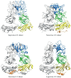
FIG 1
FIG 1
Chimeric sapovirus capsids. (A) Sequence alignment of Yokote and Mc114 showing the predicted N-term (orange), S domain (yellow), and hinge (H) region (olive), the P1-1 and P1-2 subdomains (green), and the P2 subdomain (marine). The 289th residue in Yokote (red X) shows the site where the constructs were switched. Mc114/Yokote contained Mc114 N-term/S/P1-1 (residues 1 to 289) and Yokote P2/P1-2/VP2 [residue 290 to the poly(A)], while Yokote/Mc114 contained Yokote N-term/S/P1-1 (residues 1 to 289) and Mc114 P2/P1-2/VP2 [residue 290 to the poly(A)]. (B) Schematic of the chimeric Mc114/Yokote and Yokote/Mc114VP1 constructs.
FIG 2
FIG 2
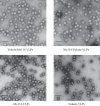
Sapovirus VLPs. Electron micrographs of negatively stained sapovirus VLPs, chimeric Yokote/Mc114, chimeric Mc114/Yokote, parental Mc114, and parental Yokote are shown. Scale bars, 100 nm.
FIG 3
FIG 3
Chimeric sapovirus VLP cross-reactivities. The cutoff (dashed line) was at OD492 = 0.15 as previously determined (13, 14). (A) Antigen ELISA reactivities of Mc114, Mc114/Yokote, Yokote, and Yokote/Mc114 VLPs with Mc114 VLP antiserum. (B) Antigen ELISA reactivities of Mc114, Mc114/Yokote, Yokote, and Yokote/Mc114 VLPs with Yokote VLP antiserum. (C) Summary of the cutoff titers (OD492 = 0.15) of the parental and chimeric VLP reactivities to Mc114 and Yokote antisera.
FIG 4
FIG 4
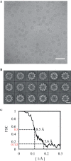
Single-particle cryo-EM of chimeric-sapovirus Yokote/Mc114 VLPs. (A) Representative raw micrograph of frozen-hydrated chimeric VLPs. Scale bar, 100 nm. (B) Reference-free 2D class averages of VLPs used directly in initial model building. Scale bar, 200 Å. (C) Fourier shell correlation (FSC) curve for the final refined VLP structure. The resolution of the structure was estimated to be ∼8.5 Å using the 0.5 FSC criterion.
FIG 5
FIG 5
Cryo-EM map of the chimeric-sapovirus Yokote/Mc114 VLP determined at 8.5-Å resolution. (A) Stereo view of the icosahedral capsid. The graded color coding represents the radius, 125 Å (orange), 144 Å (yellow), 160 Å (green), and 190 Å (marine). The graded color coding also corresponds to the partial N-term (orange), S domain (yellow), P1 subdomain (green), and P2 subdomain (marine). Scale bar, 100 Å. The 3-fold and 5-fold axes are indicated. (B) A sliced section of the chimeric Yokote/Mc114 VLP (the inner core was removed for clarity). The S domain was partially surface exposed, and the P domain dimers were raised from the S domain by ∼5 Å (arrow). (C) Density map of the data in panel B.
FIG 6
FIG 6
Capsid C/C VP1 structures of the caliciviruses. (A) Cutaway cryo-EM map of Yokote/Mc114 C/C VP1 dimer. The graded color coding corresponds to the partial N-term (orange), S domain (yellow), P1 subdomain (green), and P2 subdomain (marine). The upper half of the P domain was involved in homodimeric interactions, whereas the lower half of the P domain did not make observable homodimeric contacts. The S domain was connected to the P domin monomer by a narrow hinge region. (B) The cryo-EM map of the C/C dimer of human GI.1 norovirus (PDB ID: 1IHM), vesivirus (PDB ID: 2GH8), and lagovirus (PDB ID: 3J1P) at 8.5-Å resolution (FSC = 0.5).
FIG 7
FIG 7
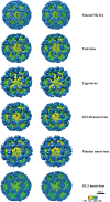
Cryo-EM and X-ray crystal structures of calicivirus particles. Stereo view of sapovirus Yokote/Mc114, vesivirus (feline calicivirus; EMD-1942), lagovirus (EMD-5410), GII.10 human norovirus (EMD-5374), murine norovirus (8-Å virion map provided by Thomas Smith), and human GI.1 norovirus (1IHM). The 3-fold and 5-fold axes are shown, and the particles are color coded by radius as 125 Å (orange), 144 Å (yellow), 160 Å (green), and 190 Å (marine).
FIG 8
FIG 8
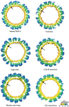
Cross-sectional view of calicivirus particles. Sapovirus Yokote/Mc114, vesivirus, lagovirus, human GII.10 norovirus, murine norovirus, and human GI.1 norovirus (the inner cores removed for clarity) were color coded by radius as 125 Å (orange), 144 Å (yellow), 160 Å (green), and 190 Å (marine). The arrows show the P domain raised from the S domain (i.e., sapovirus, vesivirus, lagovirus, GII.10 norovirus, and murine norovirus) or the P domain resting on the S domain (i.e., GI.1 norovirus).
FIG 9
FIG 9
Homology modeling of the chimeric-sapovirus Yokote/Mc114 VLP. Multiple-sequence alignments of human GI.1 norovirus (Norwalk virus), SMSV (vesivirus), and chimeric sapovirus (Yokote/Mc114) were performed in which the conserved secondary structural elements (alpha helices and beta-sheets) and sequence similarities (s.s.) were incorporated to improve the alignment accuracy. The secondary structures of S (67–234), P1-1 (235–280), P2 (281–447), and P1-2 (448–567) domains were predicted by comparing the amino acid sequence with those of norovirus and vesivirus.
FIG 10
FIG 10
The initial and refined homology models. The CCF was improved from 0.74 (A) to 0.75 (B). The Yokote/Mc114 VP1 model showed a better fit into the densities of S domain and P1 subdomains than into that of the P2 subdomain.
FIG 11
FIG 11
A comparison of four different calicivirus capsid (C/C) VP1 dimers. The cartoon shows a representation of C/C dimers of sapovirus (homology model), vesivirus (X-ray structure of VLP; EMD-1942), human GI.1 norovirus (X-ray structure of native virion; 1IHM), and lagovirus (X-ray structure of a chimeric VLP [S domain, 4EJR; P domain, 4EGT]). The sapovirus P dimer appeared more similar to vesivirus than to human GI.1 norovirus or lagovirus. The approximate domains are colored as the sapovirus S domain (yellow: 67–222), hinge (olive: 223–228), P1-1 and P1-2 (green: 229–280 and 448–567, respectively), and P2 (marine: 281 to 447); as the norovirus N-term (orange: 1–49), S domain (yellow: 50–225), hinge (olive: 216–222), P1-1 and P1-2 (green: 226–277 and 406–520, respectively), and P2 (marine: 278 to 405); as the vesivirus N-term (orange: 160–200) S domain (yellow: 201–355), hinge (olive: 356–361), P1-1 and P1-2 (green: 362–413 and 590–703, respectively), and P2 (marine: 414–589); and as the lagovirus N-term (orange: 1–66), S domain (yellow: 67–224), hinge (olive: 225–235), P1-1 and P1-2 (green: 236–287 and 449–557, respectively), and P2 (marine: 288–448).
FIG 12
FIG 12
Distribution of sequence variations in the S and P1-1 region of the capsid VP1 protein between the Yokote and Mc114 strains of sapovirus. A stereo view and surface representation of the Yokote/Mc114 homology model (dimer), viewed from the top (A) and the side (B), are shown. The top view corresponds to the view from the viral surface, and the side view is a 90° rotation of the top view. The S, P1, and P2 (sub)domains are colored dark gray, medium gray, and light gray, respectively. Regions of sequence diversity and surface-exposed residues (see Fig. 1A) are colored in the S domain (orange) and P1-1 subdomain (red). Residues that were not surfaced exposed and not indicated on the model were residues 115, 134, 166, 188, and 247.

References

    1. Hansman GS, Jiang XJ, Green KY. 2010. Caliciviruses: molecular and cellular virology. Caister Academic Press, Norfolk, United Kingdom.
    1. Hansman GS, Oka T, Sakon N, Takeda N. 2007. Antigenic diversity of human sapoviruses. Emerg Infect Dis 13:1519–1525. doi:10.3201/eid1310.070402. - DOI - PMC - PubMed
    1. Hansman GS, Oka T, Katayama K, Takeda N. 2007. Human sapoviruses: genetic diversity, recombination, and classification. Rev Med Virol 17:133–141. doi:10.1002/rmv.533. - DOI - PubMed
    1. Prasad BV, Hardy ME, Dokland T, Bella J, Rossmann MG, Estes MK. 1999. X-ray crystallographic structure of the Norwalk virus capsid. Science 286:287–290. doi:10.1126/science.286.5438.287. - DOI - PubMed
    1. Chen R, Neill JD, Prasad BV. 2003. Crystallization and preliminary crystallographic analysis of San Miguel sea lion virus: an animal calicivirus. J Struct Biol 141:143–148. doi:10.1016/S1047-8477(02)00583-X. - DOI - PubMed

Publication types

MeSH terms