. 2016 Mar 4;118(5):856-66.
doi: 10.1161/CIRCRESAHA.115.307918. Epub 2015 Dec 23.

Critical Role for Telomerase in the Mechanism of Flow-Mediated Dilation in the Human Microcirculation

Affiliations

Critical Role for Telomerase in the Mechanism of Flow-Mediated Dilation in the Human Microcirculation

Andreas M Beyer et al. Circ Res. .

Abstract

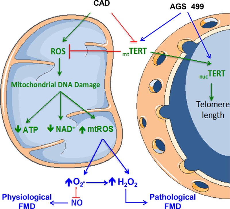
Rationale: Telomerase is a nuclear regulator of telomere elongation with recent reports suggesting a role in regulation of mitochondrial reactive oxygen species. Flow-mediated dilation in patients with cardiovascular disease is dependent on the formation of reactive oxygen species.

Objective: We examined the hypothesis that telomerase activity modulates microvascular flow-mediated dilation, and loss of telomerase activity contributes to the change of mediator from nitric oxide to mitochondrial hydrogen peroxide in patients with coronary artery disease (CAD).

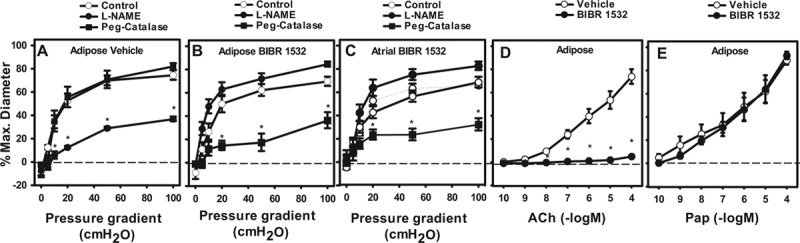
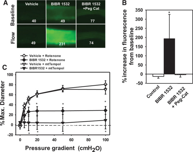
Methods and results: Human coronary and adipose arterioles were isolated for videomicroscopy. Flow-mediated dilation was measured in vessels pretreated with the telomerase inhibitor BIBR-1532 or vehicle. Statistical differences between groups were determined using a 2-way analysis of variance repeated measure (n≥4; P<0.05). L-NAME (N(ω)-nitro-L-arginine methyl ester; nitric oxide synthase inhibitor) abolished flow-mediated dilation in arterioles from subjects without CAD, whereas polyethylene glycol-catalase (PEG-catalase; hydrogen peroxide scavenger) had no effect. After exposure to BIBR-1532, arterioles from non-CAD subjects maintained the magnitude of dilation but changed the mediator from nitric oxide to mitochondrial hydrogen peroxide (% max diameter at 100 cm H2O: vehicle 74.6±4.1, L-NAME 37.0±2.0*, PEG-catalase 82.1±2.8; BIBR-1532 69.9±4.0, L-NAME 84.7±2.2, PEG-catalase 36.5±6.9*). Conversely, treatment of microvessels from CAD patients with the telomerase activator AGS 499 converted the PEG-catalase-inhibitable dilation to one mediated by nitric oxide (% max diameter at 100 cm H2O: adipose, AGS 499 78.5±3.9; L-NAME 10.9±17.5*; PEG-catalase 79.2±4.9). Endothelial-independent dilation was not altered with either treatment.

Conclusions: We have identified a novel role for telomerase in re-establishing a physiological mechanism of vasodilation in arterioles from subjects with CAD. These findings suggest a new target for reducing the oxidative milieu in the microvasculature of patients with CAD.

Keywords: coronary artery disease; flow-mediated dilation; microvascular dysfunction; mitochondria; reactive oxygen species; telomerase activity; vascular biology.


Figures

Figure 1.
Figure 1.
Telomerase inhibition replicates the coronary artery disease (CAD) vascular phenotype in human adipose and atrial vessels from non-CAD subjects. Flow-mediated dilation (FMD) was evaluated in isolated microvessels treated with vehicle (A; adipose), BIBR-1532 (B; adipose), and BIBR-1532 (C; atrial). In BIBR-treated vessels, the mechanism of FMD changed from nitric oxide (NO) to hydrogen peroxide (H2O2). D, Acetylcholine (ACh)-induced dilation was virtually eliminated after telomerase inhibition. E, No change in endothelium-independent dilation to papaverine was observed. N=4 to 12 adipose, N=5 to 9 atrial. *P<0.05 2-way analysis of variance (ANOVA) RM Tukey post hoc analysis. L-NAME indicates Nω-nitro-L-arginine methyl ester; and Peg-Catalase, polyethylene glycol-catalase.
Figure 2.
Figure 2.
Coronary artery disease (CAD) does not cause telomere shortening in microvessels or left ventricular tissue but decreases expression of the catalytic subunit catalytic subunit of human telomerase complex (TERT). A, Total average telomere length was evaluated in genomic DNA from microvessels (adipose) and left ventricular tissue from subjects with and without CAD. B, Expression of the TERT was evaluated in left ventricular tissue from subjects with and without CAD by Western blot. Values are normalized to β-actin loading control and expressed as fold change compared with non-CAD control. N=4 to 7. *P<0.05 t test.
Figure 3.
Figure 3.
Mitochondrial hydrogen peroxide (H2O2)–mediated flow-mediated dilation (FMD) after inhibition of telomerase. H2O2 levels were analyzed using the H2O2-specific florescent probe PYI (peroxy yellow 1) targeted to mitochondria (MitoPYI) in vessels from non–coronary artery disease (CAD) subjects. A, Representative image; numbers represent florescent intensity above background. B, Summary of florescence intensity at 5 minutes after initiation of flow. Specificity of the probe for H2O2 was confirmed using polyethylene glycol-catalase (Peg-Catalase). C, An inhibitor of electron transport chain complex I (rotenone) or a mitochondrial-targeted reactive oxygen species (ROS) scavenger (MitoTempol) inhibited FMD after telomerase inhibition. N=4. *P<0.05 2-way analysis of variance (ANOVA) RM (dose response curve) or t test (fluorescence data) with Tukey post hoc.
Figure 4.
Figure 4.
Mitochondrial translocation of catalytic subunit of human telomerase complex (TERT) after exposure to acute oxidative stress. A, Treatment of normal human fibroblasts (HF) with hydrogen peroxide (H2O2) resulted in translocation of TERT-GFP (green fluorescent protein) from the nucleus to the mitochondria (colocalized with Mitotracker Red). Cells were imaged using a 63× oil immersion objective. B, To confirm purity of isolated mitochondria of cells either transfected with whole cell (WC) TERT or R3E/R6E TERT (nucTERT) Western blots for tubulin (cytoplasmic marker), mitochondrial heat shock proteins 70 (mtHSP70), and Ku80 (nuclear isoform [higher molecular weight] and mitochondrial isoform) were performed. C, Expression of nucTERT increased mitochondrial superoxide production as measured with MitoSox. D, Expression of nucTERT decreased cellular adenosine triphosphate (ATP) production after external stress. n=3 to 5. *P<0.05 t Test.
Figure 5.
Figure 5.
Change in vascular phenotype is independent of changes in gene expression. Flow-mediated dilation (FMD) was evaluated in isolated adipose microvessels from subjects without coronary artery disease (CAD) treated with actinomycin D, a transcriptional inhibitor. A, Act D+vehicle. B, Act D+BIBR-1532. Mechanism of FMD changed from NO (Act D)→H2O2 (Act D+BIBR-1532). C and D, Act D was sufficient to prevent transcriptional activation of heat shock proteins 27 and 70 after acute exposure to 42°C. Sample blot shows 2/5 total treatments. *P<0.05 2-way analysis of variance (ANOVA) RM Tukey post hoc analysis (functional studies). N=4 to 5. *P<0.05 vs untreated control t Test Tukey post hoc analysis (protein levels). CO indicates control; HS, heat shock; L-NAME, Nω-nitro-L-arginine methyl ester; and Peg-Catalase, polyethylene glycol-catalase.
Figure 6.
Figure 6.
Pharmacological activation of telomerase restores nitric oxide (NO)–mediated dilation in response to flow. Flow-mediated dilation (FMD) was evaluated in isolated adipose and atrial microvessels from subjects with coronary artery disease (CAD) treated with the selective transcriptional telomerase activator AGS 499 (22 nmol/L). Vehicle-treated vessels are historic controls. A, CAD adipose+AGS 499. B, CAD atrial+AGS 499. The mechanism of FMD changed from polyethylene glycol-catalase (Peg-Catalase)–sensitive (H2O2 in CAD) to Nω-nitro-L-arginine methyl ester (L-NAME) and c-PTIO–sensitive (NO mediated in CAD+AGS 499). C, Inhibition of telomerase activity in vessels from subjects with CAD did not alter mechanism or overall dilator capacity in response to flow. D, AGS 499 resulted in transcriptional increase of catalytic subunit of human telomerase complex (TERT) mRNA. *P<0.05 vs AGS 499 control 2-way analysis of variance (ANOVA) RM Tukey post hoc analysis. N=5 to 8 adipose, N=3 to 4 atrial. *P>0.05 vs AGS 499 only–treated t Test Tukey post hoc analysis (RNA levels). #AGS 499+c-PTIO N=3 (2 adipose and 1 atrial). HUVECs indicates human umbilical vein endothelial cells.
Figure 7.
Figure 7.
Proposed mechanism of TERT in regulating balance of endothelial mitochondrial ROS and NO to maintain FMD in human microvessels. AGS 499 indicates telomeres activator; ATP, adenosine triphosphate; CAD, coronary artery disease; FMD, flow-mediated dilation; H2O2, hydrogen peroxide; mt, mitochondrial; nuc, nuclear; NAD+, nicotinamide adenine dinucleotide; NO, nitric oxide; O2–·, superoxide; ROS, reactive oxygen species; and TERT, catalytic subunit of human telomerase complex.

Comment in

  • The Secret Life of Telomerase.
    Welsh DG. Welsh DG. Circ Res. 2016 Mar 4;118(5):781-2. doi: 10.1161/CIRCRESAHA.116.308290. Circ Res. 2016. PMID: 26941421 No abstract available.

References

    1. Aviv H, Khan MY, Skurnick J, Okuda K, Kimura M, Gardner J, Priolo L, Aviv A. Age dependent aneuploidy and telomere length of the human vascular endothelium. Atherosclerosis. 2001;159:281–287. - PubMed
    1. Chang E, Harley CB. Telomere length and replicative aging in human vascular tissues. Proc Natl Acad Sci U S A. 1995;92:11190–11194. - PMC - PubMed
    1. Brouilette S, Singh RK, Thompson JR, Goodall AH, Samani NJ. White cell telomere length and risk of premature myocardial infarction. Arterioscler Thromb Vasc Biol. 2003;23:842–846. doi: 10.1161/01.ATV.0000067426.96344.32. - PubMed
    1. Minamino T, Miyauchi H, Yoshida T, Ishida Y, Yoshida H, Komuro I. Endothelial cell senescence in human atherosclerosis: role of telomere in endothelial dysfunction. Circulation. 2002;105:1541–1544. - PubMed
    1. Shaik S, Inuzuka H, Liu P, Wei W, Wang Z. Endothelium Aging and Vascular Diseases: INTECH Open Access Publisher. 2013.

Publication types