. 2016 Mar;15(3):1032-47.
doi: 10.1074/mcp.M115.055723. Epub 2015 Dec 23.

Reactive Oxygen Species (ROS)-Activated ATM-Dependent Phosphorylation of Cytoplasmic Substrates Identified by Large-Scale Phosphoproteomics Screen

Affiliations

Reactive Oxygen Species (ROS)-Activated ATM-Dependent Phosphorylation of Cytoplasmic Substrates Identified by Large-Scale Phosphoproteomics Screen

Sergei V Kozlov et al. Mol Cell Proteomics. 2016 Mar.

Abstract

Ataxia-telangiectasia, mutated (ATM) protein plays a central role in phosphorylating a network of proteins in response to DNA damage. These proteins function in signaling pathways designed to maintain the stability of the genome and minimize the risk of disease by controlling cell cycle checkpoints, initiating DNA repair, and regulating gene expression. ATM kinase can be activated by a variety of stimuli, including oxidative stress. Here, we confirmed activation of cytoplasmic ATM by autophosphorylation at multiple sites. Then we employed a global quantitative phosphoproteomics approach to identify cytoplasmic proteins altered in their phosphorylation state in control and ataxia-telangiectasia (A-T) cells in response to oxidative damage. We demonstrated that ATM was activated by oxidative damage in the cytoplasm as well as in the nucleus and identified a total of 9,833 phosphorylation sites, including 6,686 high-confidence sites mapping to 2,536 unique proteins. A total of 62 differentially phosphorylated peptides were identified; of these, 43 were phosphorylated in control but not in A-T cells, and 19 varied in their level of phosphorylation. Motif enrichment analysis of phosphopeptides revealed that consensus ATM serine glutamine sites were overrepresented. When considering phosphorylation events, only observed in control cells (not observed in A-T cells), with predicted ATM sites phosphoSerine/phosphoThreonine glutamine, we narrowed this list to 11 candidate ATM-dependent cytoplasmic proteins. Two of these 11 were previously described as ATM substrates (HMGA1 and UIMCI/RAP80), another five were identified in a whole cell extract phosphoproteomic screens, and the remaining four proteins had not been identified previously in DNA damage response screens. We validated the phosphorylation of three of these proteins (oxidative stress responsive 1 (OSR1), HDGF, and ccdc82) as ATM dependent after H2O2 exposure, and another protein (S100A11) demonstrated ATM-dependence for translocation from the cytoplasm to the nucleus. These data provide new insights into the activation of ATM by oxidative stress through identification of novel substrates for ATM in the cytoplasm.


Figures

Fig. 1.
Fig. 1.
ATM kinase activation by ionizing radiation (IR) and H2O2 in subcellular fractions. Normal human fibroblasts (NFF) were irradiated with 6 Gy or treated with 0.5 mm H2O2. NFF and A-T fibroblasts total, cytoplasmic, and nuclear extracts were prepared as described in the Methods and immunoprecipitated with anti-ATM antibody. Immunoprecipitated ATM was resolved on biphasic 5%/12% SDS-Polyacrylamide gel and Western blotting was performed with phospho-specific ATM antibodies. Variation in total ATM protein levels represents the different amount of ATM kinase present in subcellular fractions after immunoprecipitation. (A) ATM kinase activation by ionizing radiation (IR) and H2O2 in nuclear fractions prepared from normal human fibroblasts (NFF). Activation was determined by ATM autophosphorylation using antibodies against different active forms. (B) Monitoring of subcellular fractionation using specific markers. Cytoplasmic proteins: tubulin and GAPDH; Nuclear proteins: histone H3, SMC1, and KAP1 (C) Active ATM kinase is detected in total extracts, nuclear and cytoplasmic fractions from NFF using phospho-specific antibodies against ATM. (D) ATM kinase activation induced either by IR or H2O2 in total cell extracts, cytoplasmic and nuclear extracts.
Fig. 2.
Fig. 2.
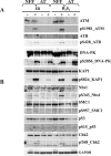
(A) Activation of DNA damage response kinases by IR and H2O2 in normal human fibroblasts and A-T primary fibroblasts. Normal human fibroblasts (NFF) and A-T fibroblasts were irradiated with 6 Gy or treated with 0.5 mm H2O2. Total extracts were prepared as described in the Methods, and samples were run on NuPAGE 3–8% tris-acetate mini gels. Western blotting was performed with phospho-specific ATM, ATR, and DNA-PK antibodies as indicated. (B) ATM-dependent signaling in response to IR and H2O2 in normal human fibroblasts and A-T primary fibroblasts. Normal human fibroblasts (NFF) and A-T fibroblasts were irradiated with 6 Gy or treated with 0.5 mm H2O2. Total extracts were prepared as described in the Methods and samples were run on NuPAGE 4–12% Bis-Tris mini gels. Western blotting was performed with phospho-specific antibodies against known ATM substrates as indicated.
Fig. 3.
Fig. 3.
Phosphoproteomics workflow and identified phosphopeptides. (A) Cultures of NFF and A-T cells were mock-treated or treated with H2O2. Cytoplasmic extracts of each were prepared, digested, and duplex stable isotope dimethyl labeled. Phosphopeptides were enriched and fractionated using the TiSH method and analyzed by LC-MS/MS. (B) Localization probability distribution of phosphosites identified using MaxQuant. (C) Counts of phosphopeptides and proteins at the thresholds used; p indicates localization probability. (D) Enrichment of cytoplasm and nucleus GO terms for the A-T and normal cells.
Fig. 4.
Fig. 4.
Analysis of phosphoproteomics data set by rank product statistics. (A) Overview of normalization process for Set 1; quantile global normalization was followed by missing value imputation, surrogate variable analysis, and rank product analysis. (B) Principle component analysis projecting the first and third components of Set 1 data after normalization. (C) Heat map illustrating overall patterns for the peptides identified in Set 1. (D) Heat maps for Sets 2–4. The number of solid boxes indicates the number of times a phosphopeptide must be detected at to be included in the set. The number of boxes with diagonal lines indicates the number of times a that missing values for a particular phosphopeptide was permitted.
Fig. 5.
Fig. 5.
Potential protein kinase substrate motifs and regulated p(S/T)Q sites. (A) MotifX analysis of Sets 1–3. (B) Heatmap showing log2 ratio values of p(S/T)Q containing phosphopeptides in Sets 1–4.
Fig. 6.
Fig. 6.
Detection of candidate proteins from phosphoproteomics screen in phospho-(S/T)Q ATM/ATR substrate antibody immunoprecipitates. Extracts from untreated, irradiated (3 Gy) and hydrogen-peroxide-treated normal human fibroblasts (NFF) and A-T fibroblasts were prepared as described in the Methods and immunoprecipitated with p-(S/T)Q ATM/ATR substrate antibodies. Immunoprecipitates and extracts were run on 4–12% NuPage gels and gels were transferred to nitrocellulose membrane and stained with Ponceau S to estimate the loading. Western blotting was performed with antibodies against a selection of candidate proteins from the phosphoproteomics screen. listtext(A) (B) HDGF and ccdc82. * Western blotting with antibodies against known substrates of ATM kinase (Mre11, Rad50) was performed to access the efficiency of p(S/T)Q antibodies imunnoprecipitations.
Fig. 7.
Fig. 7.
Translocation of S100A11 to the nucleus in response to H2O2 treatment is defective in A-T fibroblasts. Normal control fibroblasts (NFF) and A-T fibroblasts were grown on coverslips, treated with hydrogen peroxide, or left untreated as described in the Methods. IF microscopy was performed using antibodies against EEA1 and S100A11 (A) or γH2AX and S100A11 (B). Cells were imaged on a Zeiss AxioImager M1 fluorescent microscope using x63 oil lens. Images were taken with an Axiocam503 mono camera and processed using ZEN software (Zeiss). Separate channels and merged images are shown. The scale bar is 10 μm.

References

    1. Lavin M. F. (2008) Ataxia-telangiectasia: From a rare disorder to a paradigm for cell signalling and cancer. Nat. Rev. Mol. Cell Biol. 9, 759–769 - PubMed
    1. Derheimer F. A., and Kastan M. B. (2010) Multiple roles of ATM in monitoring and maintaining DNA integrity. FEBS Lett. 584, 3675–3681 - PMC - PubMed
    1. Shiloh Y. (2014) ATM: Expanding roles as a chief guardian of genome stability. Exp. Cell Res. 329, 154–161 - PubMed
    1. Shiloh Y., and Ziv Y. (2013) The ATM protein kinase: Regulating the cellular response to genotoxic stress, and more. Nat. Rev. Mol. Cell Biol. 14, 197–210 - PubMed
    1. Paull T. T. (2015) Mechanisms of ATM activation. Annu. Rev. Biochem. 84, 711–738 - PubMed

Publication types