. 2016 Mar;173(5):937-50.
doi: 10.1111/bph.13413. Epub 2016 Feb 10.

Involvement of Rac1 signalling pathway in the development and maintenance of acute inflammatory pain induced by bee venom injection

Affiliations

Involvement of Rac1 signalling pathway in the development and maintenance of acute inflammatory pain induced by bee venom injection

Yan Wang et al. Br J Pharmacol. 2016 Mar.

Abstract

Background and purpose: The Rho GTPase, Rac1, is involved in the pathogenesis of neuropathic pain induced by malformation of dendritic spines in the spinal dorsal horn (sDH) neurons. In the present study, the contribution of spinal Rac1 to peripheral inflammatory pain was studied.

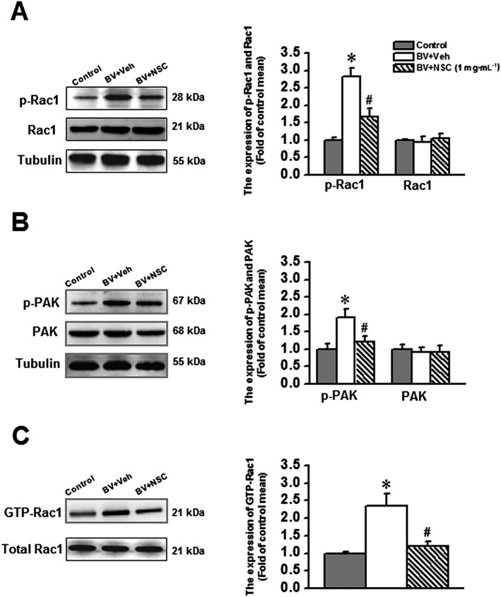
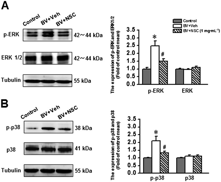
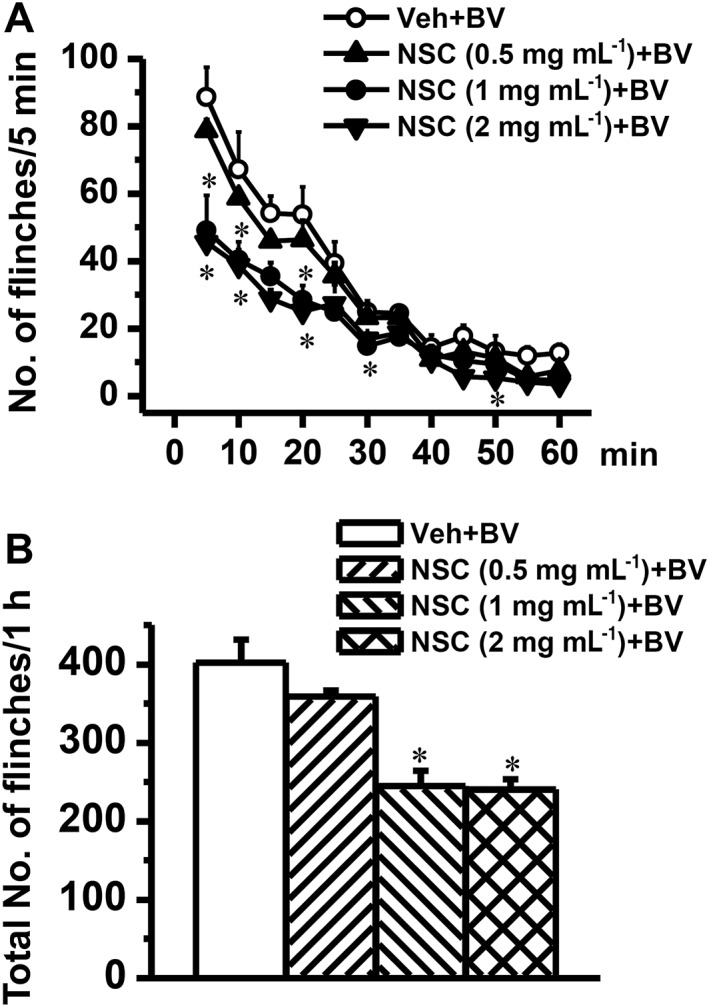
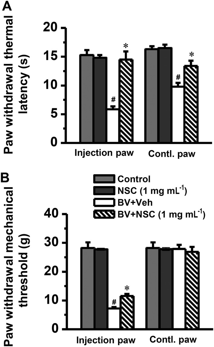
Experimental approach: Effects of s.c. bee venom (BV) injection on cellular localization of Rac1 in the rat sDH was determined with double labelling immunofluorescence. Activation of Rac1 and its downstream effector p21-activated kinase (PAK), ERKs and p38 MAPK in inflammatory pain states was evaluated with a pull-down assay and Western blotting. The preventive and therapeutic analgesic effects of intrathecal administration of NSC23766, a selective inhibitor of Rac1, on BV-induced spontaneous nociception and pain hypersensitivity were investigated.

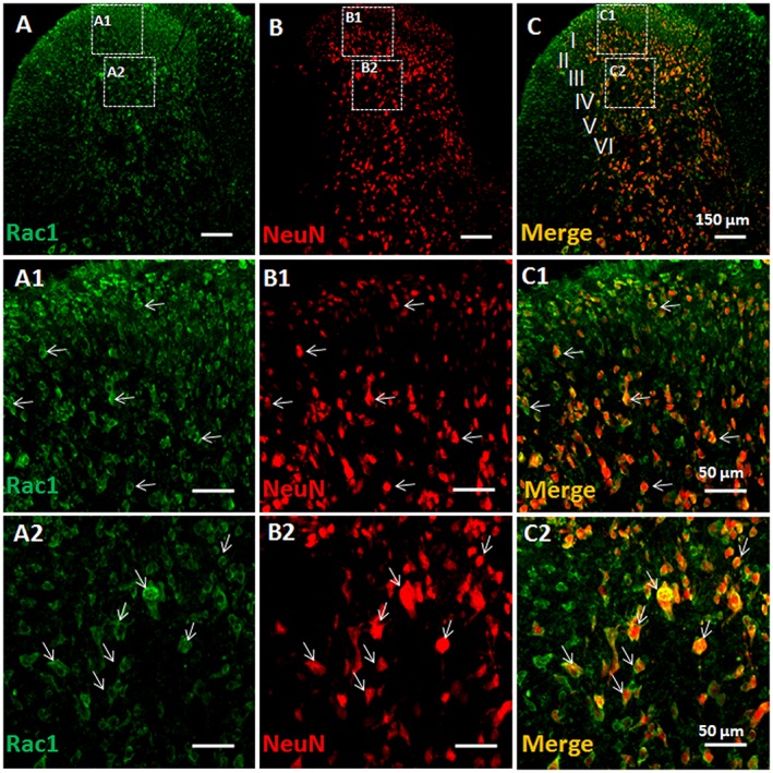
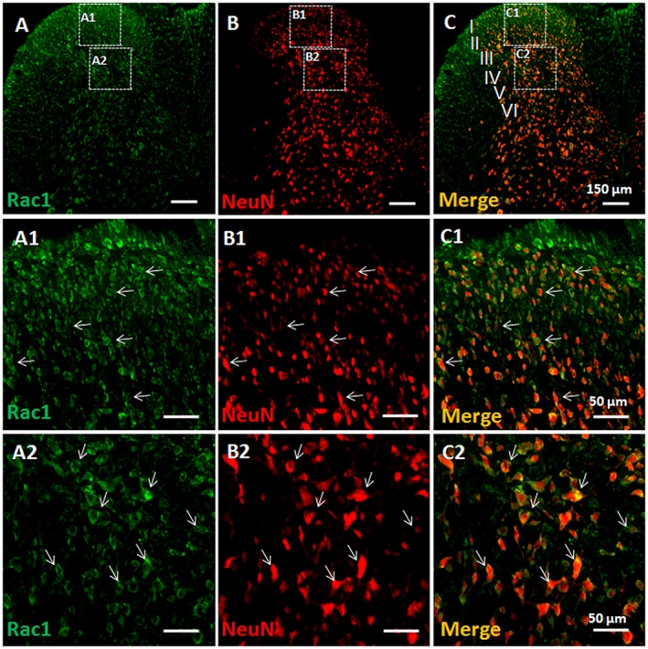
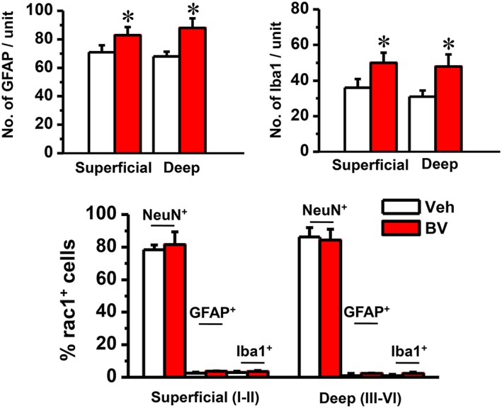
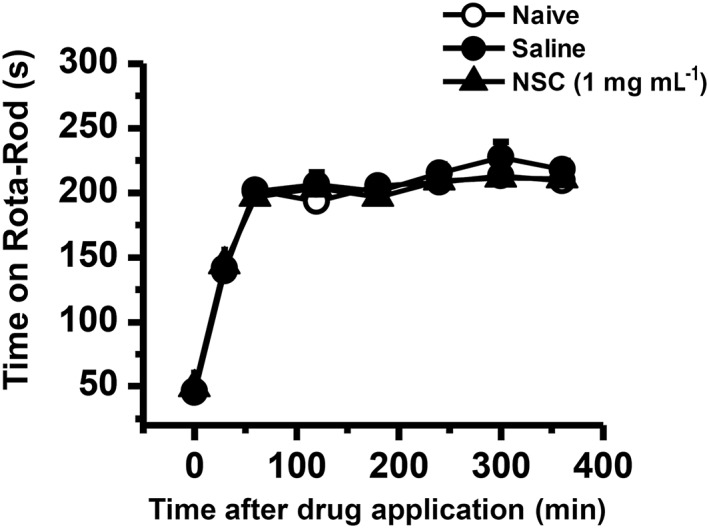
Key results: Rac1 labelling was mainly localized within neurons in both the superficial and deep layers of the sDH in rats of naïve, vehicle-treated and inflamed (BV injected) groups. GTP-Rac1-PAK and ERKs/p38 were activated following s.c. BV injection. Post-treatment with intrathecal NSC23766 significantly inhibited GTP-Rac1 activity and phosphorylation of Rac1-PAK, ERKs and p38 MAPK in the sDH. Both pre-treatment and post-treatment with intrathecal NSC23766 dose-dependently attenuated the paw flinches, primary thermal and mechanical hyperalgesia and the mirror-image thermal hyperalgesia induced by BV injection, but without affecting the baseline pain sensitivity and motor coordination.

Conclusions and implications: The spinal GTP-Rac1-PAK-ERK/p38MAPK signalling pathway is involved in both the development and maintenance of peripheral inflammatory pain and can be used as a potential molecular target for developing a novel therapeutic strategy for clinical pain.


Figures

Figure 1
Figure 1
Double immunofluorescent labelling of Rac1 and NeuN in vehicle‐treated rats. Immunofluorescent labelling of Rac1 (A) and NeuN (B) in the injection side of spinal dorsal horn of rats that received s.c. saline (vehicle) into a hind paw. (C) Merged images of A and B. A1–A2, B1–B2 and C1–C2 show enlarged images of the insets in A, B and C respectively. Scale bars, 150 μm (A–C), 50 μm (A1–C1, A2–C2).
Figure 2
Figure 2
Double immunofluorescent labelling of Rac1 and NeuN in BV‐treated rats. Immunofluorescent labelling of Rac1 (A) and NeuN (B) in the injection side of spinal dorsal horn of rats that received s.c. injection of BV into a hindpaw. (C) Merged images of A and B. A1–A2, B1–B2 and C1–C2 show enlarged images of the insets in A, B and C respectively. Scale bars, 150 μm (A–C), 50 μm (A1–C1 and A2–C2).
Figure 3
Figure 3
Quantification of immunofluorescent labelling of GFAP‐positive and Iba1‐ positive profiles and double immunofluorescent labelling of Rac1 with NeuN‐positive, GFAP‐positive‐ and Iba1‐positive profiles. The upper panel shows the average numbers of GFAP‐labelled astrocytes and Iba1‐labelled microglia cells in the injection side of both superficial (layers I and II) and deep layers (layers III and VI) of spinal dorsal horn of rats that received s.c. injection of saline (Veh) or BV into a hind paw respectively. The lower panel shows that the percentage of NeuN‐labelled neurons, GFAP‐labelled astrocytes and Iba1‐labelled microglial cells in Rac1‐positive profiles was calculated in both superficial (layers I and II) and deep layers (layers III and VI) of the spinal dorsal horn respectively. Data are expressed as mean ± SEM. *P < 0.05, significantly different from Veh.
Figure 4
Figure 4
Effects of intrathecal NSC23766 (1 mg·mL−1), a selective inhibitor of Rac1, on the BV‐induced phosphorylation of Rac1 and its downstream effector PAK and enhancement of GTP‐bound Rac1 activity. The left panels of A, B and C show representative immunoblotting and pull‐down assay bands of phosphorylated form and total protein level of Rac1 and PAK and GTP‐bound Rac1. The right panels of A, B and C show quantitative analysis of the level of phosphorylated form and total protein expression for Rac1 (n = 5) and PAK (n = 5) and that of GTP bound Rac1 activity (n = 5) in group of animals receiving s.c. saline injection (Control), s.c. BV injection followed by intrathecal vehicle (saline, BV + Veh) and s.c. BV injection followed by intrathecal NSC23766 (BV + NSC). The relative density for bands of phosphorylated Rac1 or PAK was normalized to the value of total Rac1 or PAK. The relative density for bands of GTP‐Rac1 activity was normalized to the total Rac1. The relative density for bands of total Rac1 and PAK was normalized to Tubulin. Data are expressed as mean ± SEM. *P < 0.05, significantly different from Control. #P < 0.05, significantly different from BV + Veh. p‐Rac1, phosphorylated Rac1; p‐PAK, phosphorylated PAK.
Figure 5
Figure 5
Effects of intrathecal intrathecal administration of NSC23766 (1 mg·mL−1), a selective inhibitor of Rac1, on the bee venomBV‐induced phosphorylation of ERK1/ERK2 and p38 MAPK. The left panels of A and B show representative immunoblotting bands of phosphorylated form and total protein level of ERK1/ERK2 and p38 MAPK. The right panels of A and B show quantitative analysis of the level of phosphorylated form and total protein expression for ERK1/ERK2 and p38 MAPK in group of animals receiving s.c. saline injection (Control, n = 5), s.c. BV injection followed by intrathecal administration of vehicle (saline, BV + Veh, n = 5) and s.c. BV injection followed by intrathecal NSC23766 (BV + NSC, n = 5).The relative density for bands of phosphorylated ERK1/ERK2 and p38 MAPK was normalized to the value of total ERK1/ERK2 and p38 MAPK. The relative density for bands of total ERK1/ERK2 and p38 MAPK was normalized to Tubulin. Data are expressed as mean ± SEM. *P < 0.05, significantly different from Control. #P < 0.05, significantly different from BV + Veh. p‐ERK1/p‐ERK2, phosphorylated ERK; p‐p38, phosphorylated p38 MAPK;
Figure 6
Figure 6
Effects of intrathecal pre‐administration of NSC23766 on BV‐induced paw flinch reflex. Graph A shows time courses of dose‐related effects of intrathecal NSC23766 (NSC) on the BV‐induced paw flinches. Graph B shows dose‐related effects of intrathecal NSC23766 on the total number of BV‐induced paw flinches. The drug was dissolved in physiological saline (vehicle; Veh) and administered 5 min prior to s.c. BV injection. Data are expressed as mean ± SEM. *P < 0.05, significantly different from Veh + BV.
Figure 7
Figure 7
Effects of intrathecal pre‐administration of NSC23766 on BV‐induced pain hypersensitivity. Graph A shows dose‐related inhibitory effects of NSC23766 (NSC) on the BV‐induced primary (injection paw) and mirror‐image (contralalteral (Contl) paw) thermal hyperalgesia. Graph B shows partial inhibitory effects of NSC23766 on the BV‐induced primary mechanical hyperalgesia. The drug was dissolved in physiological saline (vehicle; Veh) and administered 5 min prior to s.c. BV injection. Data are expressed as mean ± SEM. #P < 0.05, significantly different from Control; *P < 0.05, significantly different from Veh + BV.
Figure 8
Figure 8
Effects of i.t. post‐administration of NSC23766 on BV‐induced pain hypersensitivity. Graph A shows complete reversal by NSC23766 (NSC) of the BV‐induced primary (injection paw) and mirror‐image contralalteral (Contl) paw thermal hyperalgesia. Graph B shows partial reversal effect of NSC23766 on the BV‐induced primary mechanical hyperalgesia. The drug was dissolved in physiological saline (vehicle; Veh) and administered 2 h after s.c. BV injection. Data are expressed as mean ± SEM. ## P < 0.05, significantly different from Control; *P < 0.05, significantly different from BV + Veh.
Figure 9
Figure 9
Effects of intrathecal NSC23766 on motor coordination. Graph shows effects of NSC23766 on motor coordination of naïve rats and rats receiving intrathecal saline or NSC23766. The drug was injected 5 min before the initiation of the test. A total of eight trials were conducted, and the first three trials were considered as the training session. NSC, NSC23766. Data are expressed as mean ± SEM.

References

    1. Alexander SPH, Fabbro D, Kelly E, Marrion N, Peters JA, Benson HE, et al (2015). The Concise Guide to PHARMACOLOGY 2015/16: Enzymes. Br J Pharmacol 172: 6024–6109. - PMC - PubMed
    1. Cao FL, Liu MG, Hao J, Li Z, Lu ZM, Chen J (2007). Different roles of spinal p38 and c‐Jun. N‐terminal kinase pathways in bee venom‐induced multiple pain‐related behaviors. Neurosci Lett 427: 50–54. - PubMed
    1. Chen HS, Chen J (2000). Secondary heat, but not mechanical, hyperalgesia induced by subcutaneous injection of bee venom in the conscious rat: effect of systemic MK‐801, a non‐competitive NMDA receptor antagonist. Eur J Pain 4: 389–401. - PubMed
    1. Chen HS, Chen J, Sun YY (2000). Contralateral heat hyperalgesia induced by unilaterally intraplantar bee venom injection is produced by central changes: a behavioral study in the conscious rat. Neurosci Lett 284: 45–48. - PubMed
    1. Chen HS, He X, Qu F, Kang SM, Yu Y, Liao D, et al (2009). Differential roles of peripheral mitogen‐activated protein kinase signal transduction pathways in bee venom‐induced nociception and inflammation in conscious rats. J Pain 10: 201–207. - PubMed

Publication types

MeSH terms