. 2016 Jul;16(7):1999-2015.
doi: 10.1111/ajt.13685. Epub 2016 Mar 17.

Regulatory T Cell Infusion Can Enhance Memory T Cell and Alloantibody Responses in Lymphodepleted Nonhuman Primate Heart Allograft Recipients

Affiliations

Regulatory T Cell Infusion Can Enhance Memory T Cell and Alloantibody Responses in Lymphodepleted Nonhuman Primate Heart Allograft Recipients

M B Ezzelarab et al. Am J Transplant. 2016 Jul.

Abstract

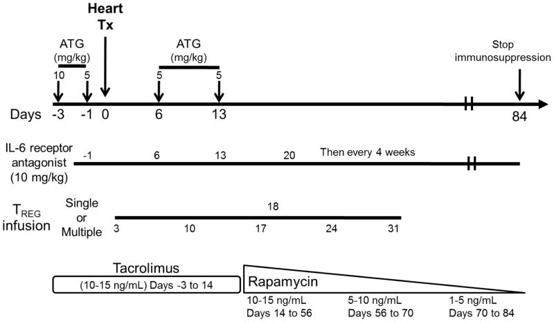
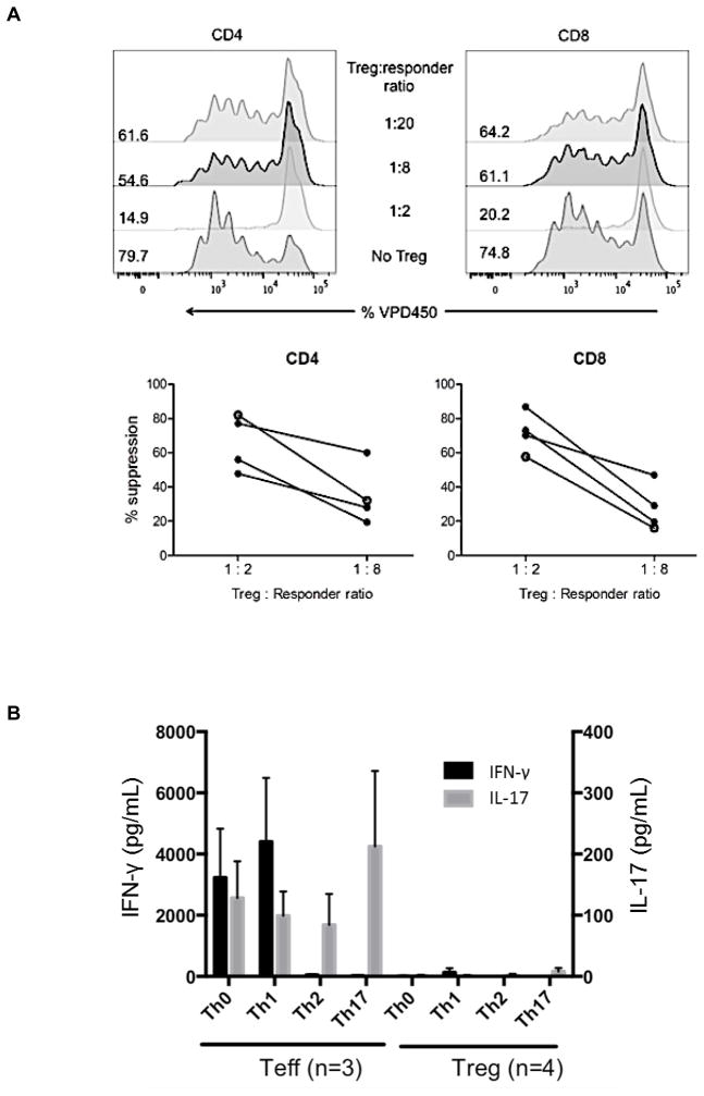
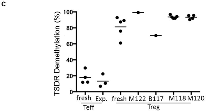
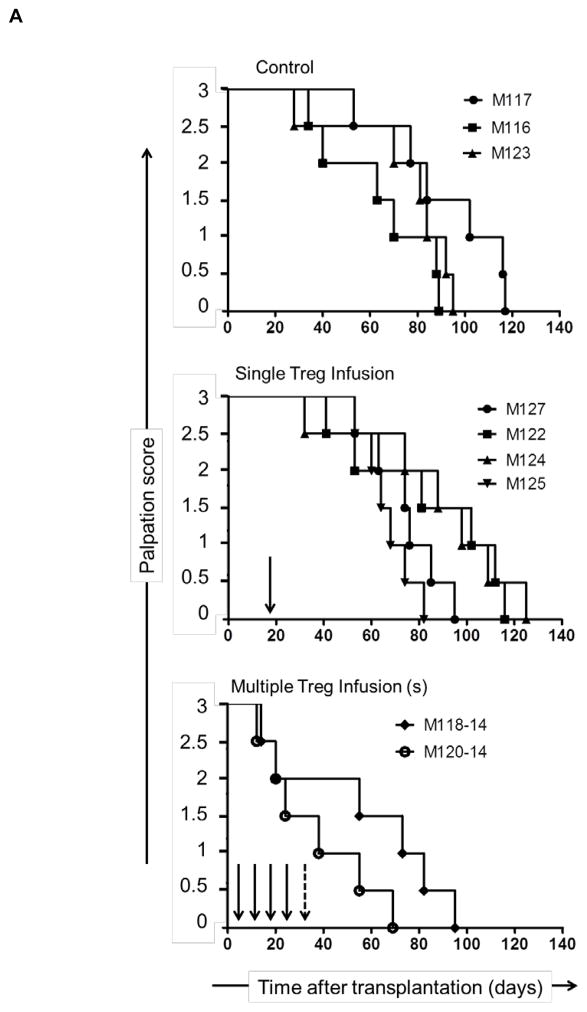
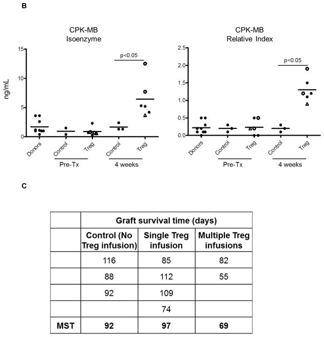
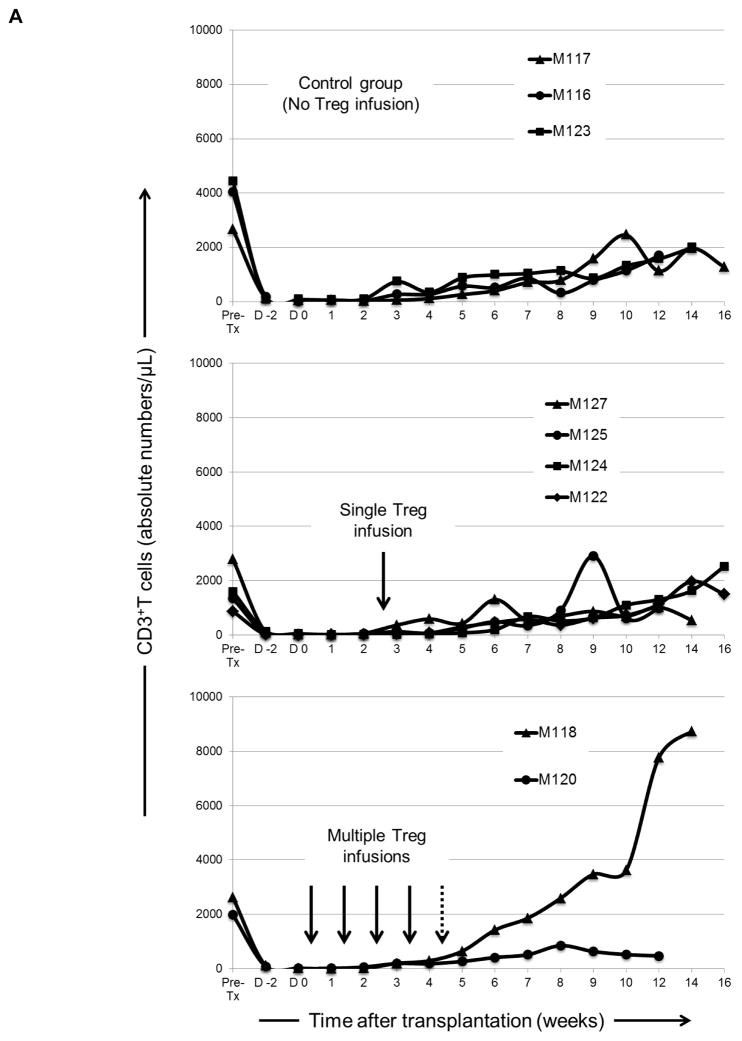
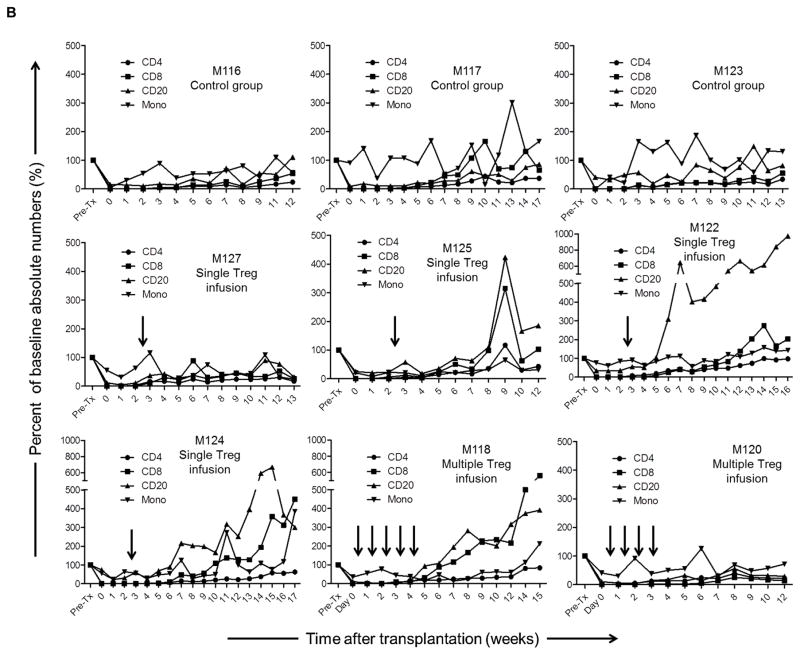
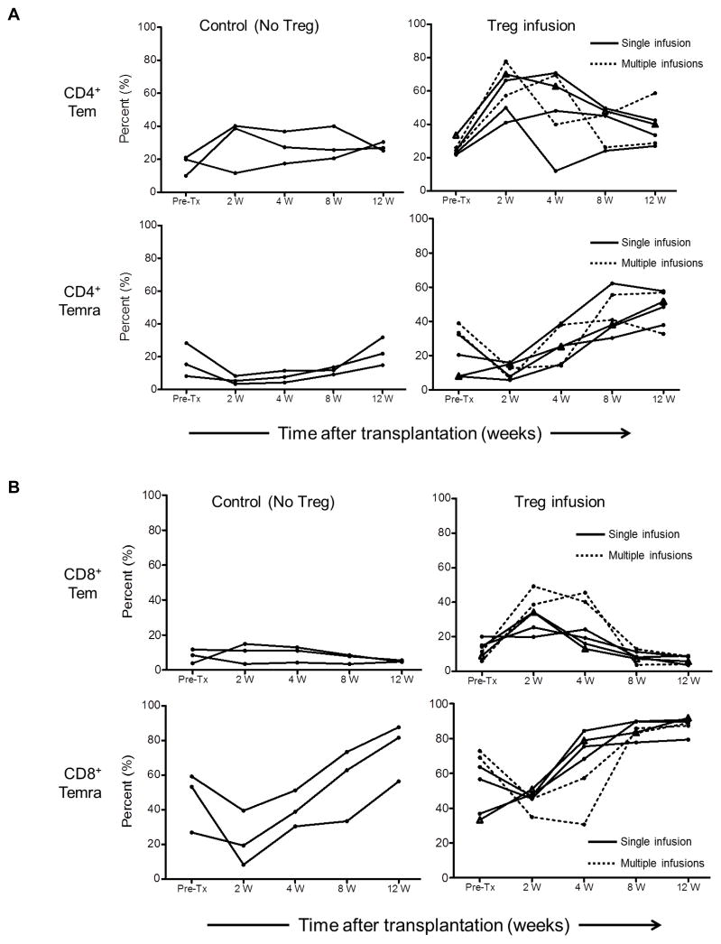
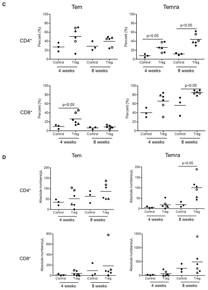
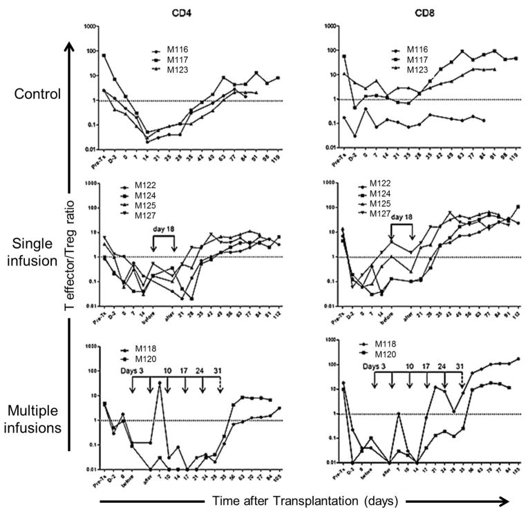
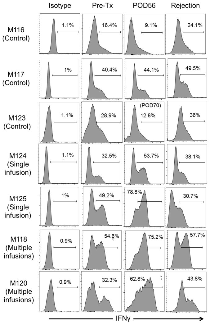
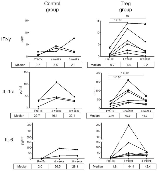
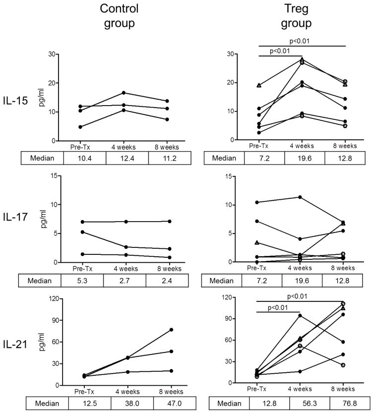
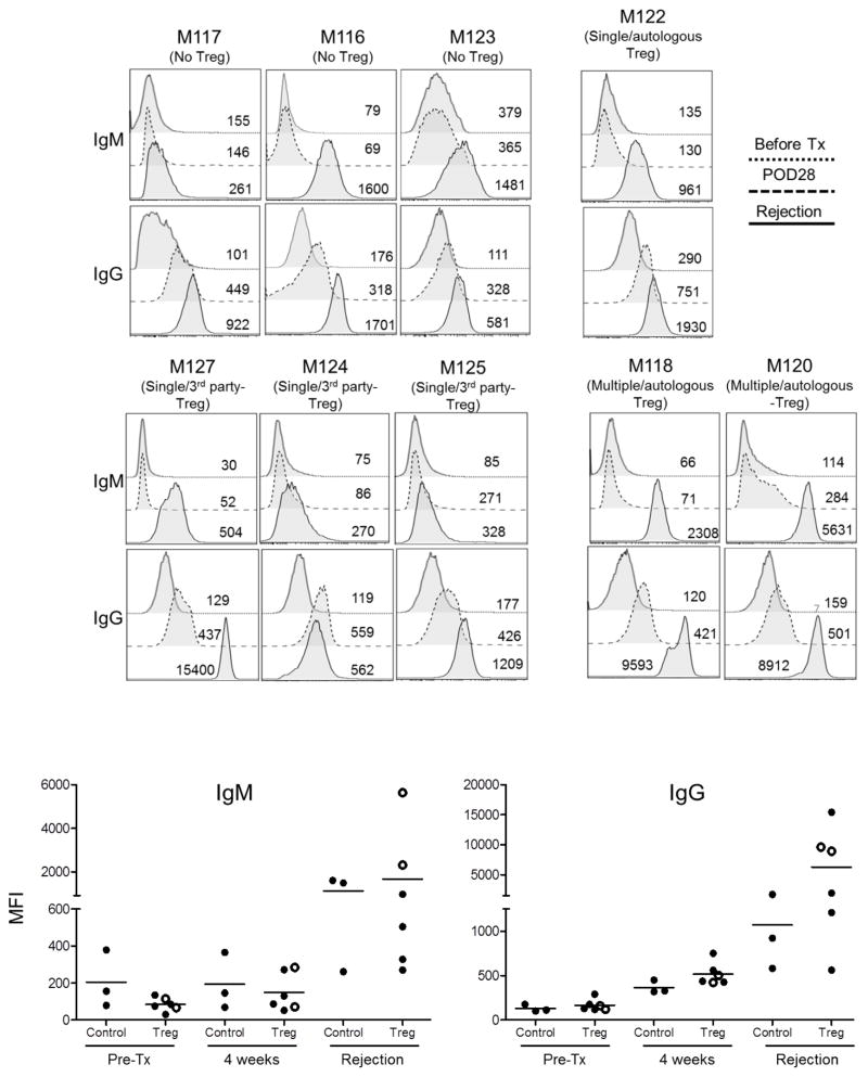
The ability of regulatory T cells (Treg) to prolong allograft survival and promote transplant tolerance in lymphodepleted rodents is well established. Few studies, however, have addressed the therapeutic potential of adoptively transferred, CD4(+) CD25(+) CD127(-) Foxp3(+) (Treg) in clinically relevant large animal models. We infused ex vivo-expanded, functionally stable, nonselected Treg (up to a maximum cumulative dose of 1.87 billion cells) into antithymocyte globulin-lymphodepleted, MHC-mismatched cynomolgus monkey heart graft recipients before homeostatic recovery of effector T cells. The monkeys also received tacrolimus, anti-interleukin-6 receptor monoclonal antibodies and tapered rapamycin maintenance therapy. Treg administration in single or multiple doses during the early postsurgical period (up to 1 month posttransplantation), when host T cells were profoundly depleted, resulted in inferior graft function compared with controls. This was accompanied by increased incidences of effector memory T cells, enhanced interferon-γ production by host CD8(+) T cells, elevated levels of proinflammatory cytokines, and antidonor alloantibodies. The findings caution against infusion of Treg during the early posttransplantation period after lymphodepletion. Despite marked but transient increases in Treg relative to endogenous effector T cells and use of reputed "Treg-friendly" agents, the host environment/immune effector mechanisms instigated under these conditions can perturb rather than favor the potential therapeutic efficacy of adoptively transferred Treg.

Keywords: T cell biology; animal models: nonhuman primate; basic (laboratory) research/science; heart transplantation/cardiology; immune regulation; immunobiology; lymphocyte biology; translational research/science.


Conflict of interest statement

Disclosure

The authors of this manuscript have no conflicts of interest to disclose as described by the American Journal of Transplantation.

Figures

Figure 1
Figure 1. Protocol for infusion of ex vivo-expanded Treg into heart-allografted monkeys treated with ATG, anti-IL-6R mAb, tacrolimus and rapamycin
Times of immunosuppressive drug administration in relation to the day of heart transplant are shown. The transplant recipients received either no Treg or single or multiple infusions at the time(s) indicated (days) in relation to heart transplantation.
Figure 2
Figure 2. Suppressive function and stability of ex vivo-expanded cynomolgus Treg infused into heart graft recipients
(A) Ex-vivo-expanded Treg were tested for their ability to suppress VPD450-labeled autologous or allogeneic anti-CD3/CD28-activated CD4+ and CD8+ T cell proliferation. Percent divided cells was calculated using FlowJo software (top). The suppressive effect of this representative batch of Treg was tested on autologous (open circles) and three different allogeneic (closed circles) activated T cells (bottom). Treg function was expressed as percent suppression of T cell proliferation calculated using the formula: (percent divided T cells without addition of Treg – percent divided T cells with Treg)/percent divided T cells without addition of Treg x 100%. The expanded Treg showed strong suppressive activity when added to bead-stimulated T cells. Data are representative of the 4 batches of expanded Treg used for infusion after transplantation. (B) Primary CD4+CD25 effector T cells (Teff) or Treg expanded ex vivo for 3 weeks were stimulated with NHP-specific anti-CD3/CD28 microbeads at a cell:bead ratio of 10:1 for 4 days under the following culture conditions: Th1 (IL-12 + anti-CD4 mAb), Th2 (IL-4 + anti-IFN-γ mAb), or Th17 (IL-6 + IL-21 + IL-23 + IL-1β + TGF-β + anti-IL-4 mAb + anti-IFN-γ mAb). Supernatants were collected and assayed for IFN-γ and IL-17 using CBA kits. Data (means ± 1SD) are from n=3 or n=4 separate individual Teff or Treg cultures. (C) Demethylation status of Foxp3 TSDR in freshly-isolated Treg and Treg expanded for individual or multiple infusions from 4 different monkeys for 3 weeks before infusion into heart allograft recipients. Data are also shown for freshly-isolated and expanded Teff.
Figure 3
Figure 3. Heart allograft function and survival in control and Treg-infused monkeys
(A) Heart graft palpation scores at various times post-transplant in monkeys that did not receive Treg (control; n=3) and in those that received single (n=4) or multiple infusions (n=2). Vertical arrows indicate time(s) of Treg infusion. (B) Markers of cardiac muscle injury (CPK-MB isoenzyme) and CPK-MB Relative index in control and Treg-infused graft recipients pre-treatment and one month post-transplant. Open circles denote monkeys that received multiple Treg infusions. Open triangles denote the monkey that received a single autologous Treg infusion. (C) Heart graft survival times in the control and Treg groups. MST= median graft survival time.
Figure 4
Figure 4. Kinetics of peripheral blood T and B cell and monocyte numbers in lymphodepleted control and Treg-infused heart allograft recipients
(A) Kinetics of CD3+ T cell depletion and recovery in individual monkeys. (B) Absolute numbers of CD4+ and CD8+ T cells, CD20+ B cells, and monocytes in individual monkeys at various times pre-transplant and post-transplant. Vertical arrows denote the time(s) of Treg infusion(s).
Figure 5
Figure 5. Levels of CD4+ and CD8+ effector memory (Tem) and terminally-differentiated effector memory (Temra) cells in peripheral blood of lymphodepleted control and Treg-infused heart allograft recipients
(A) CD4+ and (B) CD8+ Tmem and Temra at various times pre-treatment and post-transplant. (C) and (D) Mean values and significances of differences in relative and absolute numbers between control and Treg-infused monkeys at one month and two months post-transplant. Open circles denote monkeys that received multiple Treg infusions. Triangles denote the one monkey that received a single autologous Treg infusion.
Figure 6
Figure 6. T effector:Treg ratios in control and Treg-infused heart allograft recipients
Ratios of CD4+ or CD8+ T effector cells (Teff) to CD4+ Treg in peripheral blood are shown pre-treatment, one day after the initial ATG infusion, on the day of transplant and thereafter in control animals and those that received single or multiple Treg infusion(s). Values before and 30 min after the single or the first of multiple infusions are shown in the middle and lower panels, respectively.
Figure 7
Figure 7. Incidences of CD8+ IFNγ+ effector T cells in Treg-infused heart allograft recipients
Incidences of CD8+ IFNγ+ cells in control and Treg-infused monkeys determined by flow cytometry after ex vivo stimulation of circulating CD8+ T cells with PMA and for 3 h, as described in the Materials and Methods. Pre-transplant values and those obtained 8 weeks post-transplant, and at the time of rejection are shown. POD=post-operative day.
Figure 8
Figure 8. Systemic levels of proinflammatory cytokines in Treg-infused heart allograft recipients
Various cytokines were determined in serum of individual monkeys by Luminex assay (as described in the Materials and Methods) pre-transplant and at one and two months post-transplant in individual control and Treg-infused heart allograft recipients. Open circles denote monkeys that received multiple Treg infusions. Open triangles denote the monkey that received a single autologous Treg infusion.
Figure 9
Figure 9. Circulating alloAb levels in heart allograft recipients
Levels of IgM and IgG alloAbs levels were determined by flow cytometry in the serum of control (no Treg) and Treg-infused monkeys before transplant, on day 28 post-transplant and at graft rejection. Numbers on panels denote mean fluorescence intensity (MFI). Horizontal bars in lower panels denote mean values obtained from individual monkeys.

Comment in

  • Regulating T Cell Behavior.
    Knechtle SJ, Fairchild RL. Knechtle SJ, et al. Am J Transplant. 2016 Jul;16(7):1949-50. doi: 10.1111/ajt.13724. Epub 2016 Mar 17. Am J Transplant. 2016. PMID: 26792650 No abstract available.

Similar articles

Cited by

References

    1. Bluestone JA, Thomson AW, Shevach EM, Weiner HL. What does the future hold for cell-based tolerogenic therapy? Nat Rev Immunol. 2007;7(8):650–654. - PubMed
    1. Wood KJ, Bushell A, Hester J. Regulatory immune cells in transplantation. Nat Rev Immunol. 2012;12(6):417–430. - PubMed
    1. Issa F, Hester J, Goto R, Nadig SN, Goodacre TE, Wood K. Ex vivo-expanded human regulatory T cells prevent the rejection of skin allografts in a humanized mouse model. Transplantation. 2010;90(12):1321–1327. - PMC - PubMed
    1. Juvet SC, Whatcott AG, Bushell AR, Wood KJ. Harnessing regulatory T cells for clinical use in transplantation: the end of the beginning. Am J Transplant. 2014;14(4):750–763. - PubMed
    1. Brunstein CG, Miller JS, Cao Q, McKenna DH, Hippen KL, Curtsinger J, et al. Infusion of ex vivo expanded T regulatory cells in adults transplanted with umbilical cord blood: safety profile and detection kinetics. Blood. 2011;117(3):1061–1070. - PMC - PubMed

Publication types