Review
. 2016 Jan 7;11(1):137-54.
doi: 10.2215/CJN.09430915. Epub 2015 Dec 23.

B Cells, Antibodies, and More

Affiliations
Review

B Cells, Antibodies, and More

William Hoffman et al. Clin J Am Soc Nephrol. .

Abstract

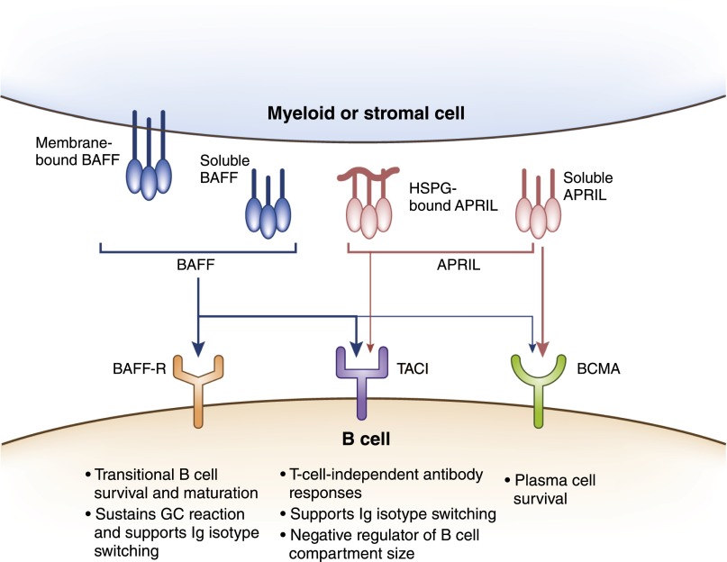
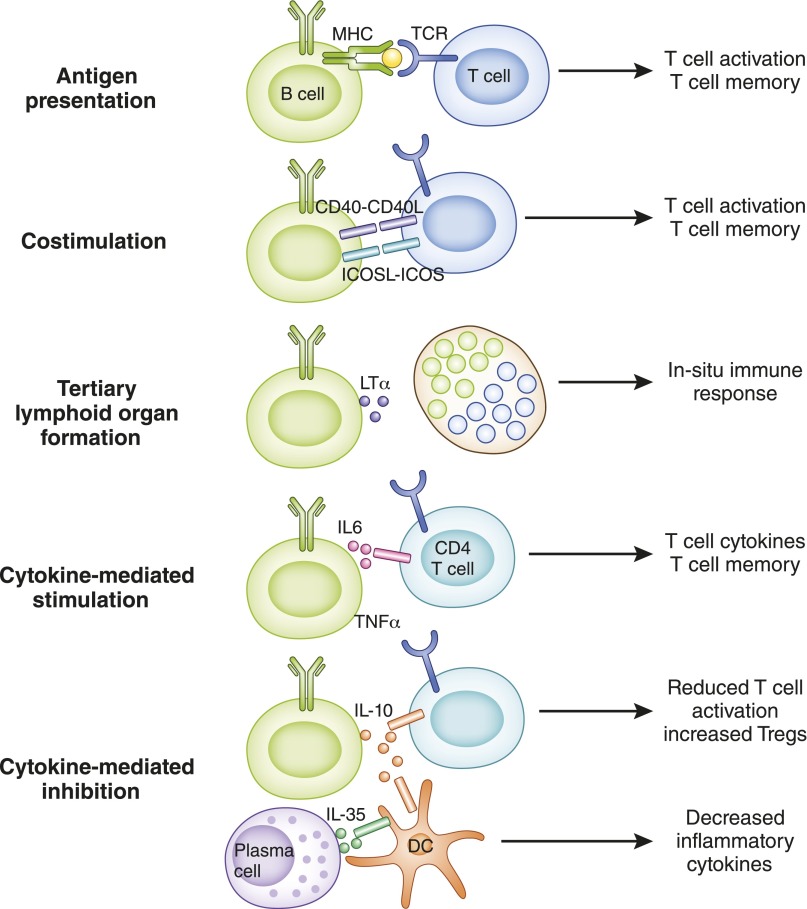
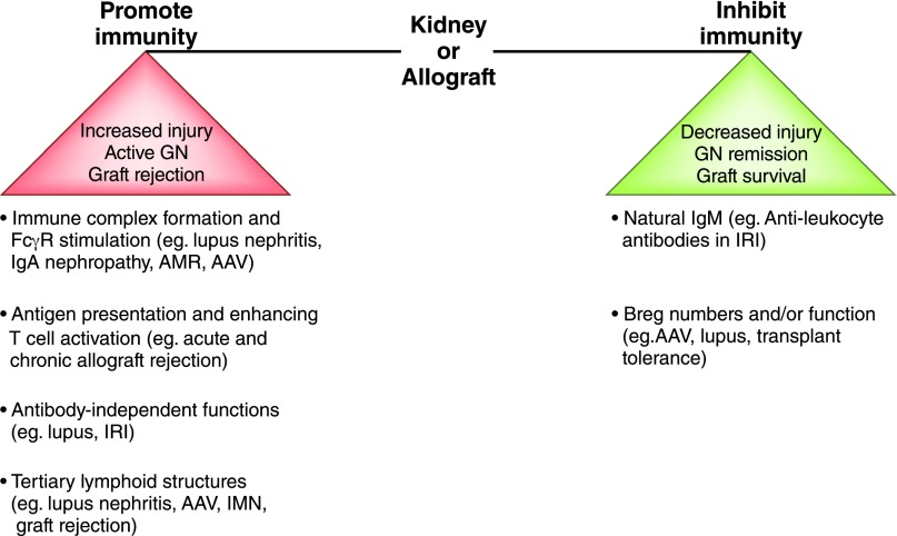
B cells play a central role in the immunopathogenesis of glomerulonephritides and transplant rejection. B cells secrete antibodies that contribute to tissue injury via multiple mechanisms. In addition, B cells contribute to disease pathogenesis in autoimmunity and alloimmunity by presenting antigens as well as providing costimulation and cytokines to T cells. B cells also play an immunomodulatory role in regulating the immune response by secreting cytokines that inhibit disease onset and/or progression. B cell-targeted approaches for treating immune diseases of the kidney and other organs have gained significant momentum. However, much remains to be understood about B-cell biology in order to determine the timing, duration, and context of optimal therapeutic response to B cell-targeted approaches. In this review, we discuss the multifaceted roles of B cells as enhancers and regulators of immunity with relevance to kidney disease and transplantation.

Keywords: Antibodies; B regulatory cells; B-Lymphocytes; T-Lymphocytes; autoimmunity; cytokines; glomerulonephritis; graft rejection; immune system diseases; plasma cells.


Figures

Figure 1.
Figure 1.
B-cell lineage subsets and functions. B lymphocytes of all lineages arise from progenitors derived from hematopoietic stem cells (HSCs). Most B1 B lymphocytes develop from B1 progenitors in the fetal liver with little input from bone marrow beyond the perinatal period. B2 B lymphocytes develop from transitional 2 (T2) B cells derived from B-cell progenitors in the bone marrow, with subsequent differentiation into marginal zone (MZ) and follicular (FO) lineages occurring in the spleen. Stronger B-cell receptor (BCR) signals induce Bruton tyrosine kinase (BTK) and support maturation to FO B cells, while weaker BCR signals allow expression of neurogenic locus notch homolog protein 2 (NOTCH2) giving rise to MZ B cells. B lymphocytes of each lineage have distinct and overlapping functions in recognizing antigens via T-independent and T-dependent pathways, production of rapid IgM, and long-lasting IgG antibody responses essential for host defense.
Figure 2.
Figure 2.
Antibody structure. Antibodies (immunoglobulins) are composed of two heavy chains (VH and CH) and two light chains (VL and CL). The antigen-binding fragment, Fab, is composed of one variable domain from each heavy and light chain (VH and VL). The variable domains contain the complementarity determining regions (CDRs) with the most sequence variations and determine antibody specificity. The constant domains CH2 and CH3 of the heavy chain make up the crystallizable fragment, Fc, which mediates effector functions through binding to Fc receptors (FcRs) on cells and to complement (C1q).
Figure 3.
Figure 3.
B-cell development and mechanisms of self-tolerance. B-cell development begins in the bone marrow and is completed in peripheral lymphoid tissues, such as the spleen. Development in the bone marrow progresses sequentially through pro-B, pre-B, and immature B cell stages and expression of surface IgM, mature B-cell receptor (BCR). Immature B cells with strong reactivity to self-antigen undergo clonal deletion or rearrange their immunoglobulin gene segments; this is called receptor editing, which eliminates self-reactivity and allows entry to the transitional B-cell pool. Transitional B cells depend on B cell–activating factor (BAFF) for survival and differentiate into mature B cells in the spleen. Those transitional 1 and 2 (T1/T2) B cells with strong self-reactivity undergo clonal deletion or remain outside splenic follicles as hyporesponsive anergic B cells that can be rescued upon receiving T cell help to enter the mature B-cell pool. Mature B cells that are activated by foreign antigen and enter germinal center (GC) reactions give rise to isotype-switched memory B cells and plasma cells. During the process of somatic hypermutation (SHM), a few memory B cells acquire self-reactivity due to random immunoglobulin gene rearrangements and persist as IgG+ self-reactive clones in the periphery.
Figure 4.
Figure 4.
B-cell activation and differentiation into memory B cells and plasma cells. B cells that have encountered antigen migrate to the T–B border by upregulating C-C chemokine receptor 7 (CCR7) and Epstein-Barr virus–induced receptor 2 (EBI2), where they first encounter cognate T cells that mature into T follicular helper cells (Tfhs). B cells can differentiate into extrafollicular plasma blasts or memory B cells independent of germinal centers (GCs). B cells that express B-cell lymphoma 6 (Bcl6) return to the follicles, where they are retained via sphingosine-1-phosphate receptor 2 (S1PR2) expression to form GCs with Tfhs. Within GCs, B cell–Tfh interactions via MHC 2–T-cell receptor, B7–CD28, CD40–CD40L, inducible costimulator ligand (ICOSL)–inducible costimulator (ICOS), programmed cell death protein ligand 1 (PDL1)–programmed cell death protein 1 (PD1), and IL-21 receptor (IL-21R)–IL-21 facilitate somatic hypermutation and immunoglobulin isotype class-switch recombination (CSR) that generate high-affinity GC-dependent memory B cells and long-lived plasma cells. Following antigen re-exposure, extrafollicular memory B cells now enter GCs to generate isotype-switched and high-affinity secondary memory B cells and plasma cells, while GC-dependent memory B cells can rapidly differentiate into secondary plasma cells or re-enter GC to produce secondary memory B cells and plasma cells. DC, dendritic cell.
Figure 5.
Figure 5.
B cell–activating factor (BAFF), a proliferation-inducing ligand (APRIL), and their receptors. BAFF and APRIL are transmembrane proteins of the TNF family that can be proteolytically cleaved to produce soluble forms. They are produced by myeloid cells, such as dendritic cells (DCs), neutrophils, monocytes, macrophages, and stromal cells. BAFF binds strongly to receptors, B cell–activating factor-receptor (BAFF-R) and transmembrane activator and cyclophilin ligand interactor (TACI), and weakly to B-cell maturation antigen (BCMA), whereas APRIL binds strongly to BCMA and moderately to TACI. APRIL can also exist bound to heparin sulfate proteoglycan (HSPG) in extracellular matrix and interacts with TACI in this form. BAFF promotes survival and maturation of transitional B cells into mature B cells, supports B cell proliferation, class-switch recombination (CSR), and plasma cell survival. APRIL is critical for T-independent responses and supports CSR and survival of plasma cells.
Figure 6.
Figure 6.
Cellular functions of B cells. B cells interact with T cells and innate cells, such as dendritic cells (DCs), via several mechanisms that influence the outcome of the immune response. Antigen presentation, costimulation (such as CD40–CD40L, inducible costimulator ligand [ICOSL]–inducible costimulator [ICOS]), and cytokine production (such as IL-6 and TNF-α) contribute to enhanced T-cell activation and differentiation (e.g., T follicular helper cells), cytokine polarization (e.g., Th1 and Th17), and formation of long-lived memory T cells. Lymphotoxin (LTα) produced by B cells contributes to formation of tertiary lymphoid organs in peripheral tissues that are sites of in situ immune responses causing tissue injury. B cells and plasma cells also secrete cytokines, such as IL-10 and IL-35, that reduce T-cell activation and cytokine production and increase T cells with regulatory properties in addition to modulating functions of innate cells, such as DCs (e.g., decreased IL-6 and IL-12), to attenuate immune responses.
Figure 7.
Figure 7.
B cells as enhancers and regulators of immunity in kidney disease and transplantation. B cells can promote or inhibit immune responses, mediating kidney injury, GN, and transplant rejection by various mechanisms of action, and the balance between these functions influences disease outcomes. Isotype-switched antibodies contribute to antibody-mediated rejection (AMR) and GN (e.g., lupus, IgA nephropathy, ANCA-associated vasculitis [AAV]) by forming immune complexes and activating FcγR while natural IgM antileukocyte antibodies are protective in ischemia-reperfusion injury (IRI). Antibody-independent functions of B cells contribute to lupus and ischemia-reperfusion injury and mediate graft rejection by presenting antigen and driving T-cell activation. B cells form tertiary lymphoid structures that are the sites of local immune responses causing tissue injury in lupus nephritis, AAV, idiopathic membranous nephropathy (IMN) and graft rejection. Various B-cell populations with regulatory functions (e.g., IL-10) contribute to graft survival and GN remission (e.g., AAV), and their disrupted numbers or function are observed in transplant rejection and GN relapse (e.g., AAV and lupus).

Similar articles

Cited by

References

    1. Montecino-Rodriguez E, Dorshkind K: B-1 B cell development in the fetus and adult. Immunity 36: 13–21, 2012 - PMC - PubMed
    1. Pillai S, Cariappa A: The follicular versus marginal zone B lymphocyte cell fate decision. Nat Rev Immunol 9: 767–777, 2009 - PubMed
    1. Baumgarth N: The double life of a B-1 cell: Self-reactivity selects for protective effector functions. Nat Rev Immunol 11: 34–46, 2011 - PubMed
    1. Cerutti A, Cols M, Puga I: Marginal zone B cells: Virtues of innate-like antibody-producing lymphocytes. Nat Rev Immunol 13: 118–132, 2013 - PMC - PubMed
    1. Springer GF, Horton RE: Blood group isoantibody stimulation in man by feeding blood group-active bacteria. J Clin Invest 48: 1280–1291, 1969 - PMC - PubMed

Publication types