. 2016 Feb;101(2):523-32.
doi: 10.1210/jc.2015-3566. Epub 2015 Dec 23.

β-Cell Deficit in Obese Type 2 Diabetes, a Minor Role of β-Cell Dedifferentiation and Degranulation

Affiliations

β-Cell Deficit in Obese Type 2 Diabetes, a Minor Role of β-Cell Dedifferentiation and Degranulation

Alexandra E Butler et al. J Clin Endocrinol Metab. 2016 Feb.

Abstract

Context: Type 2 diabetes is characterized by a β-cell deficit and a progressive defect in β-cell function. It has been proposed that the deficit in β-cells may be due to β-cell degranulation and transdifferentiation to other endocrine cell types.

Objective: The objective of the study was to establish the potential impact of β-cell dedifferentiation and transdifferentiation on β-cell deficit in type 2 diabetes and to consider the alternative that cells with an incomplete identity may be newly forming rather than dedifferentiated.

Design, setting, and participants: Pancreata obtained at autopsy were evaluated from 14 nondiabetic and 13 type 2 diabetic individuals, from four fetal cases, and from 10 neonatal cases.

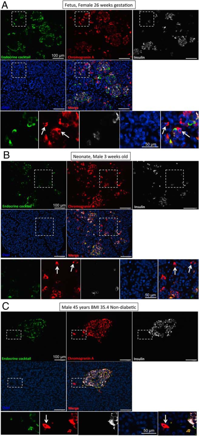
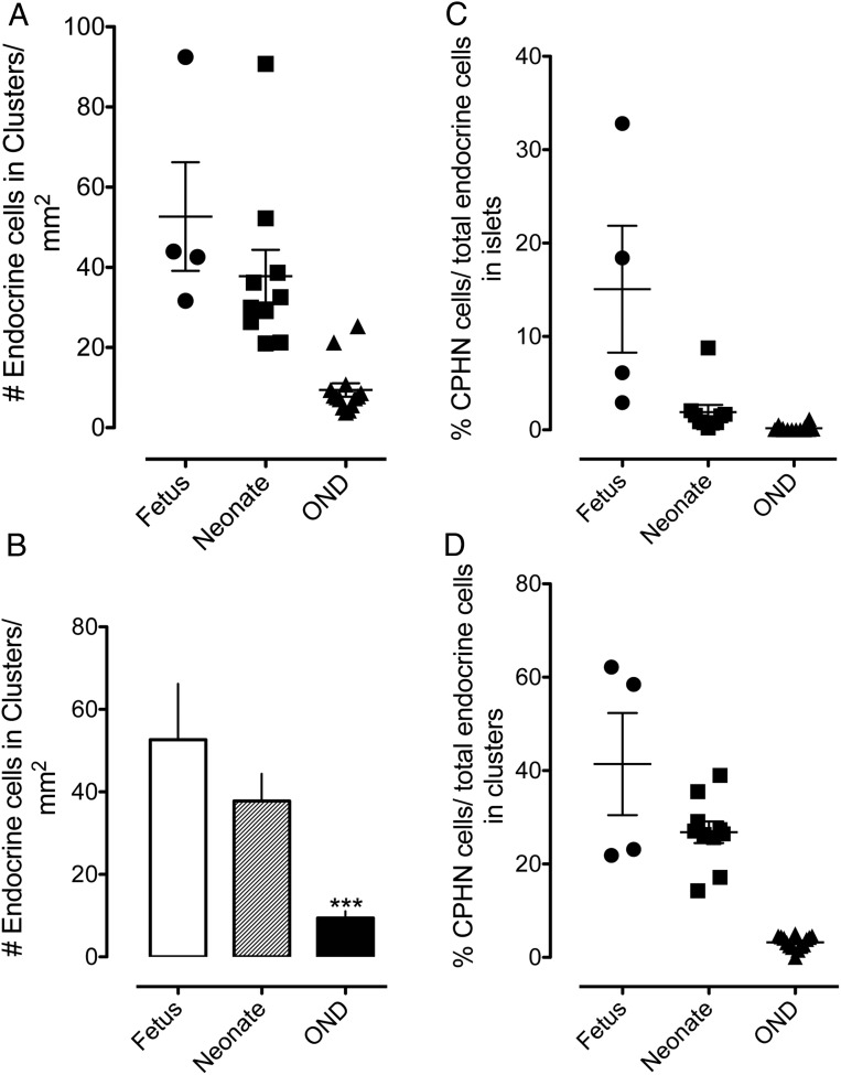
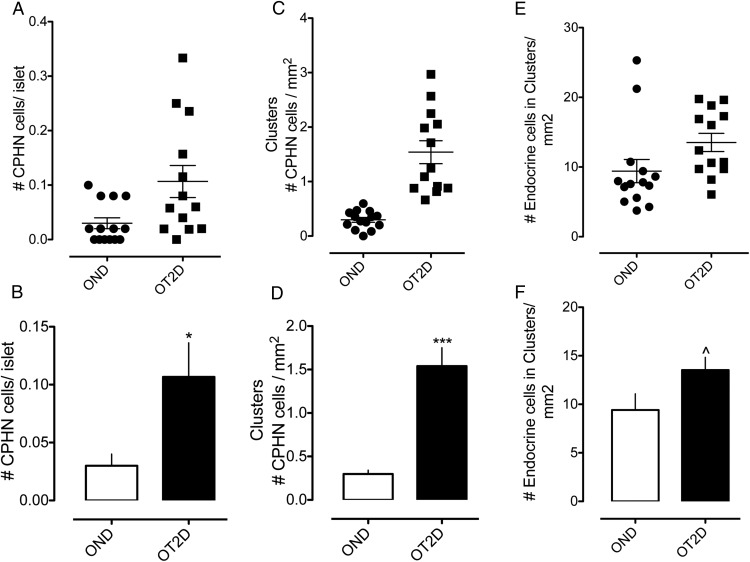
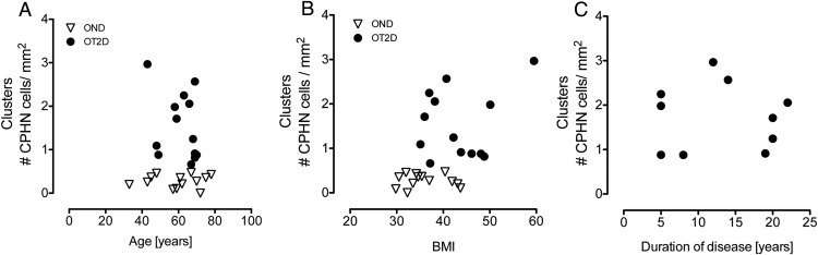
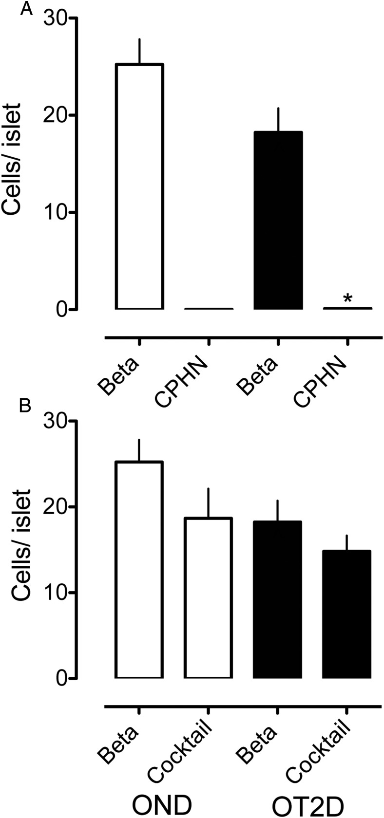
Results: Whereas there was a slight increase in islet endocrine cells expressing no hormone in type 2 diabetes (0.11 ± 0.03 cells/islet vs 0.03 ± 0.01 cells/islet, P < .01), the impact on the β-cell deficit would be minimal. Furthermore, we established that the deficit in β-cells per islet cannot be accounted for by an increase in other endocrine cell types. The distribution of hormone negative endocrine cells in type 2 diabetes (most abundant in cells scattered in the exocrine pancreas) mirrors that in developing (embryo and neonatal) pancreas, implying that these may represent newly forming cells.

Conclusions: Therefore, although we concur that in type 2 diabetes there are endocrine cells with altered cell identity, this process does not account for the deficit in β-cells in type 2 diabetes but may reflect, in part, attempted β-cell regeneration.


Figures

Figure 1.
Figure 1.
Representative images taken at low power (×10 objective) to show pancreas from a fetus at 26 weeks' gestation (A), a neonate 3 weeks after delivery (B), and a 45-year-old nondiabetic adult with a BMI of 35.4 kg/m2 (C). Pancreas sections in all cases were stained for chromogranin A (red), endocrine cocktail (glucagon, somatostatin, pancreatic polypeptide, and ghrelin) (green), insulin (white), and DAPI (blue), and the merged image is shown. Insets in all panels show cells staining for chromogranin A but none of the other endocrine hormones. These CPHN cells are frequently found in fetal pancreas, become less frequent in neonatal life, and are infrequently found in nondiabetic adult human pancreas. Scale bar, 100 μm (low power images), 50 μm in high power inset images).
Figure 2.
Figure 2.
The number of endocrine cells present in scattered clusters of from one to three cells in the exocrine pancreas parenchyma (A, B). Scattered clusters of endocrine cells are most numerous in sections of fetal pancreas, and the frequency decreases after birth. By adulthood, endocrine clusters are relatively infrequent, although they are always present in pancreatic sections (52.66 ± 13.55 vs 9.41 ± 1.67 endocrine cells/mm2; fetus vs OND, ***, P < .0001; 37.80 ± 6.56 vs 9.41 ± 1.67 endocrine cells/mm2, neonate vs OND, ***, P < .0001). The percentage of endocrine cells that are CPHN in human fetus and neonate compared with nondiabetic adult subjects. The results are grouped according to their location, either within islets (C) or in small clusters of up to three endocrine cells (D). CPHN cells are found frequently in fetal and neonatal tissue, with the frequency rapidly declining after birth. Most CPHN cells are found in small clusters of between one and three endocrine cells scattered throughout the exocrine pancreas. Islets are as follows: 15.06% ± 6.79% vs 1.89% ± 0.79% vs 0.17% ± 0.09% CPHN cells in islets, fetus vs neonate vs OND; P < .01, fetus vs neonate; P < .001, fetus vs OND; P < .01, neonate vs OND. Clusters are as follows: 41.40% ± 10.94% vs 26.79% ± 2.31% vs 3.22% ± 0.37% CPHN cells in clusters, fetus vs neonate vs OND; P < .05, fetus vs neonate; fetus and neonate vs OND, P < .0001. Panels A, C, and D: black circles, fetus; black squares, neonate; black triangles, OND; panel B, white bar, fetus; striped bar, neonate; black bar, OND.
Figure 3.
Figure 3.
Examples of CPHN cells in pancreas from an OND subject (A) and two OT2D subjects (B, C). Individual layers stained for endocrine cocktail (glucagon, somatostatin, pancreatic polypeptide, and ghrelin) (green), chromogranin A (red), insulin (white), and DAPI (blue) are shown along with the merged image. Arrows indicate CPHN cells. Scale bar, 50 μm.
Figure 4.
Figure 4.
The number of nonhormone-expressing endocrine cells (CPHN cells) in OND and OT2D. A and B, The number of nonhormone-expressing endocrine cells (CPHN cells) per islet cross-section is increased in the OT2D subjects (0.11 ± 0.03 vs 0.03 ± 0.01 cells/islet, OT2D vs OND, *, P < .01). C and D, The number of nonhormone-expressing endocrine cells (CPHN cells) present in scattered clusters of endocrine cells is increased in OT2D (1.54 ± 0.21 vs 0.30 ± 0.05 cells/mm2, OT2D vs OND, ***, P < .0001). E and F, The number of endocrine cells (both hormone expressing and nonhormone expressing) present in scattered clusters of endocrine cells is increased in OT2D (13.53 ± 1.31 vs 9.41 ± 1.67 cells/mm2 of pancreas, OT2D vs OND, ^, P < .05). Panels A, C, and E, black circles, OND subjects; black squares, OT2D subjects. Panels B, D, and F, white bars, OND subjects; black bars, OT2D subjects.
Figure 5.
Figure 5.
The relationship between the scattered clusters of nonhormone-expressing endocrine cells (CPHN cells) in OND and OT2D subjects with age, BMI, and duration of disease. There was no relationship of the CPHN cells with age (A), BMI (B), or duration of disease (C). Panels A and B show the 13 OT2D subjects included in the study; panel C shows only 10 OT2D subjects because duration of disease was not known in three OT2D subjects. In all panels: open triangles, OND subjects; black circles, OT2D subjects.
Figure 6.
Figure 6.
The impact of nonhormone-expressing endocrine cells on the deficit in β-cell mass in type 2 diabetes is minimal. The number of cells/islet of the CPHN cell fraction is very small when compared with the number of β-cells/islet in the OND, and the deficit in β-cells in the OT2D subjects cannot be accounted for by the increase in CPHN cells in type 2 diabetes. A, There is an approximately 30% deficit in β-cells in the islets from the subjects with type 2 diabetes (18.23 ± 2.49 vs 25.23 ± 2.59 β-cells/islet, OT2D vs OND, ^, P < .05). The number of CPHN cells/islet is increased in type 2 diabetes (0.11 ± 0.03 vs 0.03 ± 0.01 CPHN cells/islet, OT2D vs OND, *, P < .01), but this increase does not account for the β-cell deficit seen in type 2 diabetes. B, Islet endocrine cells expressing islet hormones other than insulin are not increased in type 2 diabetes (14.83 ± 1.83 vs 18.67 ± 3.47 endocrine cocktail cells/islet, OT2D vs OND, P = NS). Therefore, the β-cell deficit in type 2 diabetes (18.23 ± 2.49 vs 25.23 ± 2.59 β-cells/islet, OT2D vs OND, ^, P < .05) cannot be explained by conversion of β-cells to other endocrine cells. White bars, OND subjects; black bars, OT2D subjects.

Similar articles

Cited by

References

    1. Buchanan TA, Xiang AH, Peters RK, et al. Preservation of pancreatic β-cell function and prevention of type 2 diabetes by pharmacological treatment of insulin resistance in high-risk Hispanic women. Diabetes. 2002;51:2796–2803. - PubMed
    1. Xiang AH, Takayanagi M, Black MH, et al. Longitudinal changes in insulin sensitivity and β cell function between women with and without a history of gestational diabetes mellitus. Diabetologia. 2013;56:2753–2760. - PMC - PubMed
    1. Kloppel G, Lohr M, Habich K, Oberholzer M, Heitz PU. Islet pathology and the pathogenesis of type 1 and type 2 diabetes mellitus revisited. Surv Synth Pathol Res. 1985;4:110–125. - PubMed
    1. Butler AE, Janson J, Bonner-Weir S, Ritzel R, Rizza RA, Butler PC. β-Cell deficit and increased beta-cell apoptosis in humans with type 2 diabetes. Diabetes. 2003;52:102–110. - PubMed
    1. Rahier J, Guiot Y, Goebbels RM, Sempoux C, Henquin JC. Pancreatic β-cell mass in European subjects with type 2 diabetes. Diabetes Obes Metab. 2008;10(suppl 4):32–42. - PubMed

Publication types