. 2016 Feb 1;196(3):1081-90.
doi: 10.4049/jimmunol.1500719. Epub 2015 Dec 23.

LZTFL1 Upregulated by All-Trans Retinoic Acid during CD4+ T Cell Activation Enhances IL-5 Production

Affiliations

LZTFL1 Upregulated by All-Trans Retinoic Acid during CD4+ T Cell Activation Enhances IL-5 Production

Hong Jiang et al. J Immunol. .

Abstract

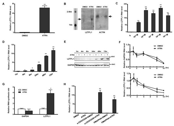
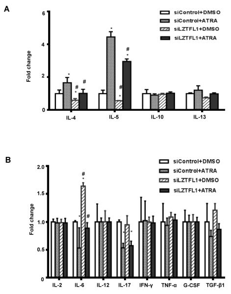
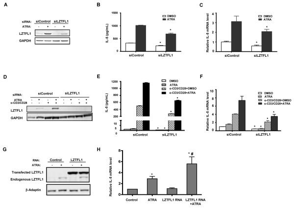
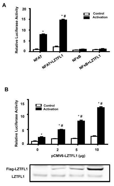
Retinoic acids, which are metabolites of vitamin A, have been shown to be involved in multiple T cell effector responses through their binding to the retinoic acid receptor, a ligand-activated transcription factor. Because the molecular mechanism of regulation by retinoic acid is still not fully uncovered, we investigated the gene expression profile of all-trans retinoic acid (ATRA)-treated human CD4(+) T cells. Leucine zipper transcription factor-like 1 (LZTFL1) was upregulated by ATRA in a dose- and time-dependent manner. The expression of LZTFL1 depended on both ATRA and TCR signaling. LZTFL1 accumulated in the plasma membrane compartment of human CD4(+) T cells, and, during immunological synapse formation, it transiently redistributed to the T cell and APC contact zone, indicating its role in T cell activation. Live-cell imaging demonstrates that at the initial stage of immunological synapse formation, LZTFL1 is concentrated at the APC contact site, and, during later stages, it relocates to the distal pole. Knockdown of LZTFL1 reduced the basal- and ATRA-induced levels of IL-5 in CD4(+) T cells, and overexpression of LZTFL1 enhanced the TCR-mediated NFAT signaling, suggesting that LZTFL1 is an important regulator of ATRA-induced T cell response. Together, these data indicate that LZTFL1 modulates T cell activation and IL-5 levels.


Figures

FIGURE 1
FIGURE 1
ATRA induces LZTFL1 expression in human primary CD4+ T cells. (A) Human CD4+ T cells isolated from healthy donors were primed with anti-CD3 and anti-CD28 Abs (α-CD3/CD28) and treated with DMSO or 1 μM of ATRA for 3 d. RNAs were extracted from these cells, and LZTFL1 RNA was quantitated by reverse transcription, followed by real-time PCR. LZTFL1 RNA expression was normalized to GAPDH RNA expression, and the relative RNA fold changes compared to those from DMSO treatment were plotted (mean ± SD). (B) LZTFL1 and actin mRNAs (indicated by arrows) were detected by Northern blotting. (C–D) LZTFL1 RNA expression levels in response to different concentrations of ATRA for 72 h (C), and at 1μM ATRA for various lengths of time (D), were analyzed. (E) LZTFL1 and GAPDH proteins were detected by Western blotting. (F) CD4+ T cells treated with DMSO or ATRA for 24 h were incubated with or without actinomycin D (5 μg/ml) for 10 h and the levels of LZTFL1 and GAPDH RNAs were analyzed by real-time PCR. (G) Nuclear run-on assay was used to analyze the effect of ATRA on LZTFL1 transcription initiation. CD4+ T cells were treated with DMSO or ATRA for 24 h. Nuclei were isolated, and biotinylated transcripts were synthesized in vitro, captured, and analyzed by real-time PCR. (H) The effect of different T cell activators on LZTFL1 RNA expression was analyzed. Results are representative of three experiments with cells from three different donors. *, p < 0.05; **, p < 0.01.
FIGURE 2
FIGURE 2
LZTFL1 localizes on the cell membrane in an F-actin-dependent manner. (A) Human CD4+ T cells isolated from healthy donors were transiently transfected with LZTFL1-Flag expression DNA. Cells were fixed and stained with anti-Flag Ab for LZTFL1-Flag (green). Cholera toxin B and DAPI were used to stain the cytoplasmic membrane marker GM1 ganglioside (red) and the nucleus (blue), respectively, and the staining was analyzed by confocal microscopy. (B) Fluorescent images of Jurkat T cells stained with anti-LZTFL1 Ab (green) and DAPI (blue). (C) Fluorescent images of Halo-LZTFL1-expressing Jurkat T cells treated with or without latrunculin B (1 μM) for 30 min. Cells were stained with phalloidin for F-actin (green), anti-halo Ab for LZTFL1 (red), and DAPI for the nucleus (blue).
FIGURE 3
FIGURE 3
LZTFL1 transiently redistributes to the contact zone between T cells and APCs. (A) Jurkat T cells (denoted by T) were copelleted with BMQC-stained, SEE-loaded Raji B cells (blue; denoted by B) and incubated at 37°C for 1.5-, 5-, and 15-min increments to induce IS formation. Cell conjugates were fixed and co-stained with Abs for LZTFL1 (green) and TCRβ (red). SEE-, no SEE-loading control, incubated for 15 min. Arrows indicate the T cell and B cell contact zone. (B) The percentage of conjugated T cells was calculated for all T cells in 10 fields chosen at random at indicated time points (mean ± SD). Results are representative of three independent experiments. *, p-value < 0.05. (C) LZTFL1 accumulation at the IS and the distal pole (DP) was quantified, and the percentage in total conjugates was plotted for both (mean ± SD). Results are representative of three independent experiments. *, p-value < 0.05 (comparison to LZTFL1 located at the IS after 1.5 min of incubation); (D and E) Live cell imaging of LZTFL1 localization at the IS. Jurkat T cells (red; denoted by T) stably expressing Halo-STOP (D) or Halo-LZTFL1 (E) interacting with SEE superantigen–loaded Raji B cells (blue; denoted by B) and forming the IS at indicated times on live cell imaging, as analyzed by wide field microscope (Supplemental video1). Data are representative of two independent experiments. (Scale bar, 10 μm.)
FIGURE 4
FIGURE 4
Involvement of LZTFL1 in ATRA-induced Th2 cytokine expression. Human CD4+ T cells isolated from healthy donors were transfected with control siRNA (siControl) or siRNA targeting human LZTFL1 (siLZTFL1). Cells were then activated with anti-CD3 and anti-CD28 Ab-bound beads (α-CD3/CD28) and treated with DMSO or 1 μM of ATRA for 3 d. Th2 (A), Th1, and Th17 (B) cytokine expressions in the culture supernatants were quantitated by ELISA. n = 3. *, p-value < 0.05 (comparison to cells transfected with siControl and treated with DMSO); #, p-value < 0.05 (comparison to cells transfected with siControl and treated with ATRA).
FIGURE 5
FIGURE 5
LZTFL1 enhances ATRA induced IL-5 protein and RNA levels. (A) Human CD4+ T cells isolated from healthy donors were transfected with control siRNA (siControl) or siRNA targeting human LZTFL1 (siLZTFL1). Cells were then activated with anti-CD3/CD28 and treated with DMSO or 1 μM of ATRA for 1 d. LZTFL1 expression was detected by Western blotting. (B) The level of the IL-5 protein in the culture supernatants was quantitated by ELISA. (C) IL-5 RNA expression was detected by reverse transcription, followed by real-time PCR, as described in the Materials and Methods. IL-5 RNA expression was normalized to GAPDH RNA expression, and relative RNA fold changes compared to those from DMSO treatment were plotted (mean ± SD). (D) D10.G4.1 cells, a mouse Th2 cell line, were transfected with siControl or siLZTFL1 targeting mouse LZTFL1. Cells were activated and treated with 1 μM of ATRA for 1 d. LZTFL1 expression was detected by Western blotting. (E) Expression of the IL-5 protein was quantitated by ELISA. (F) IL-5 RNA expression was analyzed. (G-H) CD3/CD28 activation beads expanded human CD4+ T cells were transfected with LZTFL1 mRNA and 72 h later cells were used to prepare protein lysates for western blot analysis (G) and RNA for real-time PCR measurement of GAPDH and IL-5 RNA (H). Results are representatives of five independent experiments for A to F and three for G and H. *, p-value <0.05 (comparison with each corresponding cells transfected with siControl or control RNA); #, p-value <0.05 (comparison with control and cells transfected with LZTFL1 mRNA and treated with ATRA).
FIGURE 6
FIGURE 6
LZTFL1 enhances T cell activation by specifically upregulating NFAT activity. (A) Jurkat cells were cotransfected with DNAs encoding LZTFL1-Flag and NFAT-Luc or LZTFL1-Flag and NFkB-Luc for 2 d. Renilla luciferase, under the control of the thymidine kinase promoter, was used as a transfection control. Cells were activated with anti-CD3 and anti-CD28 Abs-bound beads for 10 h. The firefly luciferase to Renilla luciferase ratio was calculated, and levels relative to those of the pCMV6 empty vector–transfected and unactivated cells were plotted (mean ± SD). n = 5. *, p value < 0.05 (comparison to cells transfected with pCMV6 empty vector and unactivated); #, p < 0.05 (comparison to cells transfected with pCMV6 empty vector and activated). (B) Jurkat cells cotransfected with reporter NFAT-Luc and an increasing amount of LZTFL1 expression DNA. Cells were activated, and the relative firefly luciferase to Renilla luciferase ratio was plotted as in (A) (mean ± SD). n = 5. *, p value < 0.05 (comparison to cells transfected with pCMV6 empty vector and unactivated); #, p value < 0.05 (comparison to cells transfected with pCMV6 empty vector and activated). The expression of endogenous and transfected LZTFL1 was analyzed by Western blotting.

Similar articles

Cited by

References

    1. De Luca LM. Retinoids and their receptors in differentiation, embryogenesis, and neoplasia. FASEB journal : official publication of the Federation of American Societies for Experimental Biology. 1991;5:2924–2933. - PubMed
    1. Collins SJ. The role of retinoids and retinoic acid receptors in normal hematopoiesis. Leukemia : official journal of the Leukemia Society of America, Leukemia Research Fund, U.K. 2002;16:1896–1905. - PubMed
    1. Hall JA, Cannons JL, Grainger JR, Dos Santos LM, Hand TW, Naik S, Wohlfert EA, Chou DB, Oldenhove G, Robinson M, Grigg ME, Kastenmayer R, Schwartzberg PL, Belkaid Y. Essential role for retinoic acid in the promotion of CD4(+) T cell effector responses via retinoic acid receptor alpha. Immunity. 2011;34:435–447. - PMC - PubMed
    1. Hall JA, Grainger JR, Spencer SP, Belkaid Y. The role of retinoic acid in tolerance and immunity. Immunity. 2011;35:13–22. - PMC - PubMed
    1. Iwata M, Hirakiyama A, Eshima Y, Kagechika H, Kato C, Song SY. Retinoic acid imprints gut-homing specificity on T cells. Immunity. 2004;21:527–538. - PubMed

Publication types

MeSH terms