. 2016 Jan 7;529(7584):105-109.
doi: 10.1038/nature16450. Epub 2015 Dec 23.

A LAIR1 insertion generates broadly reactive antibodies against malaria variant antigens

Affiliations

A LAIR1 insertion generates broadly reactive antibodies against malaria variant antigens

Joshua Tan et al. Nature. .

Abstract

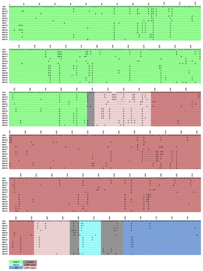
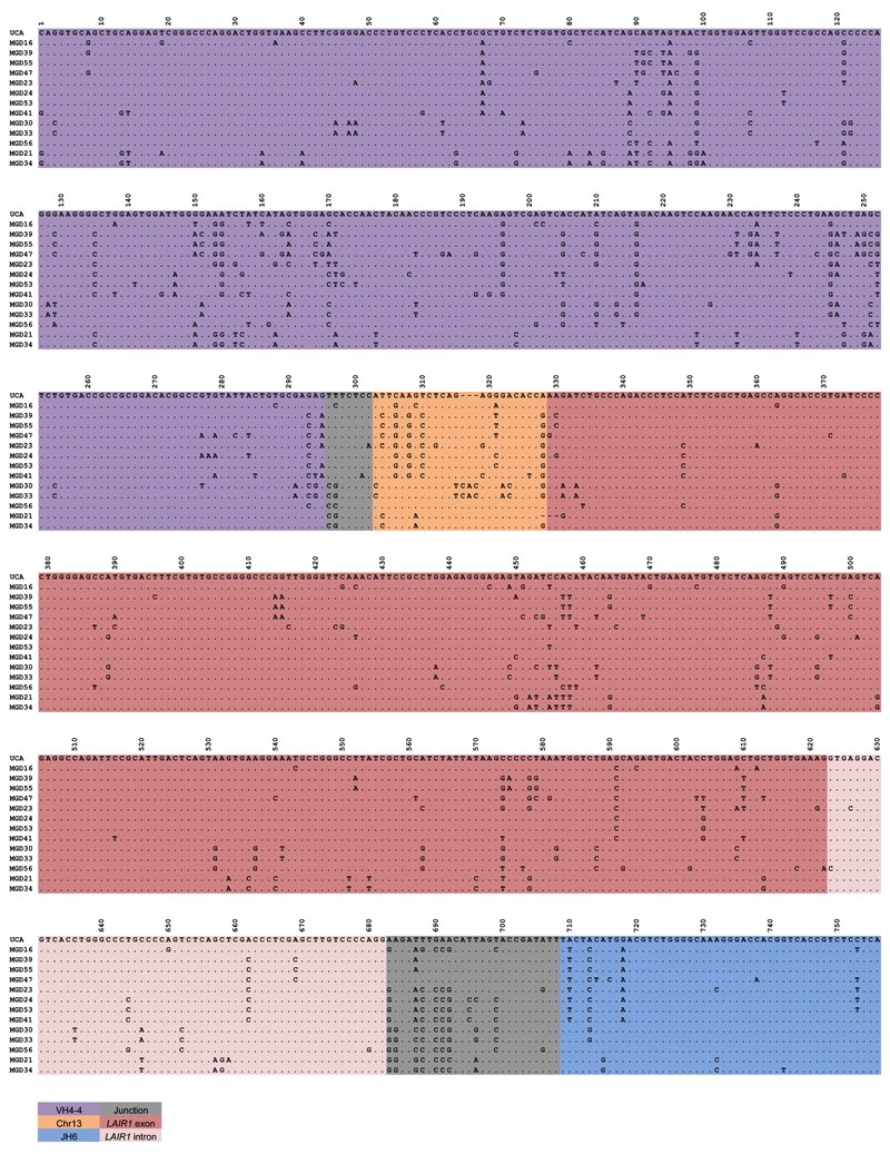
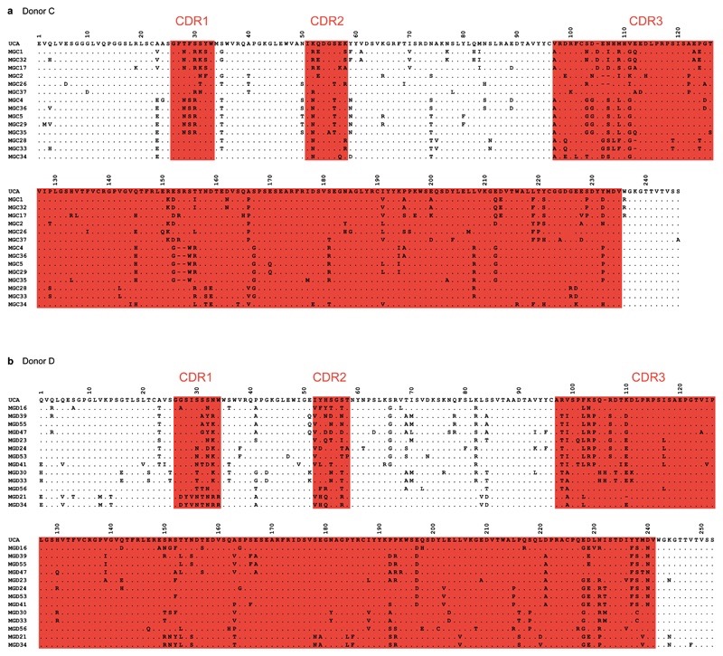
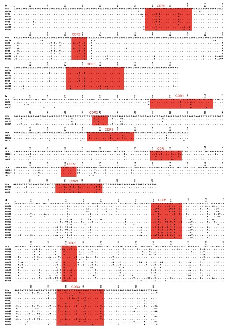
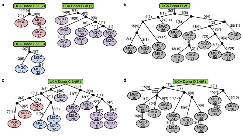
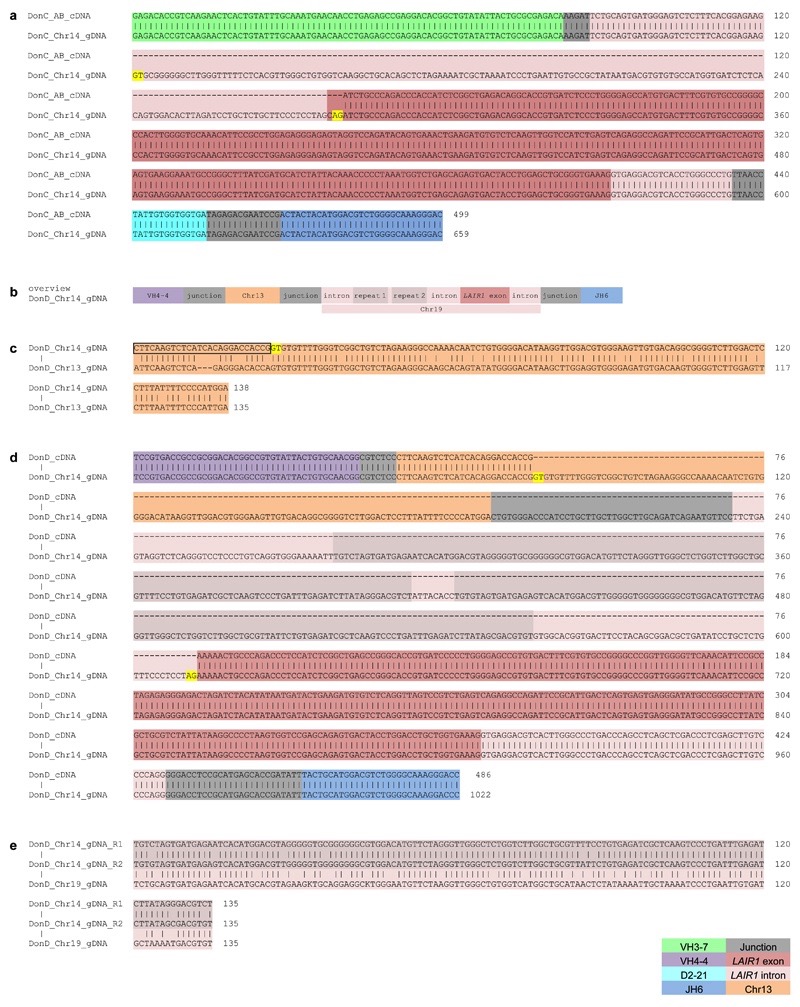
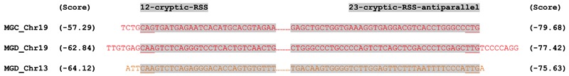
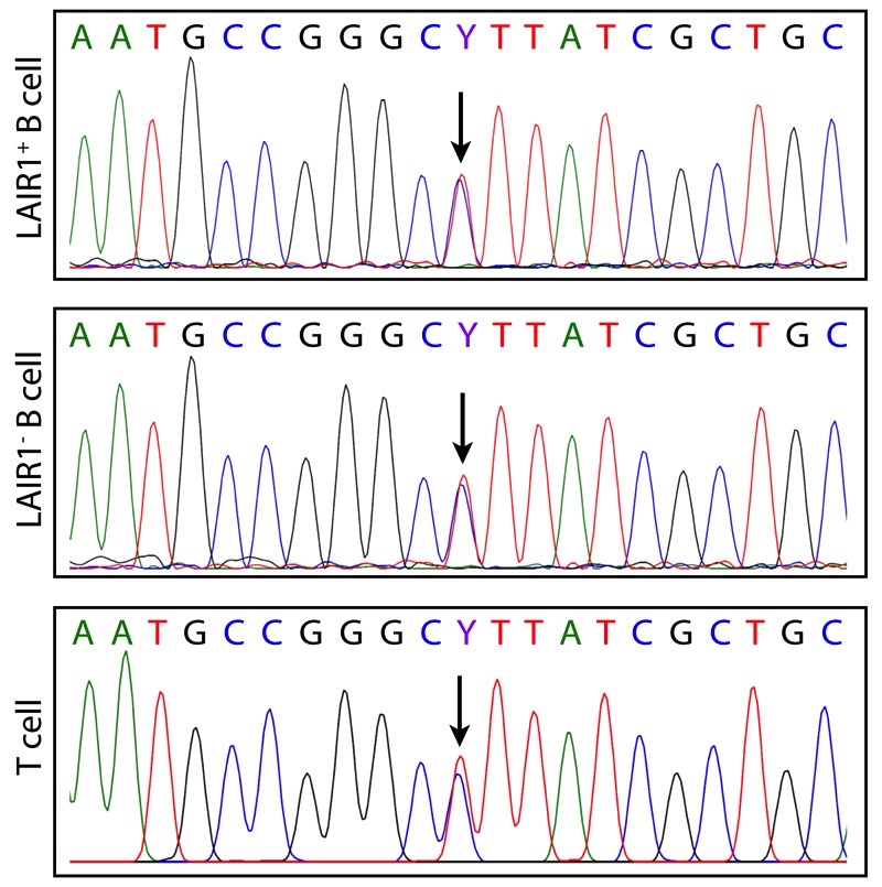
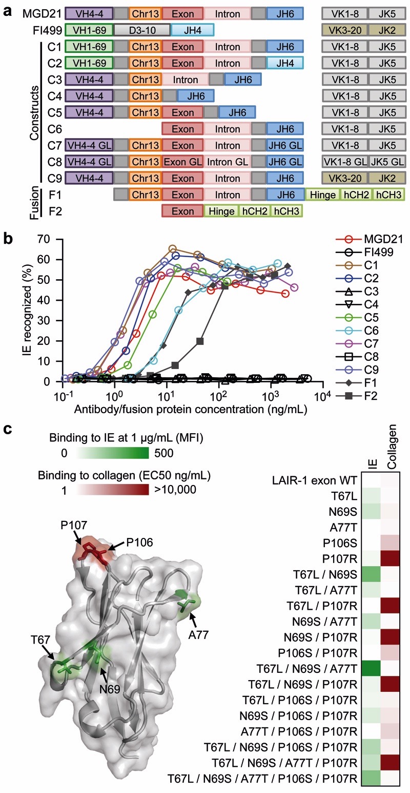
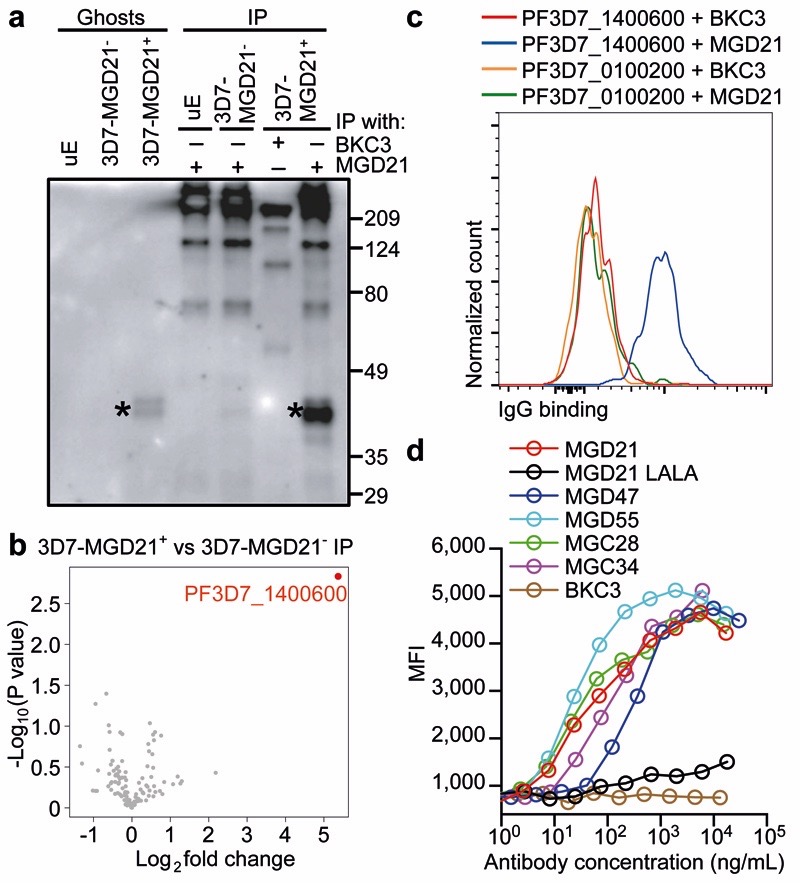
Plasmodium falciparum antigens expressed on the surface of infected erythrocytes are important targets of naturally acquired immunity against malaria, but their high number and variability provide the pathogen with a powerful means of escape from host antibodies. Although broadly reactive antibodies against these antigens could be useful as therapeutics and in vaccine design, their identification has proven elusive. Here we report the isolation of human monoclonal antibodies that recognize erythrocytes infected by different P. falciparum isolates and opsonize these cells by binding to members of the RIFIN family. These antibodies acquired broad reactivity through a novel mechanism of insertion of a large DNA fragment between the V and DJ segments. The insert, which is both necessary and sufficient for binding to RIFINs, encodes the entire 98 amino acid collagen-binding domain of LAIR1, an immunoglobulin superfamily inhibitory receptor encoded on chromosome 19. In each of the two donors studied, the antibodies are produced by a single expanded B-cell clone and carry distinct somatic mutations in the LAIR1 domain that abolish binding to collagen and increase binding to infected erythrocytes. These findings illustrate, with a biologically relevant example, a novel mechanism of antibody diversification by interchromosomal DNA transposition and demonstrate the existence of conserved epitopes that may be suitable candidates for the development of a malaria vaccine.


Figures

Extended Data Figure 1.
Extended Data Figure 1.. Nucleotide sequence alignments of VH regions of antibodies isolated from donor C.
Dots indicate positions where the nucleotide of a mature antibody is identical to that of the UCA.
Extended Data Figure 2.
Extended Data Figure 2.. Nucleotide sequence alignments of VH regions of antibodies isolated from donor D.
Dots indicate positions where the nucleotide of a mature antibody is identical to that of the UCA.
Extended Data Figure 3.
Extended Data Figure 3.. Protein sequence alignments of VH regions of antibodies isolated from donors C (a) and D (b).
Putative complementarity-determining regions (CDRs) are highlighted in red. Dots indicate positions where the amino acid of a mature antibody is identical to that of the UCA.
Extended Data Figure 4.
Extended Data Figure 4.. Nucleotide sequence alignments of VL regions of antibodies isolated from donors C (a-c) and D (d).
Antibodies from donor C use VL7-43/JL3 (a), VK4-1/JK2 (b), or VK1-5/JK2 (c), while antibodies from donor D use VK1-8/JK5. Complementarity-determining regions (CDRs) are highlighted in red. Dots indicate positions where the nucleotide of a mature antibody is identical to that of the UCA.
Extended Data Figure 5.
Extended Data Figure 5.. Genealogy trees generated from VL and LAIR-1 exon sequences.
The trees were drawn based on the somatic mutations in light chain variable regions (a, b) or LAIR-1 exons (c, d) of the antibodies isolated from donors C and D. In the donor C VL trees, VL(1), VL(2) and VL(3) refer to VL7-43/JL3, VK1-5/JK2 and VK4-1/JK2, respectively. Shown are the nucleotide and amino acid substitutions, with the latter in parentheses.
Extended Data Figure 6.
Extended Data Figure 6.. Genomic DNA analysis of LAIR-1-containing antibodies of donor C and donor D.
a, The sequence alignment of genomic DNA (gDNA) and cDNA of a LAIR-1-containing antibody from donor C reveals a 507 bp LAIR-1 insert in Chr14 and the removal of a 160 bp fragment by RNA splicing. Splice donor and acceptor sites are highlighted in yellow. b, Schematic overview of the genomic organization of a LAIR-1-containing antibody from donor D, not in scale. c, Alignment of a region of antibody-encoding DNA (Chr14) with the corresponding region of Chr13 from genomic DNA (gDNA). The sequence maintained in the mature antibody mRNA is boxed and the splice donor site is highlighted in yellow. d, Alignment of gDNA and cDNA reveals that a part of the Chr13 region and the entire inserted 5’ LAIR-1 intron are removed by RNA splicing. Splice donor and acceptor sites are highlighted in yellow. e, Alignment of the two repeated elements found in the inserted LAIR-1 intron in Chr14 with the corresponding sequence in Chr19. The repeats are named R1 and R2, and K = G/A.
Extended Data Figure 7.
Extended Data Figure 7.. LAIR-1 and Chr13 inserts are flanked by 12/23 cryptic RSS sites.
The regions on Chr19 and Chr13 of donor-derived genomic DNA corresponding to the ends of the inserts were sequenced and RSS sites were identified using the RSSsite web server. The sequences shown begin from the ends of the inserts. Cryptic RSS sites are highlighted in grey, with complementary ends underlined and prediction scores shown in parentheses.
Extended Data Figure 8.
Extended Data Figure 8.. Both LAIR-1 alleles on Chr19 are intact in B cells producing LAIR-1 antibodies.
Heterozygosity of the Chr19 LAIR-1 exon in cells from donor C showing that both LAIR-1 alleles are intact in B cells producing LAIR-1-containing antibodies. Displayed are the chromatograms obtained for B cell clones with or without a LAIR-1 insertion (LAIR-1+ or LAIR-1- B cell) and for polyclonal T cells.
Extended Data Figure 9.
Extended Data Figure 9.. Reactivity and functional assays of MGC and MGD antibodies.
a, MGD21 staining of 3D7 IE that were enriched or depleted of MGD21 reactivity (n = 3 independent experiments). b, Heat map from LC-MS analysis showing RIFIN expression levels (calculated as iBAQ scores) in erythrocyte ghosts prepared from 3D7-MGD21+ and 3D7-MGD21- IE (two experiments shown). Grey boxes indicate that expression levels are below the detection limit. c, Shown is the percentage of IE (representative of n = 2 independent experiments) or of transfected CHO cells (n = 1) stained by the antibodies. RIFINs that were enriched in 3D7-MGD21+ ghosts are highlighted blue, while RIFINs that were similarly expressed or not detected in 3D7-MGD21- and 3D7-MGD21+ ghosts are shown in red. BKC3 is a negative control antibody. d, Western blot showing MGD21 binding to IP prepared from 9605-MGD21- and 9605-MGD21+ IE (representative of n = 2 independent experiments). Specific bands are marked with a star. Anti-human IgG was used as the secondary antibody, resulting in detection of antibodies used for IP alongside antigens of interest. For gel source data, see Supplementary Figure 1. e, Percentage of 9605-MGD21- and 9605-MGD21+ IE recognized by representative MGC and MGD antibodies (representative of n = 2 independent experiments). f, Binding of MGD21 to CHO cells transfected with RIFINs (PF3D7_1400600 and PF3D7_0100200), a RIFIN chimera containing the constant region of PF3D7_0100200 and the variable region of PF3D7_1400600 (PF3D7_0100200c_1400600v), or the inverse chimera (PF3D7_1400600c_0100200v) (n = 1). g, Binding of an Fc fusion protein containing the LAIR-1 exon of MGD21 to CHO cells transfected with RIFINs or RIFIN chimeras (n = 1). h, Parasitemia of 3D7-MGD21+in vitro culture after 2 d of incubation with various concentrations of MGD21 or an irrelevant antibody (BKC3) (n = 1). i, Percentage of 3D7-MGD21+ IE recognized by MGD21 after 2 d of incubation with various concentrations of MGD21 or BKC3. The antibodies were removed after 2 d (during the ring stage of the life cycle) and the parasites were allowed to grow for 24 h to the late trophozoite/schizont stage prior to detection with MGD21 (n = 1). j, Rosetting of 9605-MGD21+ IE with blood group O+ or A+ uninfected erythrocytes (uE) after incubation with MGD21 or BKC3. Shown is the mean ± SD from n = 4 independent experiments. Statistical significance was evaluated by the Wilcoxon signed-rank test (P > 0.1 for both blood groups). k, Agglutinates of 3D7-MGD21+ or 11019-MGD21+ IE formed by MGD21 or MGC34. Scale bar, 25 μm. l, Opsonic phagocytosis of 11019-MGD21+ IE by monocytes (n = 2). The IE were stained with DAPI, which was quantified in monocytes as a measure of phagocytosis.
Extended Data Figure 10.
Extended Data Figure 10.. A schematic representation of interchromosomal LAIR-1 transposition.
Shown is the insertion of a fragment of LAIR-1 into the Ig heavy chain locus through a mechanism still to be molecularly defined, followed by the acquisition of somatic mutations that increase binding to IE and abolish binding to collagen.
Figure 1.
Figure 1.. Identification of broadly reactive monoclonal antibodies against IE.
a, Fluorescence microscopy images of single agglutinates (top) and a triple agglutinate (bottom). Scale bar, 50 μm. b-c, Plasma (pooled in groups of five) from immune adults were screened against six parasite isolates using the triple mixed agglutination assay (b). Pools that formed mixed agglutinates with at least five isolates (in red) were further investigated for individual reactivity against an extended panel of 8 isolates (c). d, Heat map showing the percentage of IE of eight parasite isolates stained by monoclonal antibodies isolated from two donors (n = 1). Closely related antibodies are grouped in alternating colors. e, Example of staining of IE by the broadly reactive antibody MGD55.
Figure 2.
Figure 2.. Broadly reactive antibodies contain a mutated LAIR-1 insert and are produced by expanded clones.
a, Protein sequence alignment of MGC1 and MGD21 with germline-encoded sequences of the corresponding VH (green or purple), DH (cyan), JH (blue) and LAIR-1 (exon in red and intronic sequences in light red). Chr13 sequences are shown in orange while grey areas show junctional sequences for which no homology was found. b-c, Genealogy trees drawn from the VH nucleotide sequences of antibodies from donors C (b) and D (c). In the donor C genealogy tree, antibodies that use different light chains are highlighted in different colors. Shown are the nucleotide and amino acid substitutions, with the latter in parentheses. d, Scheme showing genomic DNA and cDNA of LAIR-1-containing antibodies from donors C and D. Shown are the lengths of the fragments (bp in parentheses), cryptic 12 and 23 RSS sites (black and brown triangles, respectively) and splicing positions (dashed lines).
Figure 3.
Figure 3.. The mutated LAIR-1 insert is necessary and sufficient for binding to IE.
a, Design of modified MGD21 antibody constructs with selected regions replaced with counterparts from an unrelated antibody (FI499) [C1-C2, C9], deleted [C3-C6], or reverted to germline (GL) [C7-C8]. Fc fusion proteins that incorporated the LAIR-1 insert, junction and downstream sequences [F1], as well as the LAIR-1 exon alone [F2], were also designed. b, Binding of MGD21 constructs and Fc fusion proteins to IE (representative of n = 2 independent experiments). c, Selected amino acid substitutions found in MGD21 were added individually or in different combinations to the germline LAIR-1-Fc fusion protein. These mutants were tested for binding to collagen and to IE. Shown are the effect of the mutations on binding to IE or collagen (one representative of n = 2 independent experiments) and their location on the LAIR-1 structure (pdb, 3kgr). Gain of IE binding is shown in green (background mean fluorescence intensity (MFI) values subtracted). Loss of collagen binding (EC50 ELISA values) is shown in red.
Figure 4.
Figure 4.. LAIR-1-containing antibodies bind to distinct RIFINs and opsonize IE.
a, Western blot showing MGD21 binding to erythrocyte ghosts and MGD21 IP prepared from 3D7-MGD21+ and 3D7-MGD21- IE (representative of n = 2 independent experiments). Controls include uninfected erythrocytes (uE) and IP with an irrelevant antibody (BKC3). Specific bands are marked with stars. Anti-human IgG was used as the secondary antibody, resulting in detection of antibodies used for IP alongside antigens of interest. For gel source data, see Supplementary Figure 1. b, Volcano plot from LC-MS analysis of MGD21 IP prepared from 3D7-MGD21+ IE versus from 3D7-MGD21- IE (from n = 4 independent experiments). Statistical significance was evaluated by Welch tests (P< 0.01 for PF3D7_1400600). c, MGD21 and BKC3 staining of CHO cells transfected with a specific (PF3D7_1400600) or an irrelevant (PF3D7_0100200) RIFIN (representative of n = 5 independent experiments). d, Opsonic phagocytosis of 3D7-MGD21+ IE by monocytes (n = 3 for MGD21, MGD21 LALA, BKC3, n = 2 for others). The IE were stained with DAPI, which was quantified in monocytes as a measure of phagocytosis. MGD21 LALA is a mutant of MGD21 lacking Fc receptor binding.

Comment in

References

    1. Chan J-A, Fowkes FJI, Beeson JG. Surface antigens of Plasmodium falciparum-infected erythrocytes as immune targets and malaria vaccine candidates. Cell Mol Life Sci. 2014;71:3633–3657. - PMC - PubMed
    1. Scherf A, Lopez-Rubio JJ, Riviere L. Antigenic variation in Plasmodium falciparum. Annu Rev Microbiol. 2008;62:445–470. - PubMed
    1. Goel S, et al. RIFINs are adhesins implicated in severe Plasmodium falciparum malaria. Nat Med. 2015;21:314–317. - PubMed
    1. Bull PC, et al. Parasite antigens on the infected red cell surface are targets for naturally acquired immunity to malaria. Nat Med. 1998;4:358–360. - PMC - PubMed
    1. Traggiai E, et al. An efficient method to make human monoclonal antibodies from memory B cells: potent neutralization of SARS coronavirus. Nat Med. 2004;10:871–875. - PMC - PubMed

Publication types

MeSH terms

Associated data