. 2015 Dec 23;10(12):e0145239.
doi: 10.1371/journal.pone.0145239. eCollection 2015.

A Conserved GPG-Motif in the HIV-1 Nef Core Is Required for Principal Nef-Activities

Affiliations

A Conserved GPG-Motif in the HIV-1 Nef Core Is Required for Principal Nef-Activities

Marta Martínez-Bonet et al. PLoS One. .

Abstract

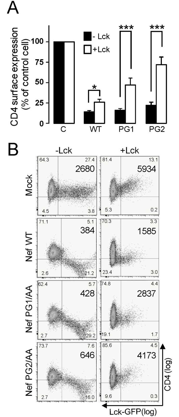
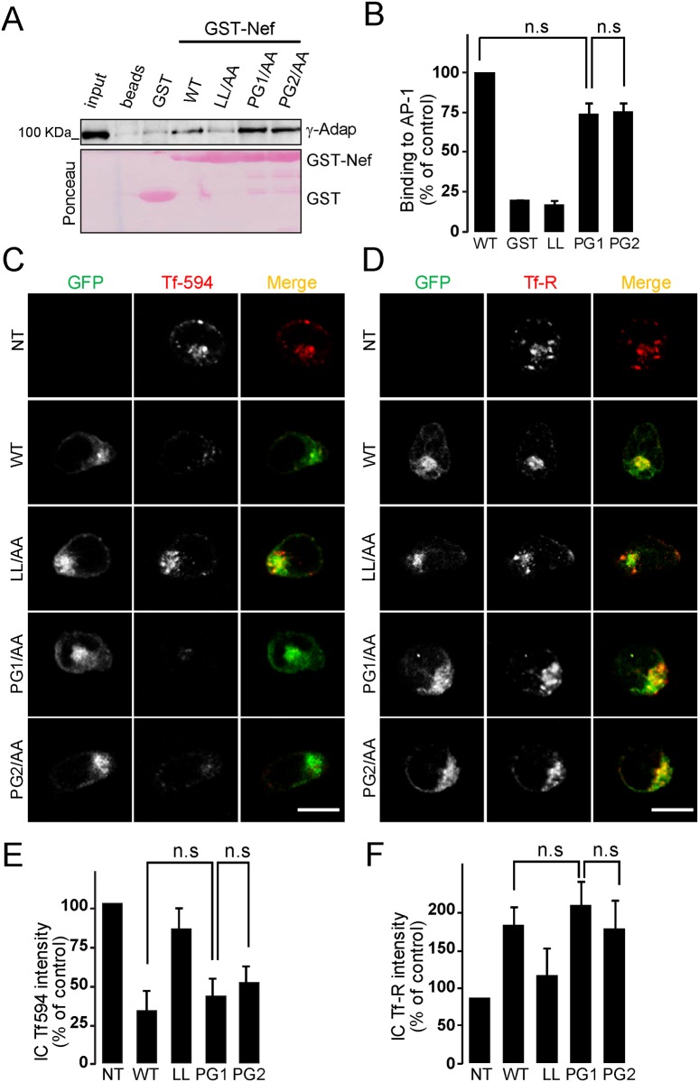
To find out new determinants required for Nef activity we performed a functional alanine scanning analysis along a discrete but highly conserved region at the core of HIV-1 Nef. We identified the GPG-motif, located at the 121-137 region of HIV-1 NL4.3 Nef, as a novel protein signature strictly required for the p56Lck dependent Nef-induced CD4-downregulation in T-cells. Since the Nef-GPG motif was dispensable for CD4-downregulation in HeLa-CD4 cells, Nef/AP-1 interaction and Nef-dependent effects on Tf-R trafficking, the observed effects on CD4 downregulation cannot be attributed to structure constraints or to alterations on general protein trafficking. Besides, we found that the GPG-motif was also required for Nef-dependent inhibition of ring actin re-organization upon TCR triggering and MHCI downregulation, suggesting that the GPG-motif could actively cooperate with the Nef PxxP motif for these HIV-1 Nef-related effects. Finally, we observed that the Nef-GPG motif was required for optimal infectivity of those viruses produced in T-cells. According to these findings, we propose the conserved GPG-motif in HIV-1 Nef as functional region required for HIV-1 infectivity and therefore with a potential interest for the interference of Nef activity during HIV-1 infection.


Conflict of interest statement

Competing Interests: The authors have declared that no competing interests exist.

Figures

Fig 1
Fig 1. The GPG-loop is conserved among Nef proteins.
A) Clustal W alignment of the 121–137 region of HIV1- Nef with homologous HIV2- and SIV-Nef sequences from Los Alamos database. The consensus sequence of the GPG-loop is highlighted to indicate the amino acid identity. X represents any amino acid residue and Ø represents a hydrophobic residue. B) Schematic representation of the domain organization of HIV-1NL4.3 Nef. The novel GPG-loop (red), conserved PxxP domain (blue), the di-Leucine based motif (dark green) as well as the membrane anchorage domain (grey), the acidic clusters (orange) and the dimerization domain (light green) are shown. C) Ribbon/surface diagram showing the HIV-1 Nef-GPG loop as generated by using the Pymol software (pdb2nef). The central GPG residues (red) are flanked by hydrophobic β2 and β3 sheets (yellow) in HIV-1 Nef structure. PxxP and di-Leucine motifs surfaces are coloured in blue or green, respectively.
Fig 2
Fig 2. Mutations on the GPG-loop abrogate HIV-1 Nef-induced CD4 down-regulation.
A-B) CEM cells were transfected with either wild type or the indicated Nef mutants as GFP-fusion proteins. Nef-induced surface CD4 down-regulation was analysed by two-colour flow cytometry in Nef-GFP expressing cells. The mean CD4 (PE) fluorescence intensity (MFI) of the GFP-positive cells was used to calculate the Nef activity on surface CD4 expression (A). Histograms represent the percentage of activity relative to wild-type Nef as the arithmetic mean ± SD of at least three independent experiments. Dot plots and the corresponding MFI are representative of three independent assays (B). ***, p< 0.001. C) The expression of wild type and the indicated Nef-GPG mutants in CEM cells was analysed by western blotting using an anti-GFP antibody. γ-adaptin levels were used as loading control staining.
Fig 3
Fig 3. The integrity of the GPG-motif is dispensable for Nef-induced CD4-downregulation in p56Lck -negative cells.
A) HeLa-CD4 cells were co-transfected with the wild type or the indicated Nef-GPG mutants (Nef-HA) along with empty or p56Lck-GFP constructs. Nef activity on CD4-downregulation was analysed by two-colour flow cytometry in GFP cells. A) Histograms represent the percentage of CD4 expression at cell surface relative to non-transfected cells. n.s, not statistically significant. ***, p< 0.001. B) Dot plots and the corresponding MFI are representative of three independent assays.
Fig 4
Fig 4. GPG mutations do not affect Nef-induced alteration of Tf-R trafficking.
(A-B) Analysis of Nef/AP-1interaction by in vitro pull-down. HeLa cell lysates were incubated with equal amounts of GSH-sepharose beads, or purified GST, wild type or the indicated Nef mutants as GST-fusions previously immobilized on GSH-sepharose beads. Bound proteins were resolved by SDS-PAGE and the association of AP-1 complex was analysed by Western blotting with anti-γ-adaptin (A) and quantified by densitometry (B). Ponceau-red staining shows the loading of the assayed Nef-GST fusion proteins on the beads. n.s not statistically significant. (C-D) Non-transfected, NT, or transfected CEM-T cells with expression plasmids for wild type or the indicated Nef-GPG mutants as GFP-fusion proteins were serum-starved and then incubated with Tfn-Alexa-594 for 15 min at 37°C before fixation (C), or analysed by indirect immunofluorescent staining for total Tf-R in permeabilized cells (D). Staining was visualized by confocal microscopy. A medial optical section of a representative cell is shown. Merged images show both GFP and Tfn-Alexa-594 or Tf-R labelling. Scale bar, 10 μm. (E-F) The intensity of the fluorescence corresponding to Tfn-594 (E) or Tf-R (F) was quantified by densitometry as described under Materials and Methods. Histograms represent the percentages of the mean fluorescence intensity relative to non-transfected cells (lower panel). Values are the arithmetic mean ± SD of at least three independent experiments in which over 50 cells were counted per condition. n.s. not statistically significant.
Fig 5
Fig 5. The GPG-motif is required for Nef-induced intracellular recruitment of p56Lck.
(A-B) The subcellular distribution of endogenous p56Lck and pTyr-Lck was analysed by indirect immunofluorescent staining of non-transfected, NT, or transfected Jurkat cells with the expression plasmids for wild type or the indicated Nef-GPG mutants as GFP-fusion proteins. A) A medial optical section of a representative cell is shown. Densitometry analysis of the distribution of endogenous p56Lck staining along the line in Jurkat cells expressing the indicated Nef-GFP proteins. Arrows indicate the position of the cell periphery (right panels). Scale bar, 10 μm. B) Quantification of the experiment shown in A. The percentage of cells with high levels of intracellular p56Lck was measured as described in Materials and Methods. Histograms represent the cells with predominant intracellular localization of p56Lck compared to control cells, respectively. Values are the arithmetic mean ± SD of at least three independent experiments in which over 100 cells were counted per condition. ***, p< 0.001. C) The expression of endogenous p56Lck and transfected Nef-GFP proteins in Jurkat cells was analysed by western blotting using an anti-p56Lck antibody and an anti-GFP antibody, respectively. γ-adaptin was used as loading control.
Fig 6
Fig 6. GPG-mutations reduce Nef-dependent inhibition of TCR-induced actin remodelling and Nef-induced down-regulation of MHCI.
(A-B) Fluorescent microscopy of Jurkat T-cells transfected with the indicated GFP or Nef-GFP expression plasmids after 5 min incubation on anti-CD3 coated cover glasses and subsequent staining for F-actin. A) Depicted pictures are a representative image at the cell-cover glass level. The upper panel presents a merge of the GFP and F-actin channels, the lower panel shows the F-actin channel only. Arrows indicate transfected cells. Scale bar, 10 μm. B) Quantification of the experiment shown in A. Shown is the percentage of cells exhibiting pronounced F-actin ring formation. Values are the arithmetic mean ± SD of at least three independent experiments in which over 100 cells were counted per condition. ***, p< 0.001. (C-D) CEM cells were transfected with the wild type or the indicated Nef-GPG mutants as GFP-fusion proteins. Nef-induced surface MHCI down-regulation was assessed by flow cytometry in Nef-GFP expressing cells. C) Dot plots and the corresponding MFI are representative of three independent assays. D) The mean fluorescence intensity (MFI) of MHCI (Alexa-555) in the GFP-positive cells was used to calculate activity relative to wild-type Nef. Values are the arithmetic mean ± SD of at least three independent experiments. **, p< 0.01; ***, p< 0.001.
Fig 7
Fig 7. Nef-GPG motif is required for Nef-enhanced infectivity of HIV-1.
Relative infectivity of HIV-1expressing Nef-GPG mutations produced in Jurkat (A) or 293T (B) cells. Infectivity was analysed 48h post-infection by Luciferase assays in TZM-bl reporter cells. Results are expressed as the percentage of infectivity relative to that of the wild type viruses normalized to 100%. Values are the mean ± SD of three independent infections, each measured in duplicate. ***, p< 0.001.

Similar articles

Cited by

References

    1. Chowers MY, Spina CA, Kwoh TJ, Fitch NJ, Richman DD, Guatelli JC (1994) Optimal infectivity in vitro of human immunodeficiency virus type 1 requires an intact nef gene. JVirol 68: 2906–2914. - PMC - PubMed
    1. Kestier HW, Ringler DJ, Mori K, Panicali DL, Sehgal PK, Daniel MD, et al. (1991) Importance of the nef gene for maintenance of high virus loads and for development of AIDS. Cell 65: 651–662. - PubMed
    1. Baur AS, Sawai ET, Dazin P, Fantl WJ, Cheng-Mayer C, Peterlin BM (1994) HIV-1 Nef leads to inhibition or activation of T cells depending on its intracellular localization. Immunity 1: 373–384. - PubMed
    1. Birch MR, Learmont JC, Dyer WB, Deacon NJ, Zaunders JJ, Saksena N, et al. (2001) An examination of signs of disease progression in survivors of the Sydney Blood Bank Cohort (SBBC). J Clin Virol 22: 263–270. - PubMed
    1. Carl S, Greenough TC, Krumbiegel M, Greenberg M, Skowronski J, Sullivan JL, et al. (2001) Modulation of different human immunodeficiency virus type 1 Nef functions during progression to AIDS. J Virol 75: 3657–3665. - PMC - PubMed

Publication types

MeSH terms

Substances