

Maternal embryonic leucine zipper kinase serves as a poor prognosis marker and therapeutic target in gastric cancer
- PMID: 26701722
- PMCID: PMC4868755
- DOI: 10.18632/oncotarget.6673
Maternal embryonic leucine zipper kinase serves as a poor prognosis marker and therapeutic target in gastric cancer
Abstract
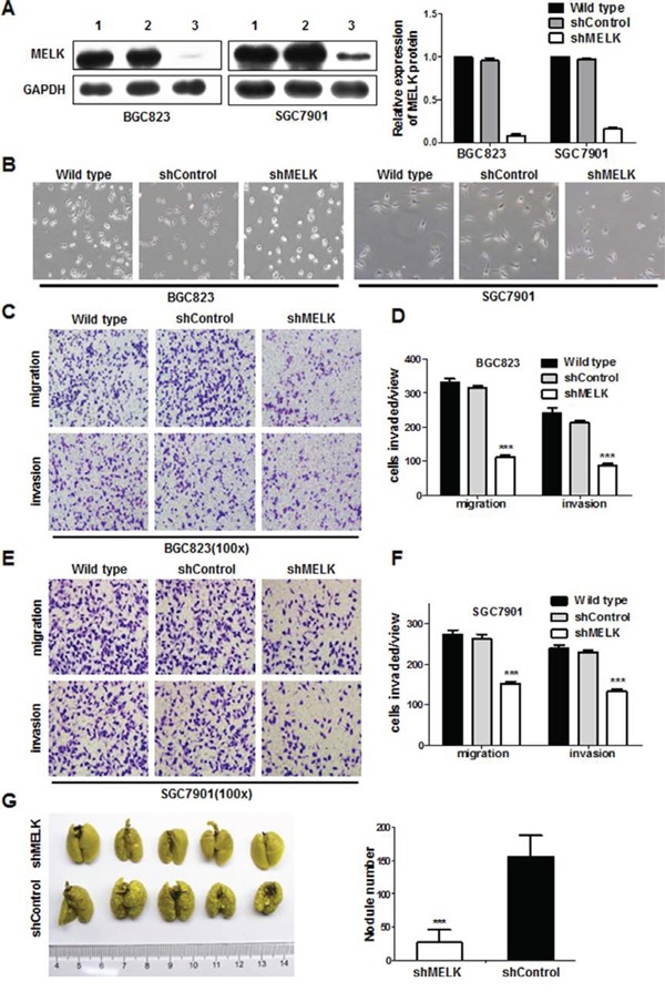
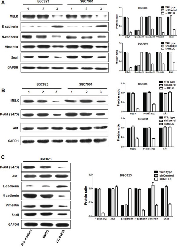
Maternal embryonic leucine zipper kinase (MELK) is upregulated in a variety of human tumors, and is considered an attractive molecular target for cancer treatment. We characterized the expression of MELK in gastric cancer (GC) and measured the effects of reducing MELK mRNA levels and protein activity on GC growth. MELK was frequently overexpressed in primary GCs, and higher MELK levels correlated with worse clinical outcomes. Reducing MELK expression or inhibiting kinase activity resulted in growth inhibition, G2/M arrest, apoptosis and suppression of invasive capability of GC cells in vitro and in vivo. MELK knockdown led to alteration of epithelial mesenchymal transition (EMT)-associated proteins. Furthermore, targeting treatment with OTSSP167 in GC patient-derived xenograft (PDX) models had anticancer effects. Thus, MELK promotes cell growth and invasiveness by inhibiting apoptosis and promoting G2/M transition and EMT in GC. These results suggest that MELK may be a promising target for GC treatment.
Keywords: MELK; PDX; gastric cancer; metastasis; prognosis.
Conflict of interest statement
The authors declare no conflicts of interest.
Figures


Similar articles
-
Maternal embryonic leucine zipper kinase serves as a poor prognosis marker and therapeutic target in osteosarcoma.Oncol Rep. 2020 Sep;44(3):1037-1048. doi: 10.3892/or.2020.7686. Epub 2020 Jul 13. Oncol Rep. 2020. PMID: 32705239 Free PMC article.
-
Maternal embryonic leucine zipper kinase enhances gastric cancer progression via the FAK/Paxillin pathway.Mol Cancer. 2014 May 4;13:100. doi: 10.1186/1476-4598-13-100. Mol Cancer. 2014. PMID: 24885567 Free PMC article.
-
Maternal embryonic leucine zipper kinase: A novel biomarker and a potential therapeutic target of cervical cancer.Cancer Med. 2018 Nov;7(11):5665-5678. doi: 10.1002/cam4.1816. Epub 2018 Oct 18. Cancer Med. 2018. PMID: 30334367 Free PMC article.
-
Maternal embryonic leucine zipper kinase in tumor cells and tumor microenvironment: An emerging player and promising therapeutic opportunity.Cancer Lett. 2023 Apr 28;560:216126. doi: 10.1016/j.canlet.2023.216126. Epub 2023 Mar 16. Cancer Lett. 2023. PMID: 36933780 Review.
-
MELK: a potential novel therapeutic target for TNBC and other aggressive malignancies.Expert Opin Ther Targets. 2017 Sep;21(9):849-859. doi: 10.1080/14728222.2017.1363183. Epub 2017 Aug 16. Expert Opin Ther Targets. 2017. PMID: 28764577 Review.
Cited by
-
Maternal embryonic leucine zipper kinase serves as a poor prognosis marker and therapeutic target in osteosarcoma.Oncol Rep. 2020 Sep;44(3):1037-1048. doi: 10.3892/or.2020.7686. Epub 2020 Jul 13. Oncol Rep. 2020. PMID: 32705239 Free PMC article.
-
G2/M checkpoint plays a vital role at the early stage of HCC by analysis of key pathways and genes.Oncotarget. 2017 Jul 18;8(44):76305-76317. doi: 10.18632/oncotarget.19351. eCollection 2017 Sep 29. Oncotarget. 2017. PMID: 29100313 Free PMC article.
-
Mutant P53 induces MELK expression by release of wild-type P53-dependent suppression of FOXM1.NPJ Breast Cancer. 2020 Jan 3;6:2. doi: 10.1038/s41523-019-0143-5. eCollection 2020. NPJ Breast Cancer. 2020. PMID: 31909186 Free PMC article.
-
Targeting Apoptosis in Cancer.Curr Oncol Rep. 2022 Mar;24(3):273-284. doi: 10.1007/s11912-022-01199-y. Epub 2022 Feb 3. Curr Oncol Rep. 2022. PMID: 35113355 Review.
-
Computational exploration of maternal embryonic leucine zipper kinase (MELK) as a cancer drug target.Saudi J Biol Sci. 2022 Jul;29(7):103335. doi: 10.1016/j.sjbs.2022.103335. Epub 2022 Jun 1. Saudi J Biol Sci. 2022. PMID: 35769060 Free PMC article.
References
-
- Jemal A, Bray F, Center MM, Ferlay J, Ward E, Forman D. Global cancer statistics. CA Cancer J Clin. 2011;61:69–90. - PubMed
-
- Wang J, Sun Y, Bertagnolli MM. Comparison of Gastric Cancer Survival Between Caucasian and Asian Patients Treated in the United States: Results from the Surveillance Epidemiology and End Results (SEER) Database. Ann Surg Oncol. 2015 - PubMed
-
- Kanat O, O'Neil BH. Metastatic gastric cancer treatment: a little slow but worthy progress. Med Oncol. 2013;30:464. - PubMed
-
- Gil M, Yang Y, Lee Y, Choi I, Ha H. Cloning and expression of a cDNA encoding a novel protein serine/threonine kinase predominantly expressed in hematopoietic cells. Gene. 1997;195:295–301. - PubMed
-
- Heyer BS, Warsowe J, Solter D, Knowles BB, Ackerman SL. New member of the Snf1/AMPK kinase family, Melk, is expressed in the mouse egg and preimplantation embryo. Mol Reprod Dev. 1997;47:148–156. - PubMed
Publication types
MeSH terms
LinkOut - more resources
Full Text Sources
Other Literature Sources
Medical
Miscellaneous
