

Concise Review: An Update on the Culture of Human Corneal Endothelial Cells for Transplantation
- PMID: 26702128
- PMCID: PMC4729556
- DOI: 10.5966/sctm.2015-0181
Concise Review: An Update on the Culture of Human Corneal Endothelial Cells for Transplantation
Abstract
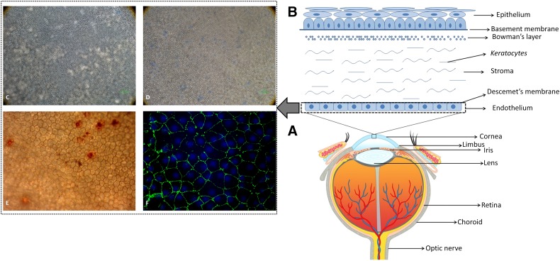
The cornea forms the front window of the eye, enabling the transmission of light to the retina through a crystalline lens. Many disorders of the cornea lead to partial or total blindness, and therefore corneal transplantation becomes mandatory. Recently, selective corneal layer (as opposed to full thickness) transplantation has become popular because this leads to earlier rehabilitation and visual outcomes. Corneal endothelial disorders are a common cause of corneal disease and transplantation. Corneal endothelial transplantation is successful but limited worldwide because of lower donor corneal supply. Alternatives to corneal tissue for endothelial transplantation therefore require immediate attention. The field of human corneal endothelial culture for transplantation is rapidly emerging as a possible viable option. This manuscript provides an update regarding these developments. Significance: The cornea is the front clear window of the eye. It needs to be kept transparent for normal vision. It is formed of various layers of which the posterior layer (the endothelium) is responsible for the transparency of the cornea because it allows the transport of ions and solutes to and from the other layers of the cornea. Corneal blindness that results from the corneal endothelial dysfunction can be treated using healthy donor tissues. There is a huge demand for human donor corneas but limited supply, and therefore there is a need to identify alternatives that would reduce this demand. Research is underway to understand the isolation techniques for corneal endothelial cells, culturing these cells in the laboratory, and finding possible options to transplant these cells in the patients. This review article is an update on the recent developments in this field.
Keywords: Cell culture; Cornea; Corneal endothelial cells; Transplantation.
©AlphaMed Press.
Figures
Similar articles
-
Advances in culture, expansion and mechanistic studies of corneal endothelial cells: a systematic review.J Biomed Sci. 2019 Jan 4;26(1):2. doi: 10.1186/s12929-018-0492-7. J Biomed Sci. 2019. PMID: 30609919 Free PMC article.
-
[Transplantation of corneal endothelial cells].Nippon Ganka Gakkai Zasshi. 2002 Dec;106(12):805-35; discussion 836. Nippon Ganka Gakkai Zasshi. 2002. PMID: 12610838 Review. Japanese.
-
Alternatives to endokeratoplasty: an attempt towards reducing global demand of human donor corneas.Regen Med. 2022 Jul;17(7):461-475. doi: 10.2217/rme-2021-0149. Epub 2022 Apr 28. Regen Med. 2022. PMID: 35481361 Review.
-
A physical biomarker of the quality of cultured corneal endothelial cells and of the long-term prognosis of corneal restoration in patients.Nat Biomed Eng. 2019 Dec;3(12):953-960. doi: 10.1038/s41551-019-0429-9. Epub 2019 Jul 22. Nat Biomed Eng. 2019. PMID: 31332343
-
Prospects for endothelial transplantation.Exp Eye Res. 2004 Mar;78(3):573-8. doi: 10.1016/s0014-4835(03)00209-4. Exp Eye Res. 2004. PMID: 15106937 Review.
Cited by
-
Phenotypic and functional characterization of corneal endothelial cells during in vitro expansion.Sci Rep. 2020 May 4;10(1):7402. doi: 10.1038/s41598-020-64311-x. Sci Rep. 2020. PMID: 32366916 Free PMC article.
-
3D in vitro model for human corneal endothelial cell maturation.Exp Eye Res. 2019 Jul;184:183-191. doi: 10.1016/j.exer.2019.04.003. Epub 2019 Apr 10. Exp Eye Res. 2019. PMID: 30980816 Free PMC article.
-
Human Corneal Endothelial Cell Cultivation From Old Donor Corneas With Forced Attachment.Sci Rep. 2017 Mar 10;7(1):142. doi: 10.1038/s41598-017-00209-5. Sci Rep. 2017. PMID: 28273942 Free PMC article.
-
Advances in culture, expansion and mechanistic studies of corneal endothelial cells: a systematic review.J Biomed Sci. 2019 Jan 4;26(1):2. doi: 10.1186/s12929-018-0492-7. J Biomed Sci. 2019. PMID: 30609919 Free PMC article.
-
PAX6, modified by SUMOylation, plays a protective role in corneal endothelial injury.Cell Death Dis. 2020 Aug 12;11(8):683. doi: 10.1038/s41419-020-02848-5. Cell Death Dis. 2020. PMID: 32826860 Free PMC article.
References
-
- Nishida T. Cornea. In: Krachmer J, Mannis M, Holland E, editors. Cornea: Fundamentals, Diagnosis and Management. Vol. 1. Philadelphia, PA: : Elsevier-Mosby; 20053–26.
-
- Polisetti N, Joyce NC. The culture of limbal stromal cells and corneal endothelial cells Methods Mol Biol 2013;1014:131–139. - PubMed
-
- Maurice DM. Davson H, editor. The cornea and sclera. In: Davson H, ed. The Eye. Orlando, FL: Academic Press, 1984:85.
-
- Bourne WM. Biology of the corneal endothelium in health and disease. Eye (Lond) 2003;17:912–918. - PubMed
Publication types
MeSH terms
Substances
LinkOut - more resources
Full Text Sources
Other Literature Sources
Medical
