

Blue reflectance in tarantulas is evolutionarily conserved despite nanostructural diversity
- PMID: 26702433
- PMCID: PMC4681340
- DOI: 10.1126/sciadv.1500709
Blue reflectance in tarantulas is evolutionarily conserved despite nanostructural diversity
Abstract
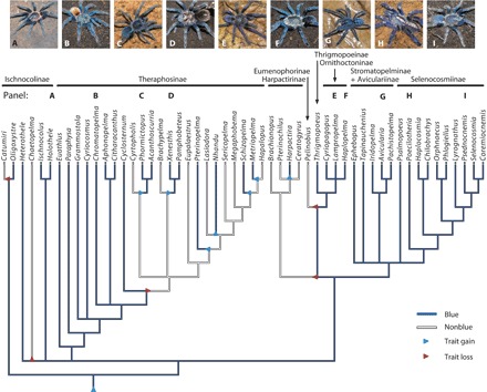
Slight shifts in arrangement within biological photonic nanostructures can produce large color differences, and sexual selection often leads to high color diversity in clades with structural colors. We use phylogenetic reconstruction, electron microscopy, spectrophotometry, and optical modeling to show an opposing pattern of nanostructural diversification accompanied by unusual conservation of blue color in tarantulas (Araneae: Theraphosidae). In contrast to other clades, blue coloration in phylogenetically distant tarantulas peaks within a narrow 20-nm region around 450 nm. Both quasi-ordered and multilayer nanostructures found in different tarantulas produce this blue color. Thus, even within monophyletic lineages, tarantulas have evolved strikingly similar blue coloration through divergent mechanisms. The poor color perception and lack of conspicuous display during courtship of tarantulas argue that these colors are not sexually selected. Therefore, our data contrast with sexual selection that typically produces a diverse array of colors with a single structural mechanism by showing that natural selection on structural color in tarantulas resulted in convergence on similar color through diverse structural mechanisms.
Keywords: Natural selection; Sexual selection; Structural color; multilayer; non-iridescence; quasi-order.
Figures


Similar articles
-
Modular color evolution facilitated by a complex nanostructure in birds.Evolution. 2015 Feb;69(2):357-67. doi: 10.1111/evo.12575. Epub 2015 Jan 16. Evolution. 2015. PMID: 25494613
-
The evolution of coloration and opsins in tarantulas.Proc Biol Sci. 2020 Sep 30;287(1935):20201688. doi: 10.1098/rspb.2020.1688. Epub 2020 Sep 23. Proc Biol Sci. 2020. PMID: 32962546 Free PMC article.
-
Nanostructural basis of rainbow-like iridescence in common bronzewing Phaps chalcoptera feathers.Opt Express. 2014 Jun 16;22(12):14625-36. doi: 10.1364/OE.22.014625. Opt Express. 2014. PMID: 24977558
-
A meta-analysis of butterfly structural colors: their color range, distribution and biological production.J Exp Biol. 2023 Nov 1;226(21):jeb245940. doi: 10.1242/jeb.245940. Epub 2023 Nov 8. J Exp Biol. 2023. PMID: 37937662 Review.
-
Gold bugs and beyond: a review of iridescence and structural colour mechanisms in beetles (Coleoptera).J R Soc Interface. 2009 Apr 6;6 Suppl 2(Suppl 2):S165-84. doi: 10.1098/rsif.2008.0354.focus. Epub 2008 Oct 28. J R Soc Interface. 2009. PMID: 18957361 Free PMC article. Review.
Cited by
-
Intermediate filaments spatially organize intracellular nanostructures to produce the bright structural blue of ribbontail stingrays across ontogeny.Front Cell Dev Biol. 2024 Jul 10;12:1393237. doi: 10.3389/fcell.2024.1393237. eCollection 2024. Front Cell Dev Biol. 2024. PMID: 39050893 Free PMC article.
-
Iridescence-controlled and flexibly tunable retroreflective structural color film for smart displays.Sci Adv. 2019 Aug 9;5(8):eaaw8755. doi: 10.1126/sciadv.aaw8755. eCollection 2019 Aug. Sci Adv. 2019. PMID: 31448332 Free PMC article.
-
A combination of red structural and pigmentary coloration in the eyespot of a copepod.J R Soc Interface. 2022 May;19(190):20220169. doi: 10.1098/rsif.2022.0169. Epub 2022 May 25. J R Soc Interface. 2022. PMID: 35611618 Free PMC article.
-
Artificial chameleon skin that controls spectral radiation: Development of Chameleon Cool Coating (C3).Sci Rep. 2018 Jan 19;8(1):1196. doi: 10.1038/s41598-018-19498-5. Sci Rep. 2018. PMID: 29352222 Free PMC article.
-
A remarkable new species of Paraparatrechina Donisthorpe (1947) (Hymenoptera, Formicidae, Formicinae) from the Eastern Himalayas, India.Zookeys. 2024 May 30;1203:159-172. doi: 10.3897/zookeys.1203.114168. eCollection 2024. Zookeys. 2024. PMID: 38855795 Free PMC article.
References
-
- D. L. Fox, Animal Biochromes and Structural Colours (Univ. of California Press, Berkeley, CA, ed. 2, 1976).
-
- Sun J., Bhushan B., Tong J., Structural coloration in nature. RSC Adv. 3, 14862–14889 (2013).
-
- Endler J. A., Variation in the appearance of guppy color patterns to guppies and their predators under different visual conditions. Vision Res. 31, 587–608 (1991). - PubMed
-
- Richards-Zawacki C. L., Cummings M. E., Intraspecific reproductive character displacement in a polymorphic poison dart frog, Dendrobates pumilio. Evolution 65, 259–267 (2010). - PubMed
LinkOut - more resources
Full Text Sources
Other Literature Sources
